


Organ-specific bioelectronics for soft tissues
 0
0 
Abstract
Organ function relies on dynamic electrical and electrochemical signaling that governs processes ranging from cardiac conduction and neural activity to gastrointestinal (GI) regulation and endocrine communication. Bioelectronic devices have demonstrated clinical impact in applications such as cardiac pacing, cochlear implants, retinal prostheses, and continuous glucose monitoring. However, when deployed on soft, wet, and continuously moving organs, the long-term stability of the device–tissue interface becomes a key challenge due to mechanical mismatch, biofouling, and degradation in physiological environments. Increasing evidence suggests that universal device architectures are insufficient for reliable long-term operation across organs with distinct mechanical, biochemical, and immunological microenvironments. Organ-specific bioelectronics has therefore emerged as a design paradigm in which materials, device structures, and system architectures are co-optimized according to the deformation modes, chemical conditions, and biological responses of individual tissues. Recent advances include ultracompliant neural interfaces that minimize inflammatory responses, GI resident devices capable of operating under strong peristalsis and chemical exposure, stretchable epidermal electronics that seamlessly integrate with skin mechanics, and epicardial or renal surface patches for monitoring visceral organs. This review summarizes recent developments in organ-specific bioelectronics from integrated perspectives of materials, device structures, and biological systems. Key material platforms, fabrication strategies, and representative applications are highlighted, followed by discussion of challenges in long-term biostability, scalable manufacturing, wireless power and data communication, and clinical translation, as well as future opportunities for organ-mimetic electronic interfaces enabling continuous monitoring and therapeutic modulation.
Keywords
INTRODUCTION
Organ function maintenance and regulation rely on the dynamic variations of electrical signals and electrochemical processes[1-3]. From cardiac conduction to central and peripheral neural activity, and to the gut-brain axis and related endocrine regulation, long-term recording and quantitative analysis of these signals are of major importance for continuous monitoring and intervention[4-6]. Over the past decades, bioelectronic devices have demonstrated clinical value in multiple settings, such as cardiac pacing, cochlear and retinal prostheses, and continuous glucose monitoring[7-9]. These practices indicate that long-term coupling between electronic systems and physiological signals is achievable from an engineering standpoint and have further propelled the development of next-generation implantable and attachable platforms that interface more intimately with organ surfaces.
When devices are deployed onto soft, wet, and continuously moving organ surfaces, the key factor limiting performance and operational lifetime often shifts from the circuitry itself to the long-term stability of the device–tissue interface[10]. Rigid or semi-rigid structures, together with relatively thick encapsulation and interconnects, can mismatch soft tissues in modulus, curvature, and dynamic deformation, thereby leading to electrode delamination, interconnect fatigue failure, and concomitant fluctuations of interfacial impedance with degraded signal quality. Meanwhile, water and ion penetration from body fluids may cause degradation of insulating performance and electrochemical corrosion[11]. Interfacial processes such as protein adsorption and cell adhesion further accelerate biofouling and drive a progressive increase in impedance over time, ultimately manifesting as reduced signal-to-noise ratio (SNR), baseline drift, and time-dependent changes in stimulation threshold[12,13].
Furthermore, the microenvironment varies substantially across organs, implying that interfacial constraints are not uniform. Brain tissue is extremely soft and highly sensitive to immune responses, where even subtle micromotion can trigger tissue injury and glial activation[14]. The gastrointestinal (GI) tract experiences vigorous peristalsis and is continuously exposed to acidic and enzymatic fluids, imposing stringent requirements on encapsulation durability and adhesive stability. Skin interfaces undergo persistent stretching, shear, sweating, and repeated attach–detach cycles. Visceral organs such as the heart, liver, and kidney reside in fluid-rich environments and experience cyclic strain or perfusion-related mechanical fluctuations[15]. Under these conditions, a universal material and structural design is often unable to simultaneously satisfy conformal stability, reliable encapsulation, and low-impedance coupling, which has driven increasing interest in organ-specific bioelectronics strategies.
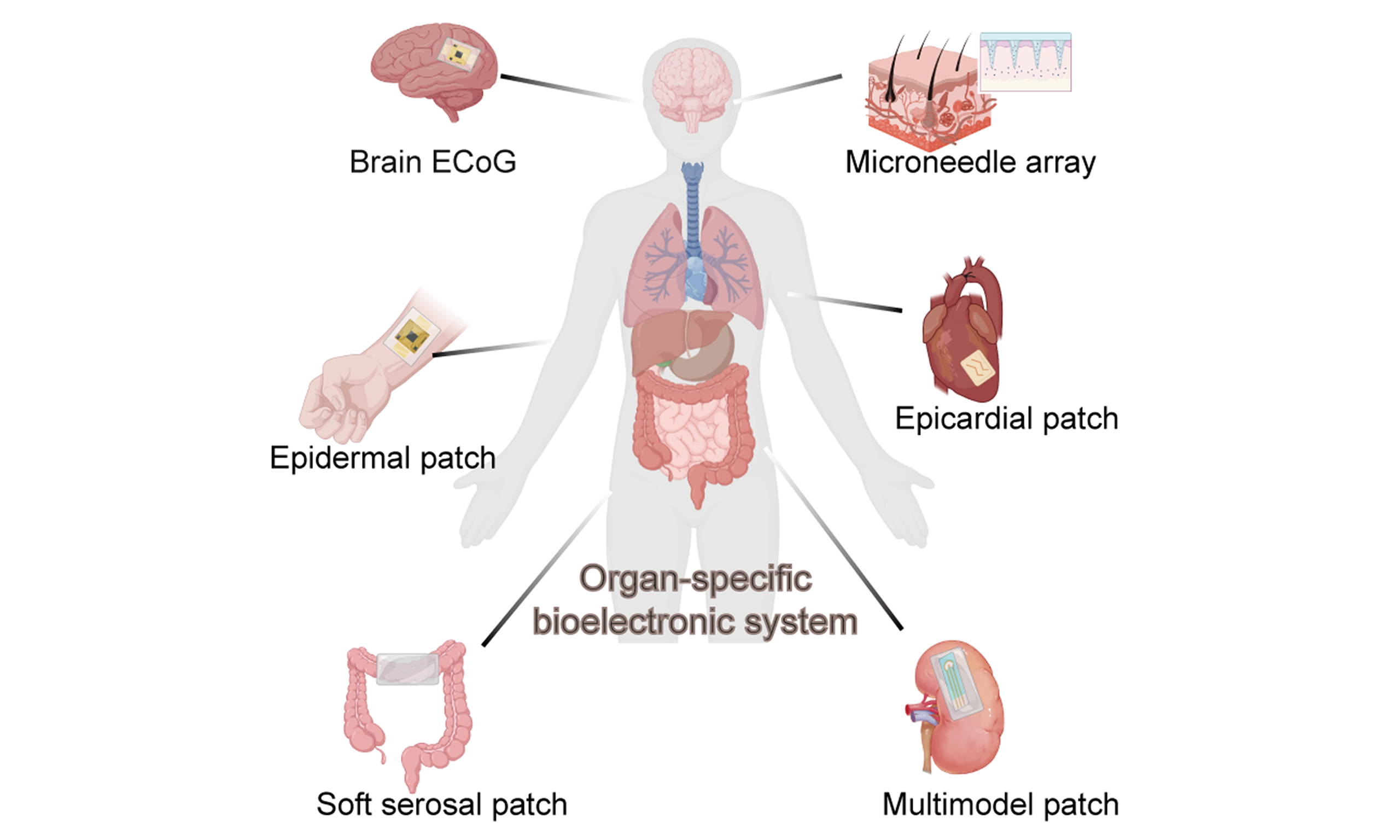
Organ-specific bioelectronics emphasizes defining design boundary conditions based on the deformation modes, chemical environment, and immune/biofouling behaviors of the target organ, and then co-optimizing the material system, structural form factor, and interfacial chemistry to achieve predictable long-term coupling stability[16]. Representative advances in recent years include ultra compliant neural interfaces that mitigate chronic inflammation and micromotion-induced damage, GI resident platforms capable of operating under weeks of peristalsis and chemical challenge, and stretchable epidermal patches that laminate onto skin with minimal mechanical loading[10,17] [Figure 1]. Despite differences in target applications, these systems exhibit a degree of commonality in materials and manufacturing, including ultrathin polymer films with multilayer encapsulation, elastomers combined with strain-relief geometric interconnects, hydrogel or mixed ionic–electronic conductor interfaces, as well as biodegradable supports and antifouling surface modifications[18].
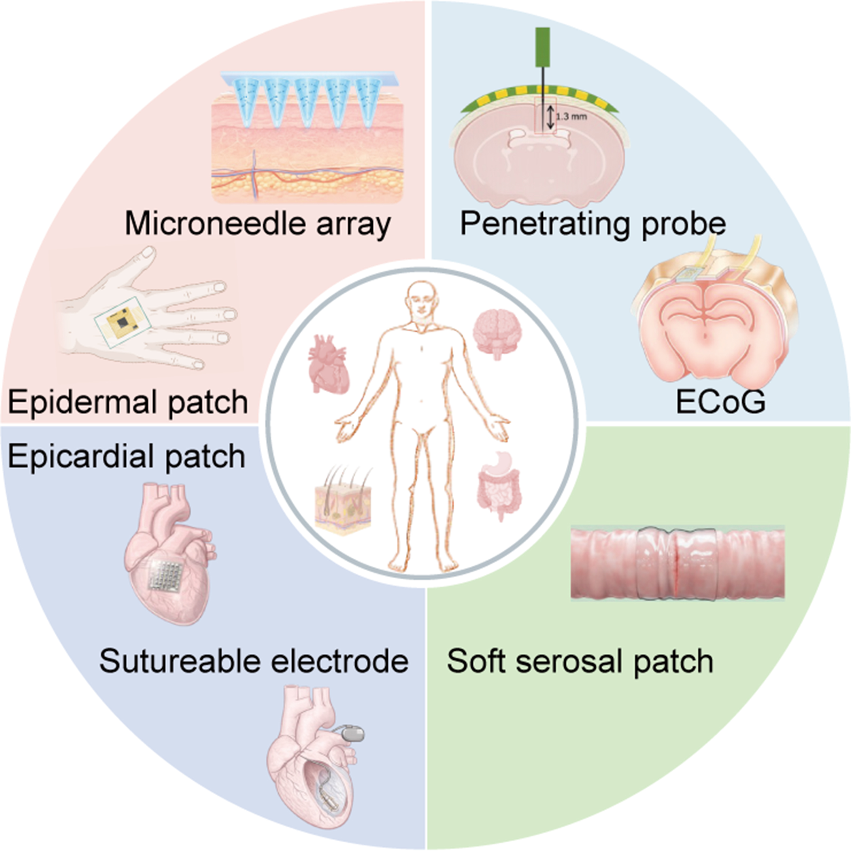
Figure 1. Representative organ-specific bioelectronic interfaces. Some schematic elements Created in BioRender. Liu, X. (2026) https://BioRender.com/w5rveah. ECoG: Electrocorticography.
In this review, we adopt a material–structure–biosystem co-design perspective to summarize structural/encapsulation substrates and conductive/interfacial materials for diverse organ interfaces and the associated key performance trade-offs, further survey relevant fabrication and integration routes and representative organ-targeted applications, and discuss open challenges in long-term stability evaluation, manufacturability, power and data links, and clinical translation[19].
MATERIALS FOR ORGAN-SPECIFIC BIOELECTRONICS
Structural and encapsulation substrates
Bioelectronic devices designed for soft-tissue organ interfaces must provide sufficient mechanical support and stress buffering to maintain stable electrical connections under bending and stretching, while also forming reliable chemical and moisture barriers that prevent water and ion penetration and protect internal circuits. In addition, minimizing inflammation and biofouling is essential for maintaining long-term biocompatibility during implantation or wearable use. A key design consideration is the mechanical matching between device substrates and target organs, since biological tissues span a wide range of Young’s moduli, ranging from a few kilopascals in the brain to megapascal levels in the epidermis and tendons, whereas commonly used bioelectronic materials include soft hydrogels and elastomers as well as high-modulus polymer films [Figure 2][20-23]. In this section, we classify the relevant material systems into four categories: non-degradable substrate–encapsulation systems, elastomeric and stretchable platforms, biodegradable and bioresorbable structural layers, and antifouling interfacial coatings, and summarize their key characteristics and representative organ-specific applications.
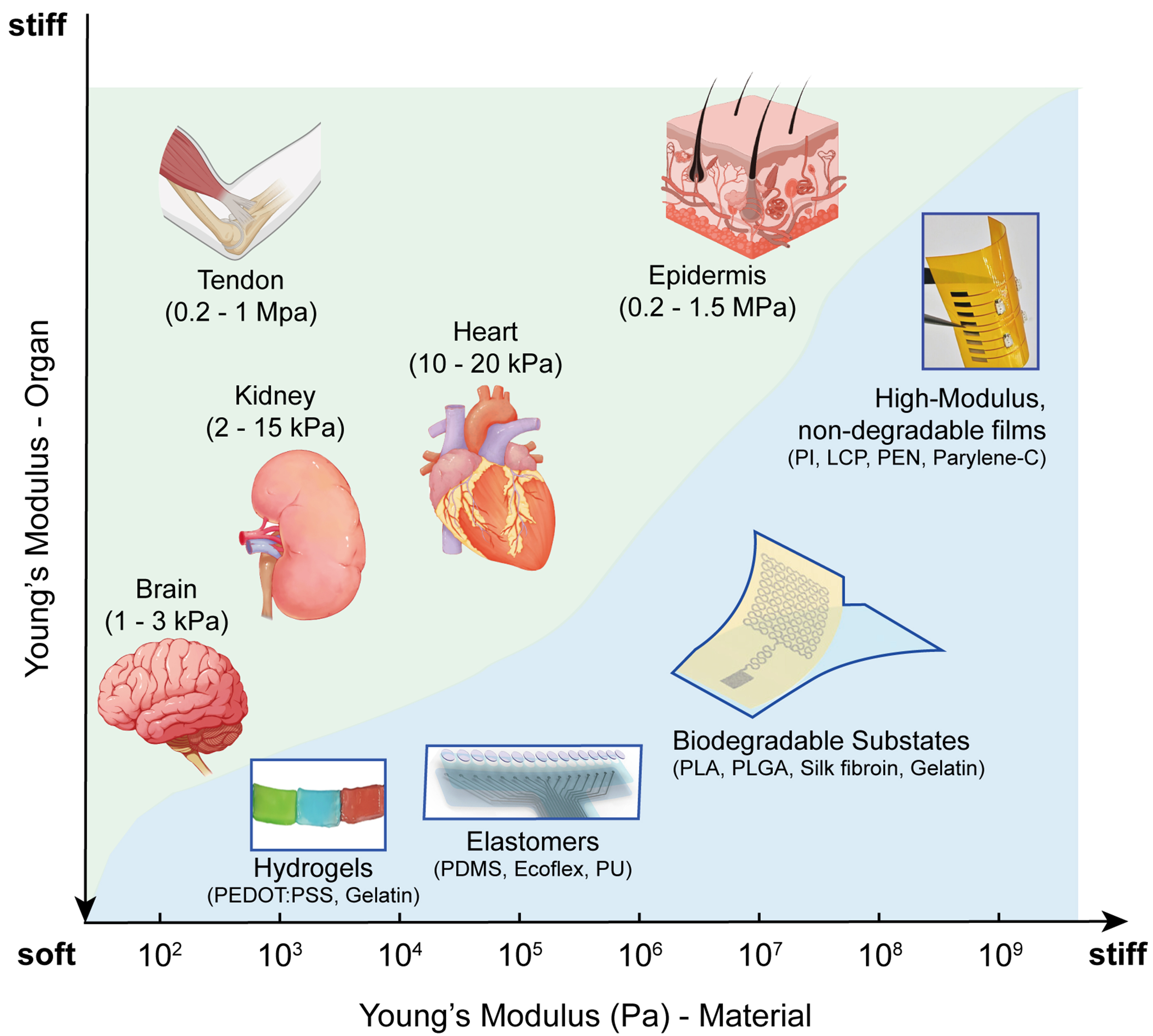
Figure 2. Mechanical matching between bioelectronic substrates and biological organs. Some elements were reproduced with permission, including hydrogels[24], Copyright © 2024, the American Association for the Advancement of Science; elastomers[21], Copyright © 2025, the American Association for the Advancement of Science; biodegradable substates[22], Copyright © 2025, WILEY-VCH Verlag GmbH & Co. KGaA, Weinheim; high-modulus, no-degradable films[25], Copyright © 2020, WILEY-VCH Verlag GmbH & Co. KGaA, Weinheim. Some schematic elements Created in BioRender. Liu, X. (2026) https://BioRender.com/tcvk8fu. PI: Polyimide; LCP: liquid crystal polymer; PEN: polyethylene naphthalate; PEDOT:PSS: poly(3,4-ethylenedioxythiophene):poly(styrenesulfonate); PDMS: polydimethylsiloxane; PU: polyurethane; PLA: polylactic acid; PLGA: poly(lactic-co-glycolic acid).
Non-degradable substrate–encapsulation systems
At organ interfaces that require high-density electrode arrays and long-term stable recording and stimulation, the structural layer must both support precise micro- and nanopatterns and maintain its geometry and mechanical properties over years[26]. To meet these demands, a class of high-modulus, non-degradable polymer films has become the standard structural material set, including Parylene-C, polyimide (PI), polyethylene naphthalate (PEN), and liquid crystal polymers (LCPs) [Figure 3A][27-31]. These materials typically exhibit Young’s moduli in the gigapascal range, far above those of soft tissues, but when the film thickness is reduced from tens of micrometres to a few micrometres or even sub-micrometre levels, their bending stiffness decreases substantially[32]. This thickness scaling allows them to conform to curved organ surfaces such as the cerebral cortex, retina, or peripheral nerves without pronounced wrinkling or structural instability, while still preserving in-plane dimensional stability and pattern fidelity.
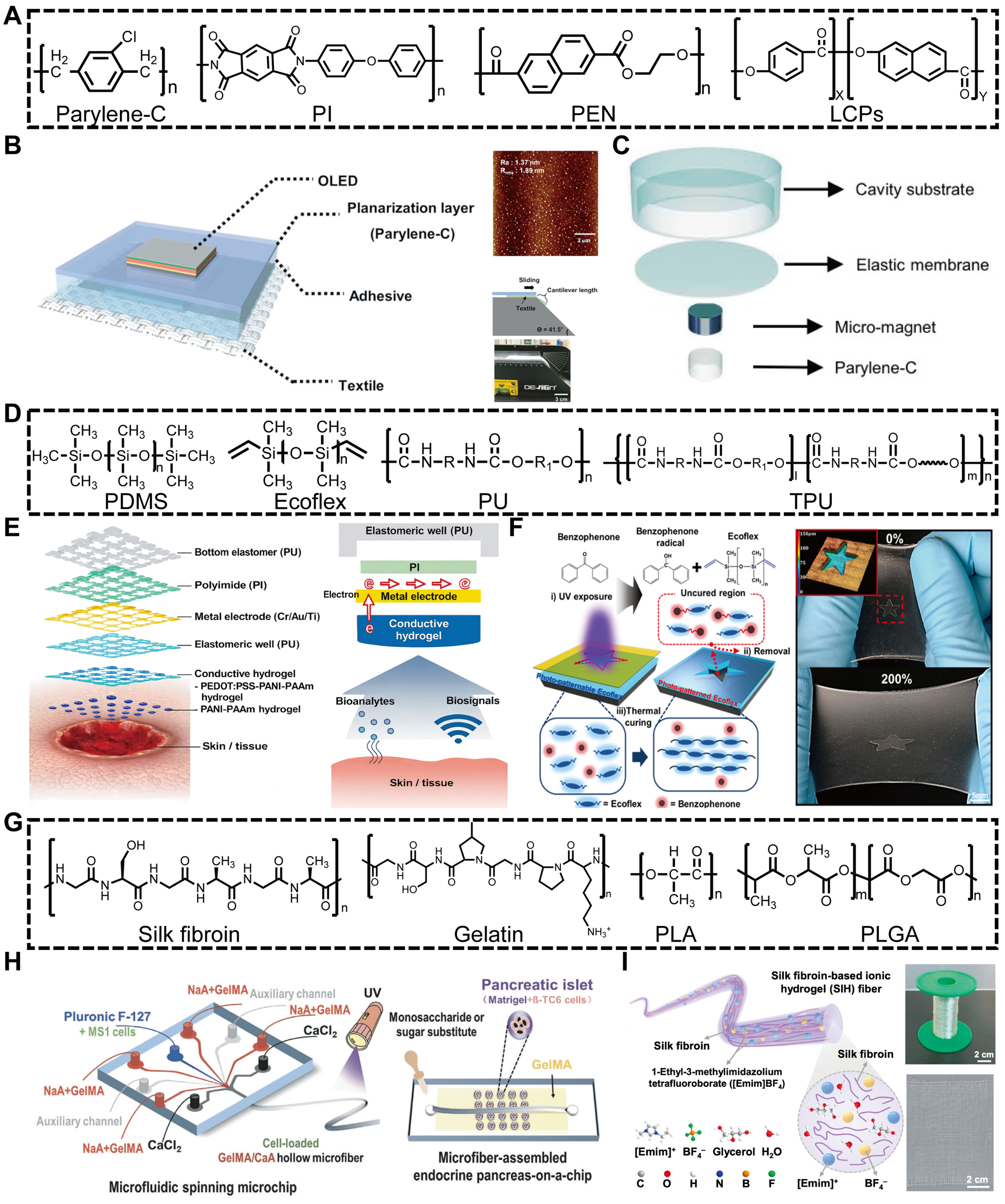
Figure 3. Representative material platforms for structural and encapsulation layers in organ-specific bioelectronics. (A) Representative non-degradable substrate and encapsulation materials; (B) Parylene-C-based planarization and device integration for flexible electronics[35]. Copyright © 2025, published by Springer Nature; (C) Parylene-C-based magnetic implant for wireless sensing[36]. Copyright © 2024, the American Association for the Advancement of Science; (D) Representative elastomeric and stretchable structural materials; (E) Stretchable bioelectronics with conductive hydrogels for low-impedance tissue interfaces[43]. Copyright © 2024, the American Association for the Advancement of Science; (F) Photo-patternable elastomeric substrates based on Ecoflex for stretchable bioelectronics[50]. Copyright © 2023, published by Springer Nature; (G) Representative biodegradable and bioresorbable structural materials. Created by the authors; (H) GelMA/CaA hollow microfiber assemblies as gelatin-derived biodegradable hydrogel microsystem[64]. Copyright © 2024, WILEY-VCH Verlag GmbH & Co. KGaA; (I) Silk fibroin-based ionic hydrogel fibers as biodegradable structural and conductive materials[56]. Copyright © 2024, published by Springer Nature. PI: Polyimide; PEN: polyethylene naphthalate; LCPs: liquid crystal polymers; OLED: organic light-emitting diode; PDMS: polydimethylsiloxane; PU: polyurethane; TPU: thermoplastic polyurethane; UV: ultraviolet; PLA: polylactic acid; PLGA: poly(lactic-co-glycolic acid).
These polymers are highly compatible with established microelectronic fabrication processes and can accommodate standard steps such as photolithography, metal interconnect patterning, and multilayer via formation. As a result, they are widely used as structural substrates for microelectrocorticography (μECoG) arrays, retinal prostheses, and cuff electrodes targeting the auditory and peripheral nerves. Ultrathin parylene-C films can further serve as transferable and biocompatible carrier substrates because of their excellent conformability, low-temperature processability, and water resistance[33,34]. For example, Cho et al. developed a textile-integrated OLED platform in which a thin parylene-C film was first formed on a guide glass and then transferred onto textile after thermal annealing, serving as a self-supporting planarization layer that replicated the smooth glass surface while preserving textile flexibility. This strategy enabled stable device fabrication and operation on rough, deformable textile substrates [Figure 3B][35]. At the same time, these materials provide good electrical insulation and moisture barrier performance, and can therefore serve as the primary encapsulation layer of the device. For example, parylene-C deposited by chemical vapor deposition can form dense, conformal, and nearly pinhole-free coatings with stable dielectric properties, making it one of the most widely used insulating and encapsulation materials for implantable neural electrodes and cardiac pacing leads [Figure 3C][36]. LCPs combine low water vapor transmission with the ability to be thermoformed and laminated, making them suitable for constructing hybrid rigid–flex encapsulation shells in intracranial or cochlear implants where higher mechanical robustness is required[37].
Overall, non-degradable high-modulus films represented by Parylene-C, PI, PEN and LCPs remain the predominant choice for organ interfaces that experience relatively small surface strain but demand high spatial resolution and long-term electrical stability, such as the cerebral cortex, retina and peripheral nerves[38-40]. Looking ahead, future efforts are likely to focus on retaining the mature processability and encapsulation reliability of these materials while introducing softer interlayers or biohybrid architectures to improve tissue integration. For example, Shi et al. developed a biohybrid multilayer interface combining PI, Au, SU-8, and living hydrogel layers to improve tissue-level compliance and interfacial integration with soft biological tissues[41].
Elastomeric and stretchable platforms
For highly deformable organs and tissues such as the skin, heart, and diaphragm, which experience substantial, multidirectional deformation during routine motion, an overly rigid structural layer can readily lead to electrode delamination, interconnect failure, or localized tissue compression. To address this, low-modulus elastomeric substrates based on polydimethylsiloxane (PDMS), Ecoflex, polyurethane elastomers (PU) and thermoplastic polyurethane (TPU) have become widely used at high-deformation organ interfaces [Figure 3D][42]. These materials combine flexible polymer backbones with relatively low crosslinking density, allowing their Young’s modulus to be tuned into the 103-107 Pa range, while sustaining 50%-200% tensile strain without plastic damage. Shin et al. reported a stretchable multichannel sensor array, illustrating how mechanically compliant elastomeric substrates can be incorporated into bioelectronic systems to enable stable impedance and pH mapping under both static and dynamic conditions [Figure 3E][43]. This tissue-like mechanical compliance allows the device to conform and move with skin, cardiac, and diaphragmatic tissues during vigorous motion, respiration, and heartbeat, thereby reducing stiffness mismatch and improving interfacial stability.
With these substrates, geometric layout further determines the effective operating range and failure mode of stretchable platforms under high strain. A widely adopted strategy is to confine rigid or semi-rigid functional units to local low-strain zones and connect them to the surrounding elastomer via deformable interconnects. Representative strategies also exploit interfacial engineering between conductive layers and elastomeric or fibrous substrates. For example, conductive coatings can be stabilized through molecular locking, pre-strain-induced wrinkling, and confined interpenetrating nanofiber networks, thereby maintaining electrical continuity and mechanical compliance under repeated deformation[44]. Such strain-engineered architectures enable graded dissipation of mechanical deformation around rigid functional components, thereby maintaining electrical continuity and signal stability during repeated stretching and release. At the material level, such interconnects are typically realized using metal thin films, metal meshes or embedded liquid-metal channels, all encapsulated within PDMS, Ecoflex or PU matrices[45-47]. By tailoring interconnect geometry, including interconnect length, linewidth, and radius of curvature, externally applied strain can be redistributed into bending, twisting, and local rotation of the interconnects, thereby reducing strain concentration in functional electrode and chip regions. This strain-isolation strategy helps preserve interfacial contact, contact impedance, and signal amplitude during prolonged body motion or repeated organ contraction[48,49].
From the standpoint of encapsulation and interfacial stability, elastomeric platforms are usually realized as a multilayer system, in which an elastic substrate is combined with an elastic encapsulation layer and tailored surface modification. Kim et al. developed a photo-patternable Ecoflex encapsulation strategy that enables process-compatible formation of patterned elastic encapsulation layers with multi-windows, thereby improving strain dissipation, electrical stability, and selective multi-analyte sensing in intrinsically stretchable wearable bioelectronics [Figure 3F][50]. The internal conductors and electrodes are first fully embedded in PDMS, PU or TPU to provide electrical insulation and to reduce stress concentrations at geometric discontinuities[20,51,52]. For devices that are worn on the skin or exposed to sweat and body fluids, an additional thin fluoropolymer overlayer, such as polytetrafluoroethylene (PTFE), can be applied to the outer surface to reduce water and ion permeation while maintaining sufficient water vapor transmission[53-55]. For devices adhering to the epicardium or diaphragm, the thickness and modulus of the encapsulation layer must also be balanced against the frictional interaction with pericardium or pleura, so as to avoid excessive shear during cardiac or respiratory cycles.
Biodegradable and bioresorbable structural layers
In many short-term or single-use implant applications, the ideal interface material should gradually degrade and be resorbed in vivo once monitoring or therapy is complete, thereby avoiding secondary removal surgery and reducing long-term foreign body reactions. With this goal in mind, protein-based materials such as silk fibroin and gelatin, together with synthetic polyesters such as polylactic acid (PLA) and poly(lactic-co-glycolic acid) (PLGA), have become representative systems for biodegradable structural layers [Figure 3G][56]. Protein materials are degraded enzymatically through cleavage of amide bonds, whereas polyesters undergo hydrolysis of ester bonds to generate lactic acid, glycolic acid and other small molecules that subsequently enter normal metabolic pathways and are cleared[57]. By pre-embedding such cleavable chemical bonds in the polymer backbone, these materials can be programmed to gradually lose mechanical integrity and disappear within a defined time window under the combined action of water and enzymes, providing the chemical foundation for fully resorbable support and encapsulation layers[58].
Accordingly, the central design challenge for biodegradable structural layers is to coordinate mechanical support lifetime, electrical stability and degradation behaviour within an appropriate range[59]. For PLA and PLGA, effective support time can be extended from several weeks to several months by tuning crystallinity, molecular weight and the copolymer ratio of lactic to glycolic acid[60]. Higher crystallinity and molecular weight slow water ingress and backbone scission, making them suitable for medium-term cardiac or neural stimulators, whereas a higher glycolic acid content usually accelerates hydrolysis and is better suited to postoperative short-term monitoring patches or temporary electrodes[61,62]. Silk fibroin and gelatin can be adjusted in an analogous way by varying β-sheet content, crosslinking density or integration with inorganic fillers, allowing stiffness and degradation rate to be tuned while maintaining good cell compatibility[63]. Beyond conventional films and bulk scaffolds, biodegradable protein matrices can also be engineered into hollow microfiber assemblies and organ-mimetic microsystems. For example, Tian et al. developed a microfiber-assembled endocrine pancreas-on-a-chip based on microfluidically spun GelMA/CaA hollow fibers, in which the biodegradable hollow microfibers were used to mimic vascular lumens and support material transport, and were integrated with a 3D pancreatic islet culture layer for islet culture and functional evaluation [Figure 3H][64]. Silk fibroin-based polymers can also be engineered into mechanically robust and ionically conductive fibers by incorporating ionic liquid components. Lu et al. reported a silk fibroin-based ionic hydrogel fiber composed of silk fibroin, ionic liquid, and glycerol, which exhibited high strength, large extensibility, and stable ionic conductivity, thereby enabling deformable conductive fibers for wearable bioelectronic and human–machine interface applications [Figure 3I][56]. In addition to structural substrates, biodegradable matrices can also be integrated with transient sensing interfaces and soft conductive layers. For example, agar- and hydrogel-based ionic interfaces combined with thin metal electrodes can provide temporary skin-compatible sensing platforms with minimized long-term foreign-body burden after use[65]. Another approach integrates ultrathin bioresorbable inorganic oxides as transient insulating or encapsulation layers and hydrolyzable conductive polymers as temporary charge-transport components, enabling the required electrical functions to be maintained over a prescribed time frame before gradual resorption[66,67].
At the level of organ interfaces, biodegradable and bioresorbable structural layers are particularly attractive in three types of scenarios. The first includes the heart and peripheral nerves in the postoperative or acute phase, where temporary electrophysiological interventions such as transient pacing, vagus nerve modulation or post-surgical rhythm monitoring are required. The second concerns wound beds, graft sites or remodelling soft tissues, where staged mechanical and electrophysiological monitoring or local electrical stimulation is needed during healing, but complete device disappearance is desired once tissue repair is complete[68]. The third involves patient groups for whom device retrieval is difficult or undesirable, such as neonates or very elderly individuals with multiple comorbidities. Recent work has begun to introduce dynamic crosslinks, inorganic fillers and multilayer gradient architectures into biodegradable frameworks, more tightly coupling mechanical support, electrical stability and controlled resorption[69]. These strategies aim to align the functional lifetime of the structural layer more precisely with organ healing timelines, while reducing issues such as swelling during degradation, fragment migration and local irritation, thereby opening broader materials and structural design space for single-use implants and resorbable physiological monitoring systems.
Interfacial coatings for antifouling control
In real physiological environments, once a device surface is exposed to plasma, cerebrospinal fluid or intestinal fluid, plasma proteins, inflammatory cells and microbes begin to adsorb and accumulate within a very short time[70]. This rapidly triggers inflammatory reactions, thrombosis and fibrotic encapsulation, which ultimately manifest as increased electrode impedance, reduced signal amplitude and, in severe cases, complete device failure[71,72]. Consequently, beyond the structural and encapsulation layers, a thin outermost interfacial coating that directly contacts body fluids and is engineered to resist protein adsorption, cell adhesion and even bacterial colonization has become a critical element for achieving long-term stable organ–device interfaces[73].
One of the earliest systematically explored strategies is the construction of a hydrated, hydrophilic layer. A representative approach is to graft or coat polyethylene glycol (PEG) or related hydrophilic polymers onto the device surface so that, in aqueous environments, a dense, nanometre-scale hydration layer is formed[74,75]. This hydrated shell introduces steric hindrance and a high energetic penalty for dehydration, thereby suppressing the residence of proteins and cells at the interface[76]. Because PEG is susceptible to oxidation and hydrolysis under long-term implantation conditions, recent work has increasingly shifted toward zwitterionic polymer systems, such as coatings bearing sulfobetaine or carboxybetaine side chains[77]. These materials carry both positive and negative charges on the same side chain, enabling the formation of more stable and tightly bound hydration shells. As a result, they exhibit more durable resistance to protein adsorption and cell adhesion in complex media such as plasma and cerebrospinal fluid, and have been applied to the surface modification of neural electrodes, vascular stents and catheters, where animal studies have shown reduced inflammation and attenuated glial scarring.
A second line of development draws inspiration from naturally “slippery” interfaces such as those of pitcher plants, through the creation of slippery liquid-infused porous surfaces (SLIPS)[78]. In these systems, a porous fluoropolymer or micro/nano-rough substrate serves as a scaffold in which a thin layer of medical lubricant is retained within pores or topographical features, forming a continuous, stable and self-healing liquid interface that offers virtually no effective anchoring sites for bacteria, proteins or cells. Multiple animal models have demonstrated that SLIPS coatings can markedly suppress bacterial biofilm formation and fibrotic encapsulation, making them attractive for high-contamination-risk devices such as urinary catheters, vascular implants and GI systems. Unlike hydrophilic coatings that rely primarily on chemical repulsion, SLIPS function mainly through physical dewetting and re-spreading of the infused lubricant, allowing damaged or abraded regions to recover their antifouling properties from the lubricant reservoir.
For brain, retinal and peripheral nerve electrodes, interfacial coatings must not only mitigate inflammation and glial scar formation but also preserve low impedance and ionic transport[79]. In intravascular or intestinal settings, additional requirements include antithrombotic and antibacterial performance and compatibility with fast-flowing fluids. Rationally matching these interfacial coatings with the non-degradable, elastic or biodegradable structural–encapsulation systems discussed above offers a route to constructing integrated interfaces that combine mechanical matching, chemical stability and biological tolerance across diverse organ environments, thereby providing a robust foundation for the long-term reliable operation of conductive layers and active electronic components[80].
Conductive layers
Conductive layers form the functional core of bioelectronic devices, enabling the conversion and transmission of electrical and ionic signals. Unlike the substrate layer, where a small number of materials can fulfill mechanical support roles, the conductive layer encompasses a diverse family of materials with fundamentally different conduction mechanisms and microstructures[81,82]. To capture this diversity and its relevance to device design, this section is organized by material families: organic mixed ionic-electronic conductors (OMIECs), conductive hydrogels, liquid metals, and two-dimensional nanomaterials.
In addition to differences in softness and processability, conductive biointerface materials differ substantially in their quantitative transport properties. OMIECs and conductive polymer hydrogels rely on coupled ionic and electronic transport, whereas liquid metals provide primarily electronic, metal-level conductivity and ionic hydrogels mainly conduct through mobile ions[83]. These differences determine interfacial impedance, charge-injection capability, sensing bandwidth, and long-term stability at wet tissue interfaces. Therefore, representative conductivity values and mixed-conduction characteristics of major conductive-layer materials are summarized in Table 1.
Conductive materials for bioelectronic interfaces
| Material | Main conduction | Representative values | Biointerface relevance | Ref. |
| OMIECs | Electronic + ionic | Pristine PEDOT:PSS < 1 S·cm-1; hydrogels/composites up to 28-4,000 S·cm-1 | Low-Z recording; OECTs | [83-85] |
| Conductive hydrogels | Ionic/mixed | Ion-dependent; tunable by salts, polymers, fillers | Wet coupling; soft electrodes | [49,86] |
| Liquid metals | Electronic | ~106 S·m-1 level | Stretchable interconnects | [87,88] |
| 2D materials | Electronic | Material-dependent; high in-plane transport | Thin electrodes; high surface area | [89,90] |
OMIECs
OMIECs are soft polymeric materials that transport both electronic and ionic charges while remaining mechanically compatible with biological tissues[91,92]. They are typically built from polymers with π-conjugated backbones combined with hydrophilic side chains or polyelectrolyte components, giving moduli in the MPa range, good bendability, and compatibility with solution processing[93,94]. Unlike purely electronic organic semiconductors, OMIECs undergo bulk doping in aqueous electrolytes so that charge is stored throughout the material volume[83]. This volumetric electrochemical coupling is particularly relevant at soft organ interfaces, where tissue impedance is dynamic and signal amplitudes are often small, requiring stable, low-impedance charge exchange rather than purely interfacial capacitive coupling[43].
Their mixed conduction can be viewed as the cooperation of electronic and ionic pathways within a single network[85]. Electrons or holes move along the π-conjugated backbone through locally ordered domains, with conductivity governed by backbone structure and doping level[95,96]. For representative poly(3,4-ethylenedioxythiophene):poly(styrenesulfonate) (PEDOT:PSS)-based OMIECs, electronic conductivity can range from below 1 S·cm-1 in pristine or structurally disordered films to 102-103 S·cm-1 after secondary doping, acid treatment, or structural optimization. Ions migrate within hydrophilic side chains or polyelectrolyte phases and compensate the charges created on the backbone during oxidation or reduction. Because this electron–ion compensation occurs throughout the bulk rather than at a confined interface, OMIECs exhibit high volumetric capacitance, typically on the order of tens to hundreds of F/cm3 for PEDOT-based systems, enabling high organic electrochemical transistor (OECT) transconductance at low operating voltages as well as low-impedance, high-charge-injection electrode interfaces. Such characteristics are advantageous in organ-specific bioelectronic chips that must operate under strict electrochemical safety limits while maintaining efficient coupling to excitable tissues such as brain and myocardium. Recent work has shown that the electrochemical doping rate in OMIECs strongly depends on film microstructure, film morphology, and electrolyte environment, which in turn determine device response speed and operational stability[97,98]. These kinetic factors are particularly important in neural and cardiac electrophysiology, where millisecond-scale temporal resolution is required[83,99].
Recent studies further underscore the importance of electrochemical kinetics in OMIEC-based organ interfaces. Keene et al. showed that electrochemical doping in conjugated polymers can be hole-limited, highlighting that OMIEC response speed depends not only on ion transport but also on electronic charge propagation within the polymer phase[100]. For organ-specific bioelectronic chips, such behavior directly links material-level transport dynamics to system-level bandwidth and energy constraints. Beyond transport kinetics, recent material-design strategies have further expanded the utility of OMIECs in soft biointerfaces. Montazerian et al. showed that replacing conventional hydrophobic PSS dopants with hydrophilic biomacromolecular AlgS dopants in PEDOT:AlgS improves aqueous dispersibility, molecular degradability, and ionic integration with hydrogel matrices, thereby enabling injectable and 3D-printable OMIEC-based bioelectronics [Figure 4A][101]. Together, these findings shift the focus from purely material characterization toward mechanism-informed optimization of kinetics, stability, and energy efficiency in physiological environments.
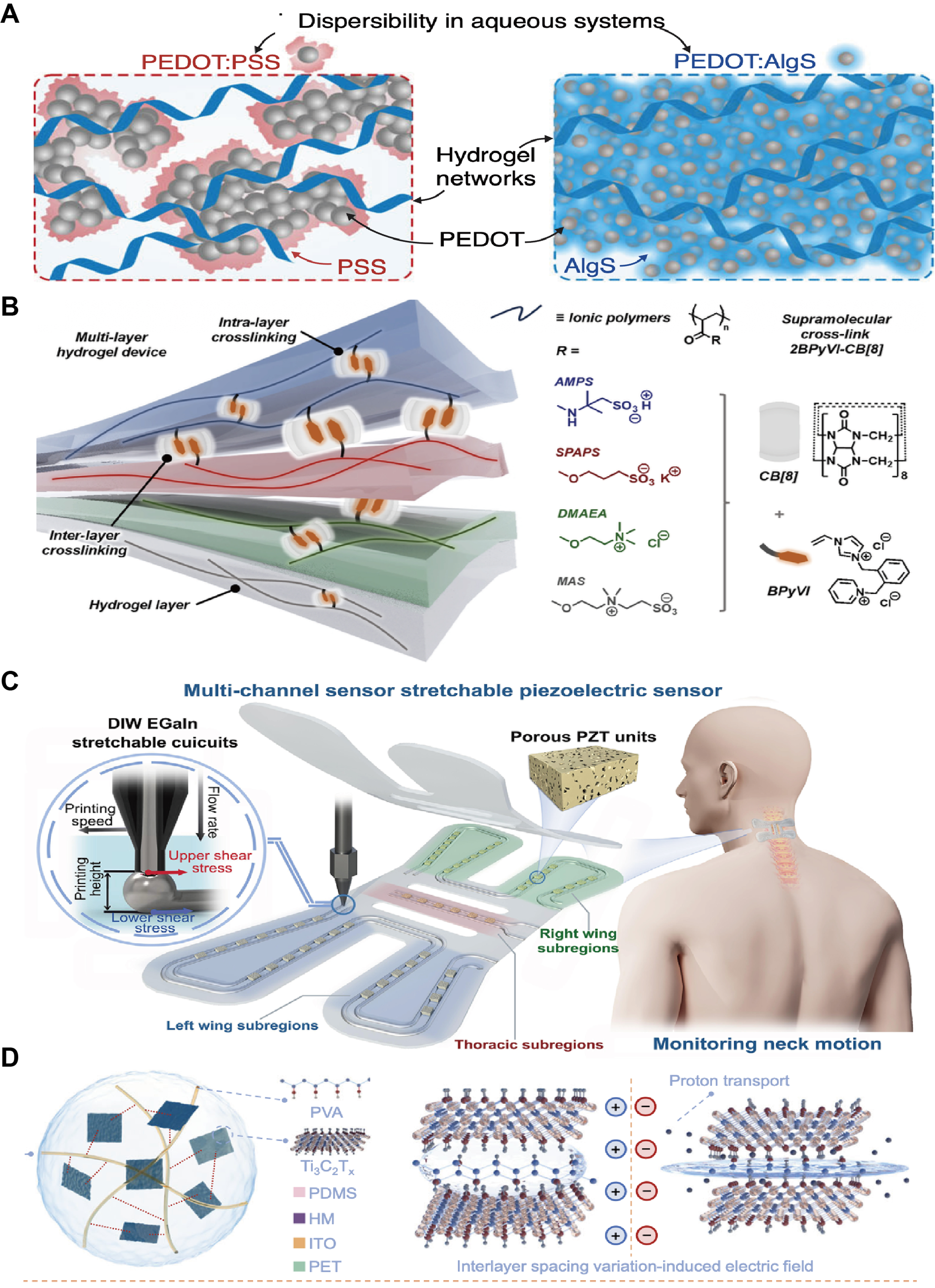
Figure 4. Representative material platforms for conductive and interfacial layers in organ-specific bioelectronics. (A) Hydrophilic-dopant-engineered PEDOT systems for degradable hydrogel bioelectronics[101]. Copyright © 2025, published by Springer Nature; (B) Supramolecular poly(ionic) networks enabling stretchable conductive hydrogels[24]. Copyright © 2024, the American Association for the Advancement of Science; (C) Topology-optimized stretchable piezoelectric sensors enabled by direct-ink-written liquid-metal circuits[128]. Copyright © 2026, WILEY-VCH Verlag GmbH & Co. KGaA; (D) Biomimetic microstructure-enabled piezoionic mechanoreceptors for ultrasensitive multimodal sensing and object recognition[150]. Copyright © 2025, published by Springer Nature. PEDOT: Poly(3,4-ethylenedioxythiophene); PSS: poly(styrenesulfonate); AMPS: 2-acrylamido-2-methyl-1-propanesulfonic acid; SPAPS: 3-sulfopropyl acrylate potassium salt; DMAEA: [2-(methacryloyloxy) ethyl] trimethylammonium chloride; MAS: [2-(methacryloyloxy)ethyl]dimethyl-(3-sulfopropyl)ammonium hydroxide; DIW: direct-ink-written; EGaIn: eutectic gallium–indium; PZT: porous lead zirconate titanate; PVA: polyvinyl alcohol; PDMS: polydimethylsiloxane; HM: hydrogel microneedles; ITO: indium tin oxide; PET: polyethylene terephthalate.
Overall, OMIECs represent a commonly adopted conductive-layer strategy in organ-specific bioelectronic chips, enabling low-impedance electrochemical coupling at wet, soft, and dynamically active tissue interfaces under low operating voltages[102,103]. Their mixed ionic–electronic transport characteristics make them particularly suitable for high-density neural interfaces in the brain and retina, as well as conformal sensing and stimulation devices deployed on mechanically deformable tissues such as myocardium and intestinal mucosa[102].
Conductive hydrogels
Conductive hydrogels are soft, water-rich polymer networks that are engineered to carry electrical signals while remaining mechanically similar to biological tissues. They are typically formed by three-dimensional crosslinking of natural or synthetic polymers such as polyacrylamide, alginate, or gelatin, which creates a stable, highly hydrated network[104]. By further introducing ionic or electronic conductive components into this matrix, conductive hydrogels can preserve softness and tissue affinity and at the same time support reliable electrical signal transmission[105-107].
Their electrical behavior is supported by two complementary mechanisms. First, the abundant water phase inside the hydrogel provides pathways for mobile ions, giving rise to ionic conduction[108,109]. Second, adding conductive elements such as metal nanowires, carbon nanotubes, graphene, or conducting polymers like PEDOT:PSS allows the formation of continuous electronic or mixed ion–electron pathways within the network[110-113]. Depending on the conductive filler, polymer composition, and percolation structure, conductive hydrogels can span several orders of magnitude in conductivity, from predominantly ion-conducting networks with conductivities typically in the 10-3-10-1 S·cm-1 range to electronically conductive composite or conductive-polymer hydrogels with conductivities reaching 1-102 S·cm-1 or higher[114]. Dynamic crosslinks based on hydrogen bonding, metal–ligand coordination, or catechol chemistry can break and reform during stretching, bending, or repeated loading. These reversible bonds help maintain the integrity of the conductive pathways and endow the material with self-healing and network reconfiguration[115-117]. Recent work has shown that supramolecular poly(ionic) networks can simultaneously achieve ionic conductivities up to 0.1 S·cm-1 and stretchabilities exceeding 1,500%, illustrating how dynamic crosslinking can reconcile efficient ionic transport with tissue-like deformability in multilayer conductive hydrogels [Figure 4B][24]. For example, metal-catechol coordination conductive hydrogels have been shown to recover both mechanical strength and electrical continuity after physical fracture, highlighting the role of dynamic bonding in sustaining cyclic stability[118]. Such dynamic bonding is particularly beneficial for organ interfaces subjected to cyclic deformation, such as myocardium and diaphragm.
Recent advances continue to expand the functional scope of conductive hydrogels toward environments and use-cases previously inaccessible to soft materials. One major direction focuses on improving environmental robustness, as exemplified by Zhang et al., who introduced a “hydro-locking” strategy that stabilizes water within double-network hydrogels, maintaining softness and conductivity from -115 to 143 °C for extreme-condition sensing[119]. Although originally demonstrated for extreme-condition sensing, such hydration-stabilization strategies may also improve long-term stability of implanted organ interfaces. A complementary direction integrates energy-harvesting and therapeutic functions, demonstrated by Xin et al., who developed a Fe2+/Fe3+-alginate thermogalvanic dressing capable of converting wound-site temperature gradients into therapeutic electrical stimulation while enabling real-time monitoring[120]. This multifunctional integration hints at future organ-resident patches capable of simultaneous sensing and localized therapy.
Overall, conductive hydrogels integrate softness, high water content, adhesion and mixed ionic-electronic transport, making them especially well-suited for electrochemical interfaces with the brain, heart and skeletal muscle, where intimate, low-modulus contact to wet, excitable tissue is essential[121]. As these materials evolve toward printable, adaptive and biodegradable architectures, they are expected to form a core platform for the next generation of implantable bioelectronics and soft robotic systems and to enable broader clinical and engineering applications[122].
Liquid metals and soft metal composites
Liquid metals and their soft composites form a class of conductors that combine metal-level electrical conductivity with fluid- or rubber-like mechanics[123]. In a narrow sense, liquid metals refer to metals or alloys that remain liquid near room temperature, most prominently gallium-based eutectic alloys such as eutectic gallium–indium (EGaIn) and Galinstan[124,125]. These alloys exhibit conductivities on the order of
Their functional behavior is governed by the interplay between metallic bonding and interfacial mechanics. Intrinsically, liquid metals are highly conductive fluids whose conductivity remains nearly unchanged under large mechanical deformation, allowing stable signal transmission under strain[126]. A nanometre-scale oxide skin and high surface tension stabilise discrete droplets and filaments and allow damaged traces to self-heal as the liquid flows and closes cracks. In soft metal composites, micron- or nanoscale droplets dispersed in PDMS, PU or hydrogels remain electrically insulated at rest; under stretching, compression or shear, the oxide shells rupture and neighbouring droplets fuse into percolating networks, producing stress-activated conductivity and inherent strain sensitivity. These features are particularly beneficial at organ interfaces subjected to continuous motion, such as skin, skeletal muscle and cardiovascular tissues, where conductive layers must tolerate repeated deformation without loss of conductivity[127]. For example, Zeng et al. showed that direct-ink-written EGaIn circuits can serve as highly stretchable electrodes in topology-optimized piezoelectric sensors for anisotropic motion monitoring, illustrating how liquid-metal interconnects enable precise patterning, stable conductivity under large deformation, and wearable bioelectronic integration [Figure 4C][128]. Beyond serving as passive fillers, gallium-based droplets can also initiate free-radical polymerisation and participate in crosslinking, as shown by Jaseem et al., who obtained tough, injectable and self-healing conductive hydrogels suitable for injectable electrodes and soft encapsulation layers on skin or muscle[129].
Building on these platforms, recent work has shifted from simply achieving conductivity and stretchability to ensuring stable operation at complex biological interfaces. A notable example is a magnetically reshapable three-dimensional liquid-metal multi-electrode array, in which EGaIn-filled deformable microtubes are folded into three-dimensional shapes under magnetic guidance and gently inserted into brain organoids for multichannel, deep electrophysiological recording and stimulation, highlighting the plasticity and compliance of liquid metals at three-dimensional brain and organoid interfaces[130]. Such demonstrations highlight the capacity of liquid metals to conform to soft, three-dimensional brain and heart while maintaining metallic-level conductivity, a property difficult to achieve with conventional rigid electrodes[131,132].
However, liquid-metal conductors also present important fabrication and stability challenges. Their high surface tension and fluidity make high-resolution and reproducible patterning difficult, while the spontaneously formed gallium oxide skin can both stabilize traces and alter wettability, adhesion, and contact resistance[133]. Poor wetting on polymer substrates may cause line retraction or discontinuous patterns, and leakage from microchannels or composite matrices under repeated deformation can compromise device reliability[134]. These issues become more pronounced in large-area or high-throughput fabrication, where ink rheology, substrate adhesion, encapsulation, and droplet stabilization must be precisely controlled[135]. Recent patterning and stabilization strategies, including surface modification, microfluidic confinement, transfer printing, direct ink writing, laser-assisted processing, and elastomer/hydrogel encapsulation, are therefore important for translating liquid-metal conductors into reliable organ-specific bioelectronic interfaces[136].
Overall, liquid metals and their soft composites combine metal-like conductivity, rubber-like softness and reconfigurable interfaces, providing a conductive-layer strategy centered on strain-invariant conductivity and mechanical robustness in highly deformable biological systems[137]. Within organ-specific bioelectronic chips, they are particularly suitable for interfaces that experience large or repetitive deformation, including epidermal and muscular electromyography (EMG) systems, cardiovascular monitoring devices, and emerging three-dimensional neural or organoid interfaces[138,139]. As encapsulation strategies, droplet stabilization methods, and biocompatible formulations continue to improve, liquid-metal-based conductors are expected to expand from wearable platforms toward more demanding implantable organ interfaces[140,141].
Two-dimensional nanomaterials
Two-dimensional conductors are atomically thin or few-layer materials in which charge carriers are largely confined to in-plane transport[142,143]. Representative systems include graphene and its derivatives, transition metal dichalcogenides (TMDs) and MXenes[144]. Compared with bulk conductors, these sheets offer very high specific surface area, tunable band structures and excellent flexibility: monolayer graphene combines high carrier mobility with nanometre-scale thickness, while MXenes provide hydrophilicity and easy dispersion through surface –O/–OH/–F terminations[145]. These characteristics enable 2D materials to act as ultrathin conductive layers that enhance interfacial conductivity and device density without significantly increasing the bending stiffness of soft, organ-facing substrates.
Their interfacial behaviour is governed by sp2-conjugated carbon networks or transition-metal d orbitals together with engineerable defects and surface groups[146,147]. Extended π or d bands support fast in-plane electronic transport, whereas edge sites, vacancies and terminations can be chemically tuned to adjust carrier density, Fermi level and charge-transfer kinetics. Reported conductivities vary widely depending on material type, flake quality, oxidation state, and film assembly; graphene-based and MXene films can reach conductivities from 103 to 105 S·m-1 or higher in optimized films, whereas oxidized or defect-rich derivatives show substantially lower values[19,148]. Unlike OMIECs, these materials do not usually exhibit bulk ionic–electronic mixed conduction, but their surface groups and electrolyte-accessible interfaces facilitate charge transfer and reduce electrode–tissue impedance[149]. For example, Ding et al. integrated MXene nanosheets into a flexible multilayer sensing architecture composed of hydrogel microneedles, PET/ITO films, and PDMS encapsulation, where the layered MXene morphology, stress-induced interlayer transport, and non-faradaic interfacial coupling enabled deformable multimodal bioelectronic sensing [Figure 4D][150]. When coated onto elastomers or hydrogels, 2D flakes form composite films that markedly reduce electrode–tissue impedance and maintain stable conduction under repeated bending and stretching, supporting long-term mechanical compatibility at dynamic tissue interfaces[104,151].
Beyond conductive 2D sheets, emerging two-dimensional polymers provide a distinct materials strategy for ultrathin encapsulation. For example, Ritt et al. showed that the molecularly impermeable 2D polyaramid 2DPA-1 can be processed into nanometre-thin, electrically insulating barrier films with exceptionally low gas permeability. A 60-nm 2DPA-1 coating markedly retarded MAPbI3 degradation by suppressing O2 and water-vapour permeation, highlighting the promise of such ultrathin 2D polymer membranes for conformal encapsulation[152]. This combination of conductive 2D layers with atomically thin barrier films offers a strategy for simultaneously improving electrical performance and protecting organ-resident devices from biofluid infiltration and molecular diffusion[153].
Overall, two-dimensional conductors combine atomic-scale thickness, high conductivity and tunable interfacial chemistry, providing a conductive-layer strategy centred on high spatial resolution, minimal mechanical footprint and chemically programmable interfaces[154,155]. In the brain and retina they can function as transparent, low-impedance electrodes and protective layers for high-fidelity recording and stimulation, whereas on the heart, skeletal muscle and intestinal mucosa, 2D conductor-elastomer or -hydrogel coatings can enhance electrical coupling and stability while minimally perturbing tissue mechanics[156]. Within organ-specific bioelectronic chips, 2D materials are particularly advantageous when high-density electrode integration and minimal structural intrusion are required, such as in retinal prostheses, cortical microarrays and conformal visceral patches[157].
Beyond mechanical and electrical matching, biocompatibility is a central design consideration in organ-specific bioelectronics[158,159]. This issue extends beyond acute cytotoxicity to include skin compatibility, foreign-body and immune responses, inflammatory remodeling, and long-term safety during chronic use[160]. These concerns depend strongly on the target organ, implantation mode, exposure duration, and local biochemical environment. Therefore, material selection should be evaluated not only by conductivity and mechanics, but also by the biological responses expected in each tissue context.
In addition to organ-specific constraints, bioelectronic materials must also be selected according to the intended modality. Recording, stimulation, biosensing, pressure sensing, temperature sensing, and delivery each impose distinct requirements for contact, impedance, charge injection, selectivity, stability, and transport. These modality-specific considerations are summarized in Table 2.
Application-specific material requirements for bioelectronic modalities
| Modality | Mechanical | Interfacial | Material | Benchmark | Devices | Ref. |
| Recording | Conformal | Low-Z | OMIECs; hydrogels | Low impedance; stable wet coupling | ECG/EEG/ECoG | [86,161] |
| Stimulation | Durable | High-CIC | PEDOT; Pt/Ir | High charge injection; electrochemical safety | Stimulator | [162-164] |
| Biosensing | Wet-operable | Selective | Enzymes; aptamers | Analyte permeability; antifouling | Sweat/ISF | [20,49,165,166] |
| Pressure | Stretchable | Low hysteresis | Elastomers; LMs | High deformability; cycling stability | E-skin | [134,167,168] |
| Temperature | Conformal | Thermally stable | Thin films | Stable thermal response | Patch | [169] |
| Delivery | Compliant | Controlled transport | Hydrogels; iontronics | Reservoir stability; biofluid compatibility | Pump/patch | [170,171] |
FABRICATION STRATEGIES FOR ORGAN-SPECIFIC BIOELECTRONICS
Developing appropriate fabrication strategies for organ-specific electrode interfaces is critical, as the fabrication process directly determines interfacial properties such as biocompatibility, stability, and functional performance[172]. Because living systems are highly sensitive to their microenvironment, bioelectronic interfaces should be designed and constructed to closely mimic the native physiological milieu of the target tissue. To ensure long-term stable operation, electrodes must achieve robust integration and conformal contact with the organ surface[173]. To enable efficient communication between bioelectronic devices and biological tissues, the fabrication method should allow precise control over interfacial structural features, including size, geometry, and spatial distribution. The manufacturing strategies should support high-resolution patterning, large-area fabrication, and 3D curved-surface structuring, while maintaining conformal contact with soft tissues[174-176]. In this part, we discuss key design considerations for constructing tissue-electrode interfaces by using stretchable conductive materials and summarize recent advances in several representative fabrication techniques.
High-resolution patterning
Organ-targeted bioelectronic interfaces commonly require micrometer-scale resolution and high-density electrode arrays to match fine anatomical structures and to support localized stimulation and recording at high spatial resolution[177]. In particular, closely spaced microelectrode grids can serve as epidermal EMG arrays capable of resolving subtle body movements with an interelectrode spacing below 500 μm, enabling discrimination of fine motor units and complex activation patterns[178]. At the organ surface, similar spatial precision is essential for mapping heterogeneous electrophysiological activity, targeting small functional units, and implementing multiplex sensing and stimulation. In this setting, high-resolution patterning is not only a question of achieving dense layouts, but also of defining sharp, well-controlled micro- and nanostructures on mechanically soft, curved, and often transient substrates that must operate stably in aqueous biological environments[179]. Meeting these requirements requires re-engineering conventional microelectronics processes, originally developed for rigid wafers, into hybrid fabrication workflows in which high-resolution patterning is decoupled from the final organ-matched mechanical support.
A common approach is to first pattern microscale structures on rigid donor wafers and then transfer them onto soft substrates such as elastomers, hydrogels, or bioresorbable polymers[179]. Standard complementary metal–oxide–semiconductor (CMOS)-compatible photolithography, e-beam lithography, and dry/wet etching are used to define metal interconnects, semiconductor membranes, and sensing elements at micrometer to sub-micrometer scales[180,181]. These ultrathin “islands” are then arranged into stretchable layouts and transferred onto compliant substrates by deterministic transfer printing or stamping[182]. Researchers have clarified the key materials and mechanics design principles needed to realize such devices, enabling stretchable circuits that retain the electrical performance of conventional silicon while matching the deformations of organs[183]. High-resolution transfer printing also allows light-emitting diodes (LEDs), OECTs, and micro-integrated circuits (micro-ICs), and various sensors to be integrated into conformal epicardial membranes, neural meshes, or epidermal patches without loss of lithographic precision.
Photolithographic microfabrication
Photolithography is a widely used microfabrication technique that provides excellent spatial resolution by using patterned light to transfer features onto thin-film substrates[184]. It is commonly applied to define metal electrodes and interconnects on polymeric substrates such as PI or SU-8, yielding flexible microelectrode arrays[185,186]. This strategy has been used to fabricate high-density arrays for neural probes, retinal implants, and other interfaces, with feature sizes on the order of single cells and finely controlled electrode spacing, allowing highly localized interaction with biological tissues[187]. A key limitation, however, is that conventional photolithography is largely restricted to planar, wafer-based processes; additional steps such as transfer printing or lamination are often required to integrate these micro-patterned structures onto organ-mimicking 3D surfaces.
Direct photopatterning offers a complementary route in which functional materials are structured in situ on a substrate without separate photoresist processing and etching [Figure 5A][188]. In this approach, light-sensitive formulations, such as photopolymerizable hydrogels containing conductive fillers or photo-crosslinkable conductive polymers, which are exposed through a mask or projected pattern to define the desired microstructures[189]. This method allows conductive pathways, electrode sites, or microelectrode arrays to be “written” directly into soft, biocompatible matrices with high precision. For organ-specific bioelectronics, direct photopatterning is particularly attractive because it can produce very soft, tissue-like electrode architectures that conform to brain, cardiac, or other organ surfaces while minimizing mechanical mismatch, thereby improving the stability and fidelity of the tissue-electrode interface.
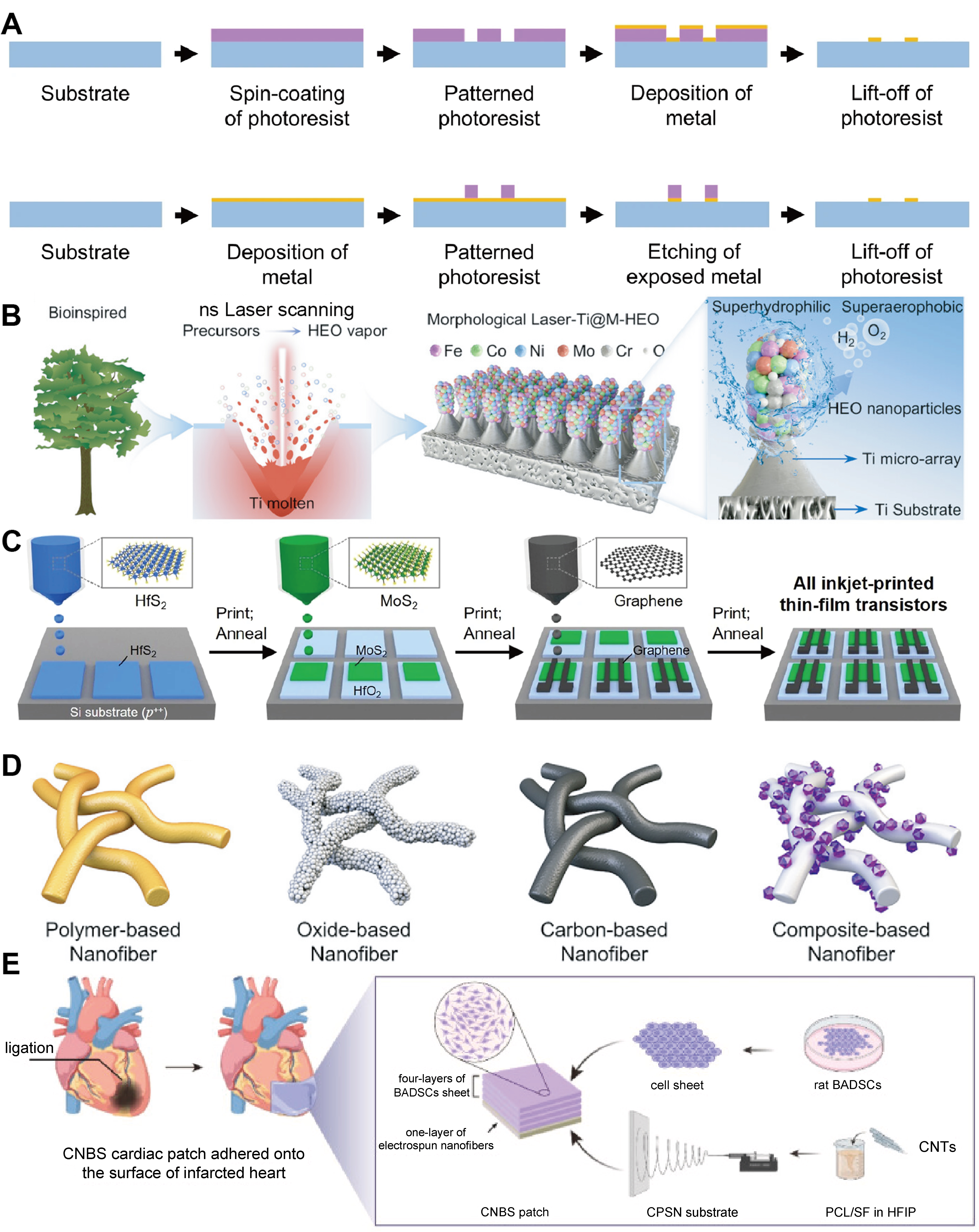
Figure 5. Fabrication strategies for organ-specific bioelectronics with high-resolution and large-area fabrication. (A) Schematic illustration of photolithographic microfabrication[188]. Copyright © 2025, published by Springer Nature; (B) LIG fabrication[191]. Copyright © 2025, WILEY-VCH Verlag GmbH & Co. KGaA; (C) Inkjet-based printing of bioelectronic devices[190]. Copyright © 2022, published by Springer Nature; (D) Electrospun nanofiber-based soft electronics[194]. Copyright © 2025, WILEY-VCH Verlag GmbH & Co. KGaA; (E) An electrospun conductive cardiac patch designed to conformally wrap around the surface of an infarcted heart[195]. Copyright © 2023, published by Elsevier. LIG: Laser-induced graphene; HEO: high-entropy oxide; BADSCs: brown adipose-derived stem cells; CNTs: carbon nanotubes; CNBS: CPSN-BADSCs sheets; CPSN: CNTs-containing electrospun polycaprolactone/silk fibroin nanofibers; PCL: polycaprolactone; SF: silk fibroin; HFIP: hexafluoroisopropanol.
Laser writing
Laser writing (or laser engraving) employs focused laser beams to locally ablate, modify, or convert materials, thereby defining conductive tracks or electrode patterns without the need for masks. It is an inherently maskless and programmable technique that lends itself well to rapid prototyping of complex layouts. A widely studied example is laser-induced graphene (LIG), in which irradiation of polymeric or biomass-derived films converts the surface into a porous, conductive graphene network that can serve as a flexible electrode[190]. This direct-write process can achieve microscale features and can be readily reconfigured at the design stage, which is advantageous for organ-specific devices that require iterative optimization or patient-specific tailoring [Figure 5B][191]. Moreover, laser writing can be performed on thin films that are later laminated onto curved tissues, or directly on preformed substrates intended to conform to skin or organ surfaces[192]. In this way, it helps bridge the gap between high-resolution patterning and practical fabrication of conformal bioelectronic interfaces.
Inkjet-based printing
Inkjet printing is a digital, additive method where conductive or functional inks are deposited in tiny droplets to form patterns. Modern inkjet systems can produce microscale features, offering fairly high printing resolution for flexible circuits. This technique allows custom electrode layouts to be printed on demand, accommodating organ-specific geometries by simply adjusting the digital design. Because it is a non-contact process, inkjet printing can pattern delicate substrates without damage, making it suitable for creating fine electrode arrays on soft, tissue-like materials [Figure 5C][190]. Its capability to deposit multiple ink materials also enables integration of sensors or stimulators tailored to organ needs. For example, printing biocompatible conductive inks for cardiac or neural patches.
Large-area fabrication
Electrospinning
Electrospinning produces nonwoven mats of micro- or nanofibers by ejecting a polymer solution (often containing conductive nanomaterials) under a high-voltage field. The collected fiber mesh can cover large areas and serves as an inherently porous, flexible substrate or conductor network. Electrospinning is a low-cost, high-throughput technique that can be scaled up for mass production[193]. The resulting fibrous networks have high surface area and can be made from biocompatible polymers, making them well-suited for interfaces with tissues and organs [Figure 5D][194]. In organ-specific bioelectronics, electrospun scaffolds can function as soft electrodes or supporting matrices that conform over an organ’s surface. For example, a cardiac patch with electrospun conductive fibers that wrap around the heart [Figure 5E][195]. Because the fiber diameter and mesh architecture can be tuned, this method allows optimization of mechanical properties (matching the compliance of an organ) while covering the target area uniformly with conductive pathways. Moreover, electrospun electronics often remain breathable and permeable, an advantage for long-term tissue integration.
Cut-and-pattern assembly
“Cut-and-paste” fabrication is a paper-crafting-inspired approach where electronic patterns are first cut out of thin conductive films or foils and then transferred onto target substrates[51,196]. In practice, this often uses a desktop vinyl cutter or laser cutter to outline circuits from metal-coated polymer sheets (such as gold on polymer). The patterned pieces (or the negative stencil) are then pasted onto surfaces like medical tape, skin patches, or organ models. This method bypasses the complexities of photolithography, achieving circuit feature sizes on the order of ~100 μm with simple equipment. It is highly suitable for large-area flexible electronics because it is not constrained by wafer size, allowing meter-long flexible circuits to be fabricated using roll-to-roll feedstocks[183,197]. In organ-specific device fabrication, cut-and-paste approaches have been used to rapidly prototype epidermal sensors and electronic tattoos that conform to the skin, as demonstrated in epidermal electronic systems (EES) and related wearable devices[198]. The same concept can also be extended to creating custom-shaped electronics for organs, such as cutting sensor meshes that match the geometry of heart or brain surfaces[199]. The main advantages of this approach are speed and low cost, although the achievable resolution is lower than that of photolithographic methods. In addition, this technique can be integrated with other large-area fabrication strategies, for instance by using a cut stencil to spray-coat or print conductive inks in subsequent steps, thereby combining pattern definition with large-area surface coverage.
Solution-based thin film coating
Chemical solution deposition involves coating a substrate with functional material from a liquid phase over a large area[200]. Techniques in this class include spin coating, dip coating, blade coating (also known as doctor blading), and similar solution-based film deposition methods. They enable uniform, thin films of conductive or active materials (polymers, nanoparticle inks, etc.) to be laid down over device areas far larger than typical wafers. For instance, one can spin-coat a conductive polymer across an entire flexible sheet, or dip-coat a 3D object to blanket it with a conformal conductive layer. These processes are simple, rapid, and cost-effective, often requiring only basic lab equipment. In organ-bioelectronics, solution deposition can be used to create continuous electrodes or sensing skins that cover whole organ surfaces (like a “electronic membrane” for an organ). While the coatings themselves are unpatterned, they can be combined with shadow masks or post-deposition patterning (laser ablation, selective etching) to define electrode regions. An example application is coating a balloon catheter or a neural probe with an even layer of conductive polymer to improve electrical interface over its entire surface area.
3D conformal fabrication
Fabricating bioelectronics for three-dimensional curved surfaces such as the epicardium of the heart or spherical organoids presents unique challenges[201]. The strategies discussed in this subsection aim to create devices that not only cover large areas but also conform closely to non-flat geometries[202]. Methods including direct three-dimensional printing, embedded printing, spray coating, and conformal molding enable the integration of electronic components with complex three-dimensional structures. These approaches ensure that sensors and electrodes maintain intimate contact with target tissues across curved surfaces and during dynamic movements, which is essential for achieving stable and reliable bioelectronic performance in vivo[203]. Accordingly, emphasis is placed on achieving mechanical conformability and three-dimensional architecture during the device fabrication process.
3D printing
3D printing (additive manufacturing) provides a direct route to build 3D device architectures that follow organ geometry. For organ-specific bioelectronics, this enables anatomically contoured implants, or electrode lattices whose overall shape and local electrode placement are defined in a single fabrication sequence. High-resolution methods such as two-photon polymerization (2PP) are particularly relevant because they produce sub-micrometer features and truly 3D structures on top of planar microelectronics. In a recent example, 2PP was used to print polymer microelectrode templates directly onto a CMOS array, followed by conformal metal coating, patterning, and passivation to yield 6,600 tissue-penetrating microelectrodes at 35 μm pitch, with independently tunable height and tip geometry[204]. These pillar electrodes penetrate the retina and place the recording sites within the retinal ganglion cell layer while avoiding the axon bundle layer, enabling high-density, large-area recordings with reduced axonal interference. This type of “direct-print” 3D electrode strategy shows that additive manufacturing can separate the design of tissue-interfacing microstructures from the underlying electronics [Figure 6A][205]. It enables organ-matched curvature, depth targeting, and heterogeneous electrode shapes within a single array. These capabilities are difficult to achieve with planar lithography alone. For instance, researchers have printed flexible electronics in a dome shape to fit a cardiac ventricle[201]. In this work, a scaled anatomical heart model is used as a 3D template for device construction. The electronic components are fabricated as serpentine metal meshes by conventional planar processing, but the overall 3D geometry and mechanical properties are set by a soft elastomeric membrane cast onto the 3D-printed heart model. After demolding, the electronics-integrated membrane can conformally wrap the entire epicardial surface, forming a stable bioelectronic interface with the heart. Beyond ex situ fabrication, recent advances have begun to extend additive manufacturing directly into living tissues. For example, Wei Gao and colleagues recently demonstrated in vivo ultrasound printing, where acoustic fields are used to localize and polymerize bioinks within deep tissues, enabling the formation of functional structures directly inside the body[206]. This approach represents a paradigm shift from pre-fabricated implants toward minimally invasive, in situ fabrication of bioelectronic components, potentially allowing device formation in anatomically constrained or otherwise inaccessible regions [Figure 6B]. By choosing biocompatible, soft materials (e.g., stretchable inks or polymer composites), printed electronics can combine precise micro-patterning with bulk 3D shapes[207]. In this way, 3D printing directly contributes to organ-interfacing devices by co-defining both device geometry and microstructure, reducing the need to bend or mechanically deform flat electronics to fit complex organ topographies[208].
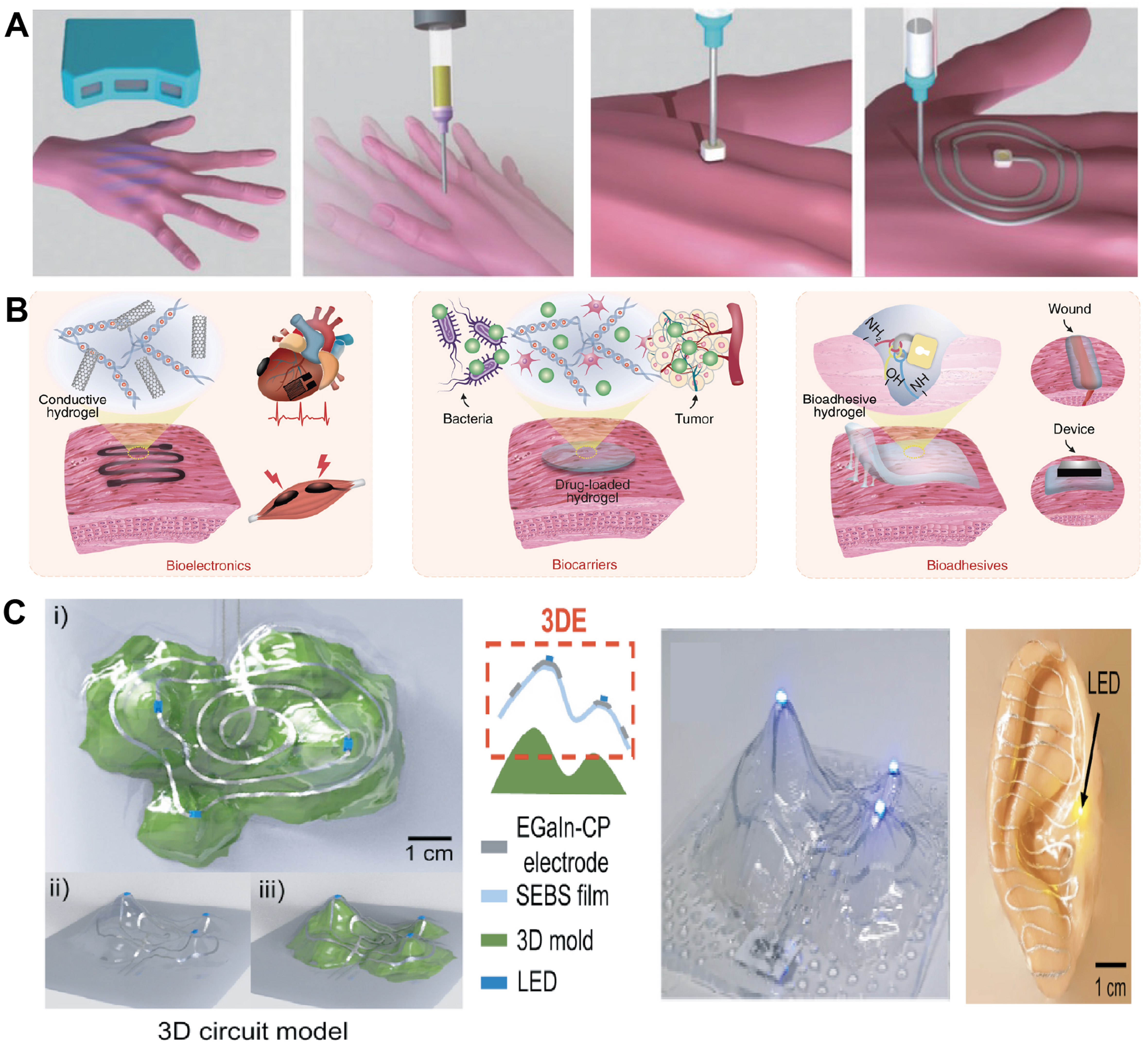
Figure 6. 3D conformal fabrication strategies for bioelectronics. (A) Adaptive 3D printing[205]. Copyright © 2018, WILEY-VCH Verlag GmbH & Co. KGaA; (B) Image-guided in vivo sound printing in deep tissues[206]. Copyright © 2025, the American Association for the Advancement of Science; (C) Conformal fabrication of 3D circuits on complex curvilinear surfaces[215]. Copyright © 2021, the American Association for the Advancement of Science. EGaIn-CP: eutectic gallium–indium with copper particles; SEBS: styrene–ethylene–butylene–styrene; LED: light-emitting diode.
Spray coating
Spray coating (or spray printing) atomizes a solution or dispersion of functional materials into fine droplets and deposits them onto a target surface. Because the spray can wrap around edges and recessed regions, this method is well suited for coating substrates with irregular or curved geometries. In soft bioelectronics, spray processes are commonly used to form thin, percolating films of conductive nanomaterials [e.g., silver nanowires (AgNWs), graphene, carbon nanotubes] or polymer blends over large areas in a single step, while allowing control over thickness and sheet resistance through the number of passes and ink concentration[209]. The technique is fast and can produce ultra-thin, uniform layers across the entire target, from convex domes to concave wells[210]. Xu et al. exemplified this strategy by spray printing AgNWs onto multiscale porous styrene–ethylene–butylene–styrene (SEBS) elastomer substrates to construct on-skin electronic devices with integrated passive-cooling capability[211]. The porous SEBS provided strong mechanical interlocking and π-π interactions with the AgNWs, allowing the sprayed networks to adhere firmly to the sponge-like surface while preserving high breathability and low thermal resistance. In this way, a single spray-coating step defined conductive traces and electrodes across complex, gas-permeable elastomer sheets, enabling multifunctional e-skin devices for electrophysiological sensing, temperature monitoring, and Joule heating without resorting to multi-step lithography. For organ-specific bioelectronics, spray coating can be applied to preformed 3D supports such as balloon catheters or molded elastomer shells[212]. A conformal conductive or sensing layer can be deposited on the entire surface or in selected regions through patterned masks, while the underlying porous or compliant substrate sets the mechanical properties and curvature[183,213]. Because spray processes are compatible with many polymers, hydrogels, and elastomers and are already mature in industrial settings[214]. They provide a practical route to scalable, conformal coatings for implantable or on-organ devices that must match complex anatomy yet remain thin, breathable, and mechanically compliant[203].
Conformal molding
Conformal molding refers to fabrication schemes in which electronic structures are formed or transferred while already constrained by a 3D target geometry. Instead of fabricating devices only on flat wafers and later forcing them to bend, the substrate or stamp is shaped to match the desired curvature, and micropatterns are created or printed in this state. For example, Choi et al. fabricated electrodes, interconnects, and LEDs on thin elastomeric films and then thermoformed the patterned circuits onto curved 3D molds, enabling conformal electronic systems on complex curvilinear surfaces [Figure 6C][215]. Alternative approaches use compliant stamps bearing micro- and nanopatterns (for example, PDMS or other elastomers); under heat and pressure, these stamps conform to spherical or highly curved surfaces and imprint conductive features directly onto them[184]. In all cases, the goal is to obtain intimate, continuous contact over the full 3D surface, which is essential for stable electrical coupling and reduction of local stress concentrations at the tissue-device interface.
Conformal additive stamp (CAS) printing provides a representative example of this concept applied with wafer-level precision. In CAS printing, ultrathin devices or “inks” are first fabricated on planar donor wafers using standard microfabrication, then retrieved by a pneumatically inflated elastomeric balloon and printed onto non-developable 3D substrates such as hemispherical shells and contact-lens molds. The balloon stamp deforms to match complex curvilinear surfaces while keeping the strain in brittle silicon elements below fracture limits, enabling high-yield transfer of photodetector arrays, antennas, hemispherical solar cells, and multifunctional smart contact lenses[216,217]. For organ-specific bioelectronics, similar conformal molding strategies allow sensor grids and electrode meshes to be fabricated in pre-curved forms that match the epicardium, ocular globe, or visceral organs[201,216,218]. Once released from the mold, these meshes “hug” the organ with minimal additional fixation and can be combined with stretchable designs to accommodate physiological motion while maintaining the imposed 3D shape.
APPLICATIONS OF ORGAN-SPECIFIC BIOELECTRONICS
The emerging landscape of bioelectronics is shifting from generic implantable or wearable devices toward organ-specific architectures engineered to match tissue stiffness, curvature, biochemical milieu, motility, and signal characteristics. Such precision interfacing is expected to improve long-term stability, reduce foreign-body responses, and enhance the fidelity of sensing or stimulation. Here, we highlight representative strategies for the brain, GI tract, skin, and visceral organs, including the heart, liver, kidney, and lung, with emphasis on how organ-level constraints guide material selection, device mechanics, and system-level design.
To clarify these design constraints, Table 3 summarizes representative mechanical environments, deformation modes, readouts, and device formats across major soft-tissue interfaces. Because interface requirements vary with application mode, device configuration, and tissue-contact depth, organ-specific bioelectronics should be viewed as a coupled design problem rather than a fixed organ-by-organ material selection.
Organ- and application-specific requirements for bioelectronics
| Organ | Modulus | Strain | Readout | Devices | Ref. |
| Brain/surface | 0.1-10 kPa | Micromotion | EEG/LFP | ECoG/patch | [85,219] |
| Brain/penetrating | 0.1-10 kPa | Micromotion | Spikes/LFP | Probe | [162,220] |
| Heart | 8-15 kPa | 20%-60% | ECG/EGM | Patch/mesh | [202,221-223] |
| Gut | 0.1-3 MPa | 50%-100% | EGG/pH | Patch/capsule | [170,224-226] |
| Skin/epidermis | 0.4-20 MPa | 20%-30% | ECG/EMG | Patch/tattoo | [86,167,227] |
Bioelectronics for the brain
The brain is among the most demanding application areas for organ-specific bioelectronics. Neural interfaces must accommodate extremely soft tissue that is mechanically fragile and highly immunoreactive, while detecting low-amplitude electrical signals with long-term stability. Traditional silicon or metaxl electrodes have a modulus order of magnitude higher than brain tissue, so even micromotion of the brain leads to micro-injury, glial scar formation, and progressive signal loss. Thus, organ-specific neural bioelectronics focus on minimizing mechanical and biochemical perturbation, essentially designing the interface as a tissue-like, long-lived “electronic scaffold” rather than a rigid probe. This means building electrodes and wires that more closely match the brain’s softness, curvature, and microstructure to achieve in vivo “stealth”. For example, the device becomes mechanically and structurally invisible to the host tissue over time.
To achieve this, mesh and injectable electronics employ sub-micrometer-thick, microporous architectures that can be delivered through fine needles and then unfold within neural tissue, reaching bending stiffness comparable to brain and allowing cellular infiltration with greatly reduced chronic inflammation and gliosis. Zhou et al. showed that syringe-injectable mesh electronics can integrate seamlessly with brain tissue while eliciting minimal chronic immune response, thereby enabling stable long-term neural interfacing[228]. In follow-up studies, researchers showed that such ultra-flexible mesh probes produce virtually no gliosis even months after implantation. Neurons and neurofilaments grow through the mesh structure by 12 weeks post-implantation, and the distribution of astrocytes and microglia becomes nearly uniform at the probe-tissue interface. This seamless tissue integration allowed stable neural recordings over three months with negligible signal loss. These findings highlight that making implants as soft and open as the brain is a viable route to chronic biocompatibility and signal stability. Researchers are complementing this approach with ultrathin inorganic conductors and soft packaging: for example, thin gold nanomembranes in serpentine (fractal) layouts, liquid-metal interconnects, and conductive polymer hydrogels have been used to create electrodes and wires that deform readily with brain movement. Such designs drastically lower the effective stiffness of the device and reduce interfacial stress, further mitigating foreign-body response.
Another organ-specific innovation for brain interfaces is the use of organic electronic materials that can directly amplify neural signals on-site in the aqueous, ion-rich environment of the brain. Traditional metal electrodes pick up microvolt-level local field potentials that then must be amplified by distant circuitry, which limits SNR. OECTs, made from mixed ionic–electronic conductors like PEDOT:PSS, have been developed as flexible, biocompatible amplifying electrodes. OECTs operate at low voltages and use ions from the tissue to modulate their conductivity, effectively acting as local amplifiers at the tissue interface. Polymer OECT-based electrode arrays on ultrathin plastic films can record brain activity with much higher SNR than conventional electrodes, due to the transistors’ built-in amplification[229]. In vivo tests of these transistor arrays on the cortical surface showed they could detect low-amplitude neural oscillations and even epileptiform spikes that were previously indiscernible with passive electrodes. Such OECT-based brain interfaces have been used for electrocorticography with excellent signal quality, and they represent a promising route to multiplexed neural sensors that record both electrical and neurochemical signals from the cortex[230].
The convergence of these organ-matched innovations is yielding a new generation of neural interfaces that can potentially operate stably for years. In the coming years, such networks of organ-specific bioelectronic devices could enable closed-loop neuromodulation therapies and high-bandwidth brain-computer interfaces that remain reliable over the patient’s lifetime. However, translating these experimental systems into clinical practice will require overcoming several key challenges. Scalable manufacturing and deployment of micro-scale, polymeric devices must be achieved to cover large brain areas or thousands of channels. Soft packaging is an unsolved problem, which can protect electronics from corrosion by body fluids, typically requires rigid, glass-like encapsulants, so new strategies (e.g., thin-film coatings) are needed to robustly seal circuits while preserving elasticity[231]. Fully wireless operation is another challenge: integrating onboard power sources or developing safe wireless power and data links is crucial for untethered implants[232]. Despite these challenges, the progress in brain-specific bioelectronics demonstrates a clear path forward: by engineering devices to physically and functionally blend into the brain’s own environment, we can improve the longevity and fidelity of neural interfaces, unlocking new capabilities in neuroscience research and clinical neuromodulation.
Beyond the mechanically matched mesh electrodes and OECT arrays discussed above, recent work has also introduced biologically assisted routes for delivering soft electronic devices into the brain. Sheng et al. reported an implantation approach during early brain development: ultra-soft mesh electronics were laminated onto the embryonic neural plate and subsequently carried inward as the plate folded and expanded into the three-dimensional brain[233]. Liang et al. developed a silk-enabled conformal intraventricular interface that can self-unfold and conformally adhere to periventricular brain structures, enabling minimally invasive and chronically stable neural recordings [Figure 7A][234]. These “developmentally integrated” implants enabled long-term recording of neural activity from single cells to populations, while markedly reducing gliosis and scar formation, indicating a relatively gentle and durable structural and functional integration with the host tissue during brain formation. Building on this concept, Yadav et al. used immune cells as carriers: subcellular wireless stimulators were covalently attached to circulating monocytes, which then trafficked to inflamed brain regions via endogenous immune surveillance pathways[235]. Following intravenous injection, the resulting cell-electronics hybrids provided targeted, wirelessly addressable focal neuromodulation without craniotomy or conventional leads [Figure 7B]. Taken together, these “development-driven” and “cell-driven” implantation modes illustrate an important direction in which soft neural interfaces are deployed by leveraging intrinsic biological processes, with the goal of reducing surgical trauma and improving long-term stability of the brain-device interface.
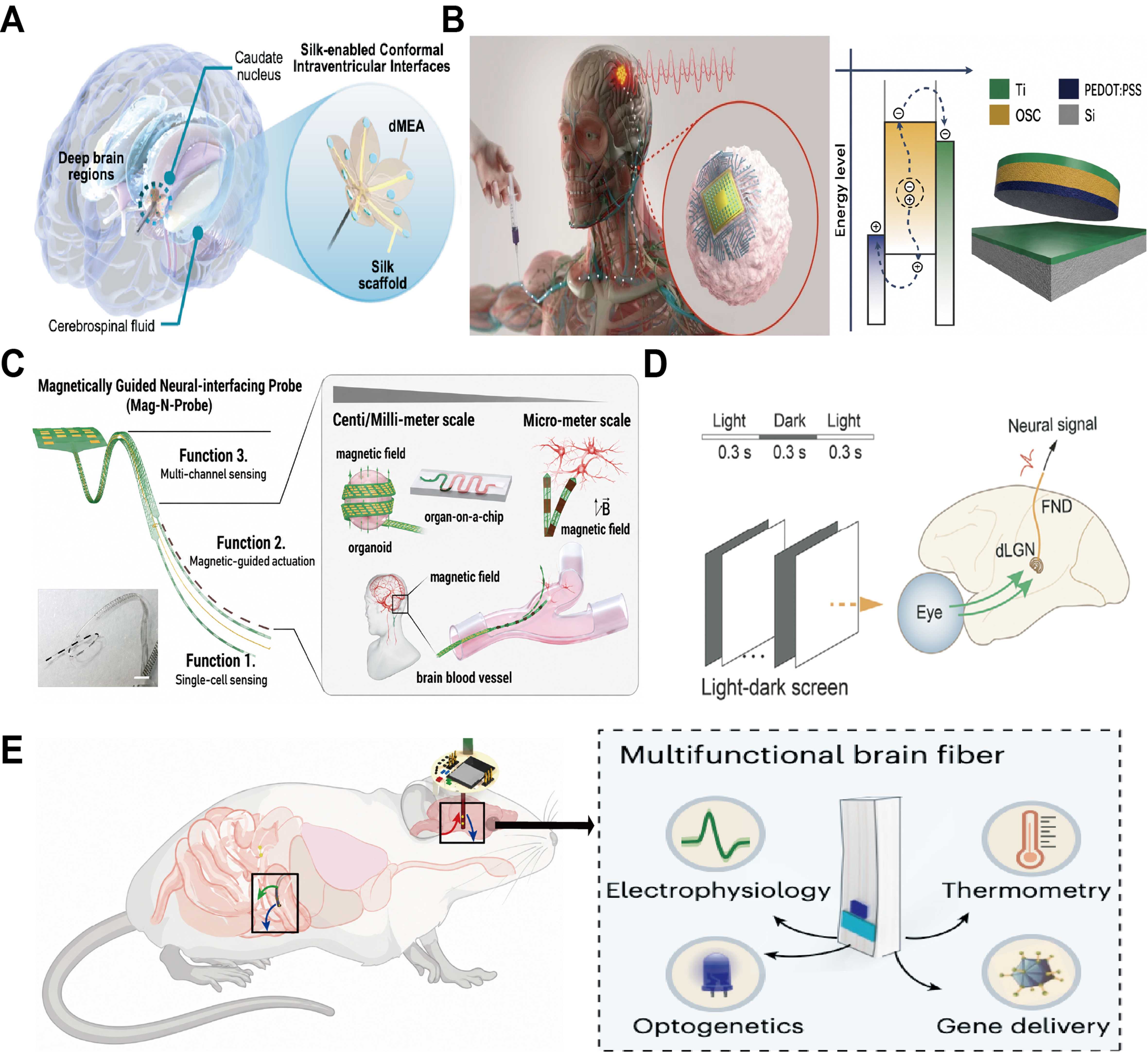
Figure 7. Bioelectronics for the Brain. (A) Silk-enabled conformal intraventricular interface for minimally invasive and chronically stable neural recordings[234]. Copyright © 2025, published by Springer Nature; (B) Cell–electronics hybrid enabling nonsurgical and targeted focal neuromodulation[235]. Copyright © 2025, published by Springer Nature; (C) Magnetically guided neural-interfacing probe for steerable implantation and multifunctional sensing[236]. Copyright © 2025, WILEY-VCH Verlag GmbH & Co. KGaA; (D) Soft-fiber neural interface for stable long-term recording under dynamic in vivo conditions[237]. Copyright © 2024, WILEY-VCH Verlag GmbH & Co. KGaA; (E) Multifunctional microelectronic fibers for multimodal brain interfacing and wireless neural modulation[238]. Copyright © 2023, published by Springer Nature. dMEA: Deformable microelectrode array; PEDOT:PSS: poly(3,4-ethylenedioxythiophene):poly(styrenesulfonate); OSC: organic semiconductor; FND: fiber neural device; dLGN: dorsal lateral geniculate nucleus.
In parallel with these advances in implantation, the physical form of neural interfaces is evolving from locally fixed probes toward fiber-like systems whose position can be adjusted in vivo. Kim and colleagues embedded magnetic nanoparticles into ultra-flexible conductors to create magnetically responsive probes that can be steered by external fields, enabling centimetre-scale navigation in tissue and near single-cell positioning for high-resolution electrophysiology [Figure 7C][236]. Tang et al. reported an axon-like soft-fiber bioelectronic device that enables reliable in vivo neural recording even under vigorous activities; the representative visual-stimulation experiment further demonstrated stable recording of light-evoked neural activity from the dLGN [Figure 7D][237]. Further extending this concept, Sahasrabudhe et al. developed multifunctional microelectronic fibers that enable wireless modulation of both gut and brain neural circuits, highlighting the potential of soft fiber-based bioelectronics for multimodal interfacing across distributed organ systems
Bioelectronics for the GI tract
The GI tract’s continuous peristaltic motion and harsh chemistry (stomach acid, enzymes) presents a unique challenge for bioelectronic devices. Any implant or ingestible in the GI system must combine robust mechanical compliance with corrosion-resistant encapsulation to survive acidic gastric fluids. At the same time, these devices should not interfere with normal GI motility or cause obstruction. Researchers are therefore developing organ-specific GI bioelectronics that can function reliably in this extreme milieu while remaining biocompatible with the digestive system’s motions and secretions.
One representative approach uses ingestible hydrogel systems that swell into soft, stomach-conforming spheres, enabling weeks-long gastric residence for continuous sensing (e.g., core temperature) while avoiding premature expulsion or mucosal irritation seen with rigid capsules; these devices can be actively deswelled on demand for safe retrieval[239]. More broadly, recent ingestible platforms combine miniaturized electronics, wireless links, and advanced materials to realize gastric- or intestinal-resident devices: star- or cage-like geometries, tough hydrogels, and elastomeric arms unfold post-swallow to resist pyloric passage yet disassemble on cue for clearance[240-242]. In parallel, micro-bio-electronic capsules integrate engineered bacteria with low-power electronics for in situ detection of GI bleeding and inflammatory biomarkers[243]. To protect such complex devices through the GI tract, researchers are exploring specialized encapsulation materials. These include biodegradable polymers like silk fibroin and PLGA, as well as pH-responsive enteric coatings and hydrogel coatings that remain intact in stomach acid but dissolve or become permeable in the intestines, thereby releasing drugs or exposing sensor surfaces only at the desired GI location[244]. A growing body of work from Traverso, Langer and colleagues has outlined design principles for these ingestible systems, emphasizing that devices must balance mechanical durability (to withstand gastric contractions), triggerable disassembly or transit, and careful biocompatibility with GI physiology to avoid irritation[245]. In essence, the materials and designs are tuned to match the GI environment. In parallel, researchers are innovating ways to power GI devices without batteries, harnessing the body’s own chemical energy. Battery-free and biochemically powered capsules have been demonstrated that harvest energy from intestinal fluids using onboard biofuel cells. A recent ingestible sensor capsule used a glucose-fueled biofuel cell to generate power from sugars in the small intestine, allowing it to continuously monitor gut glucose levels and transmit data via a wireless (magnetic) telemetry link[244]. All in a pill-sized form factor with no internal battery. By exploiting biofuel or even mechanical energy from gut peristalsis, these designs eliminate the need for bulky batteries and toxic battery materials, making the capsules smaller and safer for long-term use. Such self-powered capsules have successfully performed real-time chemical sensing in animal models, proving the feasibility of sustained metabolite monitoring using only the energy available inside the GI tract.
These innovations accelerate gut-specific bioelectronics that are now capable of comprehensive, real-time monitoring and intervention. Swallowable devices have been built to map a wide range of GI parameters: they can measure local pH, temperature, pressure, gas composition, and even sense biomarkers of infection or inflammation in different sections of the gut. Despite the rapid progress, significant challenges remain before GI bioelectronics can be broadly deployed. A key challenge is ensuring that devices can maintain secure adhesion or positioning at a target site under the constantly moving, contracting conditions of the gut. Even for devices designed to stick to the mucosal wall, the GI tract’s continual peristalsis and mucus turnover can make long-term attachment difficult. Recent work on bioadhesive hydrogel interfaces is tackling this by improving electrode-tissue contact in the stomach, but robust retention over weeks is still an open problem[225]. Another challenge is engineering encapsulation and coatings that can withstand months of corrosive attack by gastric acid and enzymes. By surmounting these challenges, GI bioelectronics will be poised to revolutionize how we monitor and treat diseases of the digestive tract, offering a future of smart pills that can diagnose, report, and respond from within our own bodies.
The first is chronically resident platforms for large-area physiological mapping. Kong et al. used 3D-printed gastric-resident frameworks with deployable arms to keep electronics in the stomach for weeks, addressing the long-standing problem that conventional capsules are cleared within hours and cannot follow slow disease dynamics [Figure 8A][242]. Building on this concept, Boys et al. developed implantable bioelectronic platforms for in vivo gut electrophysiology, demonstrating stable recording of GI electrical activity in mice, rats, and pigs, thereby highlighting the translational potential of chronic bioelectronic interfaces for the digestive tract [Figure 8B][246]. Gopalakrishnan et al. reported a wireless smart capsule that integrates pH and oxidation–reduction potential sensing to profile inflammatory status throughout the GI tract, providing a promising strategy for in situ monitoring of reactive oxygen species (ROS)-related gut inflammation and inflammatory bowel disease [Figure 8C][247]. Taken together, these studies show that resident geometries, soft ribbons and tissue-matched implants can convert the GI tract from a transient passage into a site for stable, organ-scale physiological monitoring.
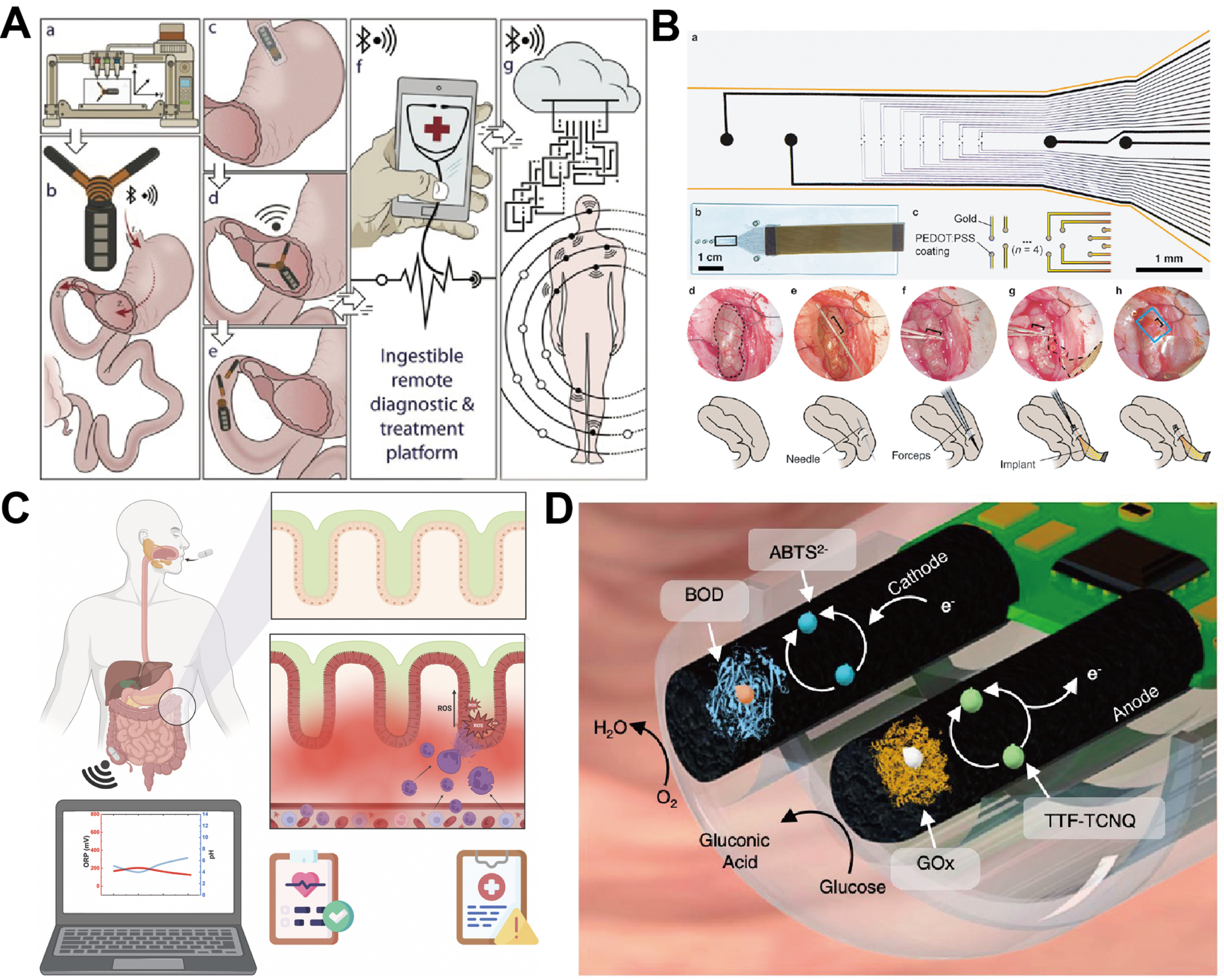
Figure 8. Bioelectronics for the GI tract. (A) Gastric-resident electronics for prolonged physiological monitoring[242]. Copyright © 2019, WILEY-VCH Verlag GmbH & Co. KGaA; (B) Implantable bioelectronics for gut electrophysiology[246]. Copyright © 2025, published by Springer Nature; (C) Smart capsule for inflammation profiling throughout the GI tract[247]. Copyright © 2023 The Authors, published by Elsevier; (D) Self-powered ingestible wireless biosensing system for in situ metabolite monitoring[244]. Copyright © 2022, published by Springer Nature. PEDOT:PSS: Poly(3,4-ethylenedioxythiophene):poly(styrenesulfonate); BOD: bilirubin oxidase; ABTS: 2,2′-azino-bis(3-ethylbenzothiazoline-6-sulfonic acid; GOx: glucose oxidase; TTF-TCNQ: tetrathiafulvalene-7,7,8,8-tetracyanoquinodiamethane.
The second emerging direction is self-powered, multimodal biochemical profiling. De la Paz et al. demonstrated an ingestible capsule in which a glucose-fuelled biofuel cell harvests energy from luminal sugars in the small intestine to power onboard electronics, allowing continuous wireless monitoring of intestinal glucose without a conventional battery [Figure 8D][244]. Building on earlier bacteria–electronics capsules for detecting GI bleeding, Inda-Webb et al. recently reported a sub-1.4 cm3 device that integrates genetically engineered probiotic biosensors with a custom photodetector and readout chip, allowing in situ detection of highly labile inflammatory mediators such as nitric oxide and hydrogen sulfide directly inside the gut[248]. By combining biochemical energy harvesting with high-density sensing, these devices directly tackle two bottlenecks of earlier capsules - short energy budget and single-analyte readout - and point toward GI systems that can profile metabolites, inflammatory mediators and therapeutic drugs over long time scales. Together with advances in adhesive interfaces and corrosion-resistant encapsulation, these two classes of technologies outline a realistic path toward future GI bioelectronics that not only survive the harsh digestive environment, but also provide continuous, organ-specific information to guide the management of motility disorders, inflammatory bowel disease and metabolic syndromes.
Bioelectronics for the epidermis
As a large, accessible and regenerating organ, skin offers a convenient interface for non-invasive bioelectronics but imposes requirements distinct from internal tissues. The epidermis is continuously renewed and subjected to multiaxial stretching, shear, perspiration and environmental exposure; contact is often intermittent and user controlled. Effective skin-mounted devices must therefore be ultrathin, compliant, breathable and hypoallergenic, forming intimate yet reversible contact without occlusion or irritation, while maintaining stable impedance during daily activities[81]. As highlighted in Bao’s recent review on skin-inspired bioelectronic materials and systems, effective epidermal devices must therefore combine ultralow bending stiffness, conformal yet gentle adhesion, gas and moisture permeability, and stable electrical performance under daily wear, while using materials that are safe for long-term contact with human skin.
In epidermal bioelectronics, skin bioimpedance can serve as a physiological readout, while skin–electrode interfacial impedance provides a practical indicator of device–skin coupling quality. Elevated interfacial impedance can reduce signal fidelity and increase susceptibility to motion artifacts during long-term wearable monitoring[249]. Therefore, soft, conformal, and hydrated contact layers are widely used to enhance skin coupling and lower contact impedance[250]. Beyond electrophysiological recording, impedance-based measurements can also provide information on skin hydration, tissue condition, and barrier-state changes, making them useful for monitoring both epidermal physiology and device–skin interface stability[251].
Pioneering EES by Kim et al. matched thickness, modulus, and bending stiffness of ultrathin serpentine metal circuits laminated on elastomer substrates to that of human skin, creating “mechanically invisible” devices that adhere via van der Waals forces alone[51]. Subsequent developments include tattoo-like electronics that can be transferred as temporary decals, large-area “electronic skin” matrices using organic semiconductors and nanomaterials for distributed pressure and temperature sensing, and conductive hydrogel or ionic patches that provide low-impedance, soft contacts for biopotential recording and neuromodulation[252]. Porous elastomers, microperforated films, nanofiber meshes, and Janus structures have been introduced to enhance breathability and moisture management, while new supramolecular and bioadhesive chemistries enable strong yet painless removal, tailored for sensitive skin and long-term wear.
Rogers and Kim’s group first demonstrated EES in which ultrathin serpentine metal interconnects on elastomer substrates were engineered to match the thickness, modulus and bending stiffness of the stratum corneum, creating “mechanically invisible” patches that adhere via van der Waals forces alone and deliver high-quality electrocardiography (ECG), EMG and electroencephalography (EEG) recordings. Building on this mechanical-matching concept, Du et al. recently developed a self-compliant ionic nanomesh: a highly porous network of elastic polymer nanofibres infiltrated with ionic conductors that laminates on skin with almost no normal stress [Figure 9A][253]. The nanomesh architecture is intrinsically gas-permeable and mechanically unconstraining, enabling stable electrophysiological recordings during sweating and motion while greatly reducing occlusion-induced irritation over multi-day wear. Together, these studies show how matching not only modulus but also normal stress and breathability is key to organ-specific epidermal interfaces.
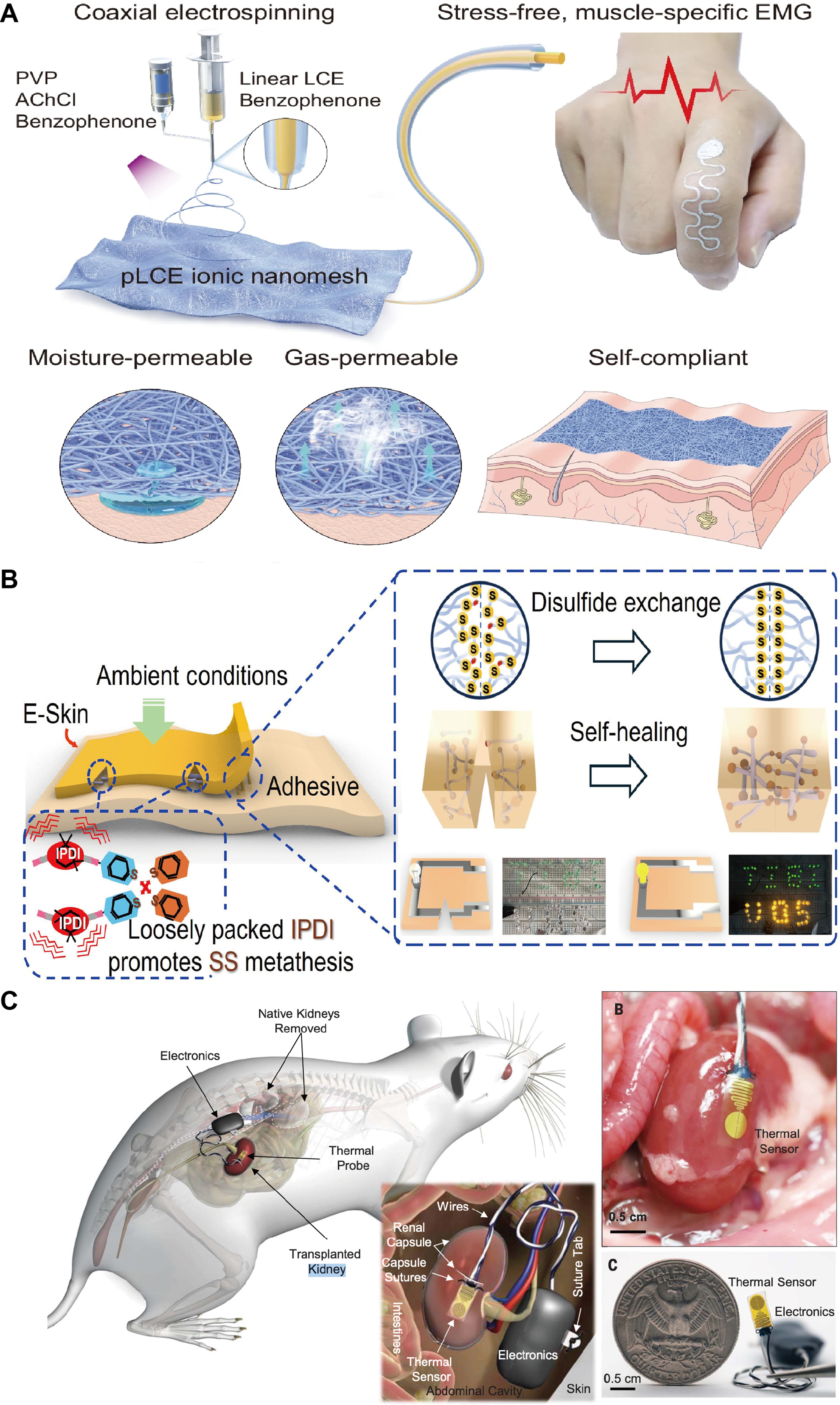
Figure 9. Bioelectronics for the epidermis and other visceral organs. (A) A self-compliant ionic nanomesh for skin electronics[253]. Copyright © 2025, published by Springer Nature; (B) A selfhealing electronic skin for movement evaluation[255]. Copyright © 2025, the American Association for the Advancement of Science; (C) A bioelectronic patch for monitoring organ transplant rejection[261]. Copyright © 2023, the American Association for the Advancement of Science. PVP: Polyvinylpyrrolidone; LCE: liquid crystal elastomers; pLCE: permeable LCE; EMG: epidermal and muscular electromyography; IPDI: isophorone diisocyanate; SS: disulfide bonds.
To improve robustness under everyday mechanical damage, Son and colleagues realized reconfigurable assemblies of self-healing stretchable transistors and circuits[254]. Their platform uses self-healing polymer dielectrics and elastomeric conductors to form transistor “islands” connected by self-healable interconnects; after cutting and rejoining, both mechanical integrity and circuit functions such as inverters and ring oscillators recover with minimal performance loss. In parallel, Lee et al. reported a rapidly self-healing electronic skin in which a disulfide-bonded TPU matrix and printed conductive networks allow the device to restore over 80% of its mechanical strength and conductivity within seconds at room temperature after being fully severed [Figure 9B][255]. Integrated with low-power wireless electronics and on-patch machine-learning algorithms, this e-skin can continuously monitor motion and classify levels of muscular fatigue in real time. These examples illustrate how molecularly engineered self-healing polymers can extend device lifetime and support intelligent, long-term epidermal monitoring.
Bao’s group has advanced skin-like active biosensors based on stretchable organic electronics to address signal drift and long-term stability. In their recent work on drift-free biosensors with stretchable diode-connected organic field-effect transistors (OFETs), they constructed diode-connected OTFT pixels on pre-strained elastomer substrates and integrated them with microfluidic sweat collectors and a soft
Skin-specific bioelectronics now underpin continuous ECG/EMG/EEG monitoring, motion and posture tracking, sweat-based biochemical sensing (electrolytes, metabolites and hormones) and transcutaneous stimulation for pain management, rehabilitation and cosmetic therapies. Beyond conventional biopotential sensing, impedance-based bioelectronic approaches have been explored for neuromuscular perception and human-machine interaction, including soft contact sensing via impedance imaging and muscle-driven joint torque estimation through voltage–torque mapping[257,258]. Recent work further demonstrates integrated impedance-based sensing platforms for enhanced neuromuscular perception in human–machine interfaces[259]. The field is converging toward multimodal patches that combine mechanical, electrophysiological and biochemical sensing with on-board processing and low-power wireless links, enabling “zero-burden” long-term monitoring in daily life. Looking ahead, integrating these mechanically invisible, gas-permeable, self-healing and drift-free device concepts with sustainable substrates, secure data architectures and haptic or AR/VR interfaces will further position epidermal bioelectronics as organ-specific platforms at the intersection of healthcare, human–machine interaction and soft robotics.
Bioelectronics for other visceral organs
Among visceral organs, the heart represents one of the most prominent examples where organ-specific bioelectronics have achieved substantial progress. The heart’s constant rhythmic beating (~1 Hz) and complex three-dimensional curvature create a challenging environment for conventional rigid implants such as metallic leads and canister pacemakers, which often introduce severe mechanical mismatch and require invasive fixation. To address these limitations, Xu et al. developed three-dimensional multifunctional integumentary membranes, thin silicone epicardial membranes conformally wrapped around the entire heart and integrated with stretchable arrays of electrodes, temperature and strain sensors, and stimulators[201]. These devices provide high-density mapping of electrical activity and mechanical deformation across the entire epicardial surface while accommodating large stroke-volume changes without delamination. Earlier electronic web architectures further demonstrated that serpentine metal interconnects embedded in elastomers can conform to highly curved cardiac geometries and tolerate large biaxial strains during beating[202]. Such systems enable simultaneous electrical mapping and pacing over large ventricular areas while maintaining stable contact with the epicardium.
Building on these epicardial platforms, soft electronics have also been integrated with interventional catheters. Recent catheter-mounted electronic arrays unfold at the catheter tip to form soft, conformal sensor patches that provide real-time multimodal feedback during procedures such as radiofrequency ablation and irreversible electroporation[260]. These systems enable co-registered measurements of temperature, contact pressure, and intracardiac electrograms at the tissue–catheter interface, illustrating how cardiac bioelectronics are evolving from point-like rigid electrodes toward organ-matched, high-coverage sensing and stimulation systems.
Beyond the heart, similar organ-specific strategies are being extended to other visceral organs. The kidney, for example, does not experience large rhythmic contractions but resides deep within the body and is subject to slow perfusion variations and motion associated with respiration. Recent studies have demonstrated that soft implantable bioelectronic interfaces can be laminated onto transplanted kidneys to continuously monitor local biophysical signatures associated with early graft rejection[261,262]. Such systems can potentially provide early warning signals of transplant rejection before conventional serum biomarkers rise. For instance, Madhvapathy et al. developed an ultrathin stretchable bioelectronic patch that is inserted beneath the renal capsule and wirelessly monitors local temperature and thermal conductivity as indicators of inflammatory activity in rat kidney transplants[261]. Continuous measurements revealed characteristic temperature variations associated with early rejection, enabling detection weeks before traditional biomarkers such as serum creatinine and BUN changed, while producing minimal foreign-body response at the organ surface [Figure 9C].
In addition to device architecture, interfacial surface properties play a critical role in determining the host response to visceral implants. Studies on soft-tissue implants have shown that surface topography can strongly influence immune cell recruitment and fibrotic encapsulation. Micro- and macro-textured surfaces, for example, can elicit distinct immune responses compared with smooth interfaces, suggesting that micron-scale topographical design may provide an additional strategy for mitigating fibrosis around long-term visceral bioelectronics[263].
Overall, bioelectronics for visceral organs highlight the importance of secure yet minimally invasive interfaces with wet, highly deformable tissues. Strategies under active development include bioadhesive hydrogel coatings that anchor devices to organ surfaces without sutures, wirelessly powered architectures that eliminate transcutaneous leads, and biodegradable electronics that function temporarily before safely dissolving. The convergence of cardiac epicardial electronics, kidney-specific monitoring systems, and therapeutic nanofluidic patches suggests that organ-tailored electronic platforms capable of sensing and intervention across internal organs are becoming increasingly feasible, opening new possibilities for proactive management of conditions such as arrhythmias, organ failure, and transplant rejection.
CONCLUSION AND OUTLOOK
Organ-specific bioelectronics is redefining soft bioelectronic interfacing from a largely generic flexible-device strategy toward a tissue-adapted design paradigm[264]. As highlighted throughout this review, recent advances in conductive and interfacial materials, structural and encapsulation platforms, and organ-matched fabrication strategies have greatly expanded the capability of bioelectronic systems to establish stable, conformal, and functionally integrated interfaces with soft tissues[49]. Across neural, GI, epidermal, and visceral-organ applications, these studies collectively show that high-performance biointerfacing depends not simply on making electronics softer, but on co-optimizing materials, structures, and device functions according to the distinct mechanical, biochemical, and immunological environments of individual organs[265].
Despite this rapid progress, the field still faces a fundamental knowledge gap in understanding what truly governs long-term device–tissue integration in different organs[161]. Soft and stretchable mechanics alone do not guarantee stable chronic performance. Instead, electrical reliability and biological acceptance are jointly shaped by interfacial hydration, ion transport, encapsulation integrity, local immune activity, fibrotic remodeling, and organ-specific modes of deformation[49,161]. These factors vary substantially across tissues such as the brain, skin, gut, heart, and kidney, making it difficult to define universal material-selection or interface-design rules[85]. As a result, the long-term interpretation of device performance, failure, and biological response remains incomplete, and more predictive frameworks that connect material properties with organ-specific in vivo behavior are still needed.
Beyond this biological complexity, major engineering challenges remain in translating promising material systems into robust organ-resident devices. In addition to passive monitoring, organ-specific bioelectronics are expected to evolve toward closed-loop systems that integrate real-time sensing, signal processing, and adaptive therapeutic modulation. Electrophysiological, chemical, mechanical, or thermal signals from the target organ could be used to guide feedback-controlled interventions, including electrical stimulation, drug or ion delivery, thermal regulation, and neuromodulation. Such closed-loop systems are particularly important for dynamic organs such as the brain, heart, gut, and skin, where pathological states evolve over time and require organ-adapted therapeutic responses.
Biocompatibility is another critical consideration for organ-specific bioelectronics, because material safety is strongly dependent on both the intrinsic chemistry of the material and the target tissue environment[159,266]. For epidermal systems, skin compatibility involves not only low cytotoxicity, but also breathability, irritation-free adhesion, sweat tolerance, and avoidance of allergic or inflammatory reactions during long-term wear[267]. For implantable interfaces, the major concerns shift toward chronic immune activation, fibrotic encapsulation, degradation products, ion or nanoparticle release, and long-term stability in wet and enzymatically active environments[268]. Different material systems therefore require different safety considerations: conductive polymers and OMIECs require evaluation of dopants, additives, and electrochemical by-products; hydrogels require control over residual monomers, crosslinkers, swelling, and degradation behavior; liquid metals and metal composites require stable encapsulation to prevent leakage or metal ion exposure; and 2D materials require careful assessment of flake size, oxidation state, persistence, and inflammatory potential[269,270]. Soft conductors, conductive hydrogels, OMIECs, liquid-metal composites, and ultrathin encapsulation layers offer attractive combinations of compliance and electrical performance, but their long-term behavior under wet, ion-rich, enzymatic, and mechanically dynamic conditions is still not fully understood[271-273]. Repeated bending, stretching, swelling, delamination, biofluid infiltration, and material fatigue can progressively degrade adhesion, impedance stability, and signal fidelity[274]. At the same time, many high-performance organ-conformal devices still depend on low-throughput or highly customized fabrication workflows, which complicates reproducibility, scaling, and integration with clinically relevant device formats[275]. Thus, improving long-term stability while preserving manufacturability remains a central engineering challenge for the field.
At the system level, organ-specific bioelectronics is expected to evolve from passive sensing interfaces toward multifunctional and closed-loop platforms. Future systems will likely integrate multimodal sensing, localized stimulation or delivery, low-power signal conditioning, wireless communication, and adaptive data analysis within unified soft architectures[276]. In this context, materials innovation will continue to play a central role, including self-healing conductors and encapsulants, biodegradable and transient structural materials, adhesive and repair-supportive interfaces, and biohybrid or living components that improve chronic tissue integration[69,277,278]. In parallel, advances in wireless power transfer, miniaturized electronics, and AI-assisted interpretation of organ-level signals may enable more autonomous bioelectronic systems capable of not only monitoring disease progression, but also actively modulating tissue function in real time[49].
Ultimately, the transition of organ-specific bioelectronics from compelling laboratory demonstrations to widespread biomedical use will depend on scalable manufacturing, rigorous long-term validation, and clear demonstration of clinical utility [Figure 10][75]. This includes reproducible large-area and high-resolution fabrication, sterilization-compatible packaging, reliable implantation or resorption strategies, and systematic evaluation in chronic animal models and human-relevant settings[279,280]. Just as importantly, future progress will require closer integration among materials scientists, device engineers, biologists, and clinicians so that organ-specific platforms are designed not only for mechanical conformity and electrical performance, but also for compatibility with real surgical workflows and therapeutic needs. With such advances, organ-specific bioelectronics is well positioned to become a foundational technology for precision monitoring, localized intervention, and next-generation bioelectronic medicine.
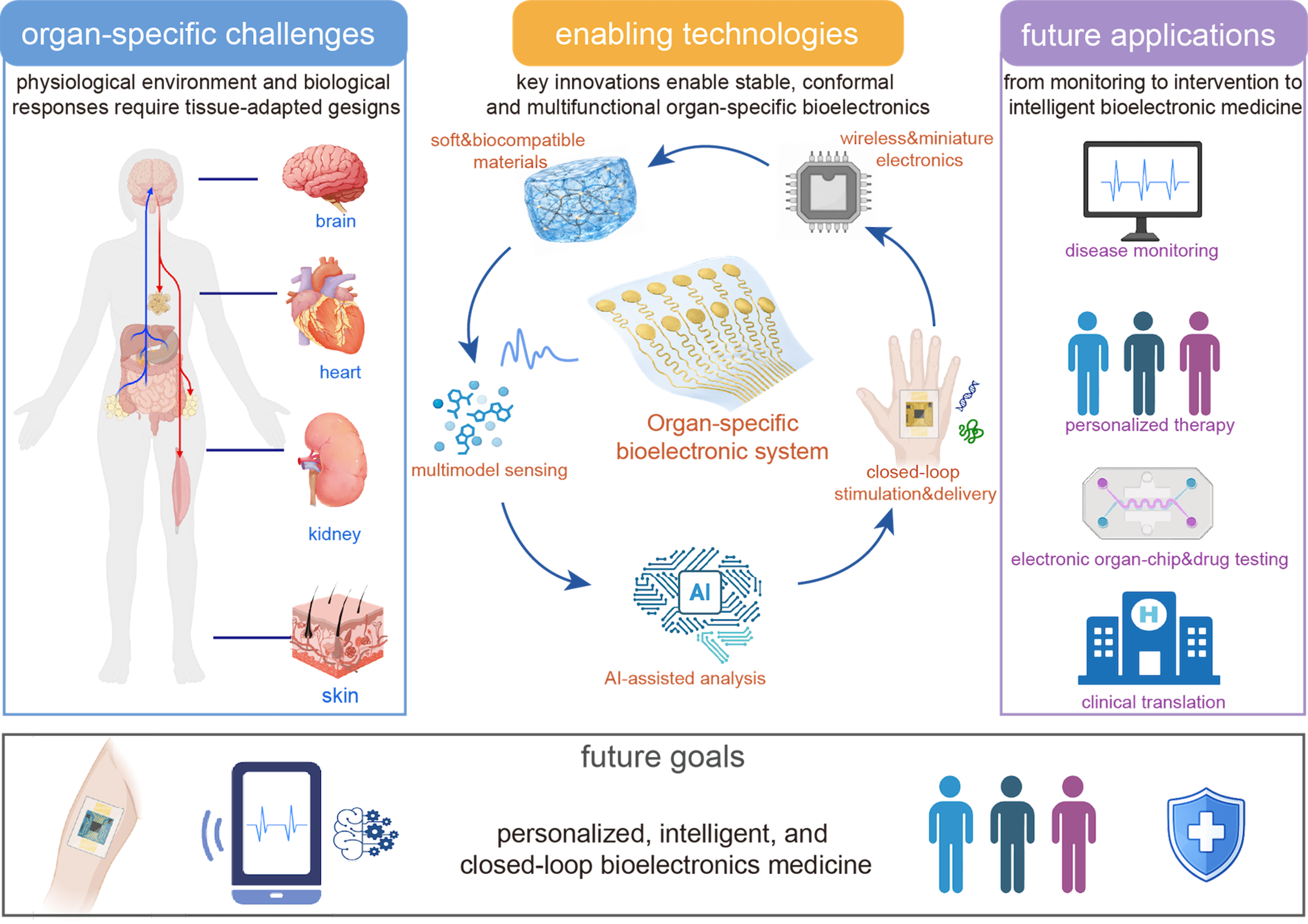
Figure 10. Future perspectives for organ-specific bioelectronics. Created by the authors using Adobe Illustrator. Some schematic elements created in BioRender. Liu, X. (2026) https://BioRender.com/k8j8vrc.
DECLARATIONS
Acknowledgments
We thank the support provided by the Institute for Health Innovation and Technology (iHealthtech), Mechanobiology Institute and the MechanoBioEngineering Laboratory at the Department of Biomedical Engineering at NUS. The graphical abstract was created in BioRender. Liu, X. (2026) https://BioRender.com/d2kjj42.
Authors’ contributions
Designed the original draft: Liu, X.; Zhang, Z.
Wrote the original draft: Liu, X.; Zhang, Z.
Reviewed and revised the manuscript: Liu, X.; Zhang, Z.; Lim, C. T.
Provided comments and suggestions on the revision of the manuscript: Li, J.; Ge, Z.; Shen, S.
Availability of data and materials
Not applicable.
AI and AI-assisted tools statement
During the preparation of this manuscript, the AI tool ChatGPT by OpenAI based on GPT-4o (released May 13, 2024) was used solely for language editing. The tool did not influence the study design, data collection, analysis, interpretation, or the scientific content of the work. All authors take full responsibility for the accuracy, integrity, and final content of the manuscript.
Financial support and sponsorship
This work was supported by the Start-Up Grant (A-0009363-06-00) from the National University of Singapore (NUS), the National Natural Science Foundation of China (No. 82372143), and the Young Science & Technology Leadership Program (2024JQPYGC04).
Conflicts of interest
Lim, C. T. is the Advisory Editor of the Soft Science journal. He had no involvement in the review or editorial process of this manuscript, including but not limited to reviewer selection, evaluation, or the final decision, while the other authors have declared that they have no conflicts of interest.
Ethical approval and consent to participate
Not applicable.
Consent for publication
Not applicable.
Copyright
© The Author(s) 2026.
REFERENCES
1. Chen, W.; Zhao, H.; Li, Y. Mitochondrial dynamics in health and disease: mechanisms and potential targets. Signal. Transduct. Target. Ther. 2023, 8, 333.
2. Li, T.; Shi, C.; Jin, F.; et al. Cell activity modulation and its specific function maintenance by bioinspired electromechanical nanogenerator. Sci. Adv. 2021, 7, eabh2350.
3. Shi, J.; Li, P.; Kim, S.; Tian, B. Implantable bioelectronic devices for photoelectrochemical and electrochemical modulation of cells and tissues. Nat. Rev. Bioeng. 2025, 3, 485-504.
4. Park, D. S.; Fishman, G. I. The cardiac conduction system: development, function and therapeutic targets. Nat. Rev. Cardiol. 2026, 23, 303-23.
5. Espinoza, D.; Seibold, F.; Stanley, S. Central and peripheral neural circuits regulating glucose homeostasis. NPJ. Biomed. Innov. 2025, 2, 34.
6. Doenyas, C.; Clarke, G.; Cserjési, R. Gut-brain axis and neuropsychiatric health: recent advances. Sci. Rep. 2025, 15, 3415.
7. Zhou, W.; Jiang, Y.; Xu, Q.; et al. Soft and stretchable organic bioelectronics for continuous intraoperative neurophysiological monitoring during microsurgery. Nat. Biomed. Eng. 2023, 7, 1270-81.
8. Xin, Y.; Sun, B.; Kong, Y.; et al. Advances in integrated power supplies for self-powered bioelectronic devices. Nanoscale 2025, 17, 2423-37.
9. Nair, V.; Dalrymple, A. N.; Yu, Z.; et al. Miniature battery-free bioelectronics. Science 2023, 382, eabn4732.
10. Lv, S.; Xu, Z.; Mo, F.; et al. Long-term stability strategies of deep brain flexible neural interface. npj. Flex. Electron. 2025, 9, 410.
11. Lim, K.; Seo, H.; Chung, W. G.; et al. Material and structural considerations for high-performance electrodes for wearable skin devices. Commun. Mater. 2024, 5, 490.
12. Weyer, H.; Roth, T. A.; Frey, E. Protein pattern morphology and dynamics emerging from effective interfacial tension. Nat. Phys. 2026, 22, 94-102.
13. Bijonowski, B. M.; Park, J.; Bergert, M.; et al. Intercellular adhesion boots collective cell migration through elevated membrane tension. Nat. Commun. 2025, 16, 1588.
14. Osetrova, M.; Tkachev, A.; Mair, W.; et al. Lipidome atlas of the adult human brain. Nat. Commun. 2024, 15, 4455.
15. Chen, Y.; Jiang, B.; Hao, H.; et al. Atomic-level regulation of cobalt single-atom nanozymes: engineering high-efficiency catalase mimics. Angew. Chem. Int. Ed. Engl. 2023, 62, e202301879.
16. Cho, I. H.; Jang, D. Y.; Kim, H. J. Adaptive conductors for organ-specific soft bioelectronic interfaces. Biomed. Eng. Lett. 2026, 16, 283-306.
17. Ghosh, A.; Li, L.; Xu, L.; et al. Gastrointestinal-resident, shape-changing microdevices extend drug release in vivo. Sci. Adv. 2020, 6, eabb4133.
18. Chaves, A.; Azadani, J. G.; Alsalman, H.; et al. Bandgap engineering of two-dimensional semiconductor materials. npj. 2D. Mater. Appl. 2020, 4, 162.
19. Thompson, B. C.; Murray, E.; Wallace, G. G. Graphite oxide to graphene. Biomaterials to bionics. Adv. Mater. 2015, 27, 7563-82.
20. Gao, W.; Emaminejad, S.; Nyein, H. Y. Y.; et al. Fully integrated wearable sensor arrays for multiplexed in situ perspiration analysis. Nature 2016, 529, 509-14.
21. Li, J.; Wu, K.; Xiao, J.; et al. Flexible multichannel muscle impedance sensors for collaborative human-machine interfaces. Sci. Adv. 2025, 11, eadv3359.
22. Mirbakht, S. S.; Golparvar, A.; Umar, M.; Kuzubasoglu, B. A.; Irani, F. S.; Yapici, M. K. Highly self-adhesive and biodegradable silk bioelectronics for all-in-one imperceptible long-term electrophysiological biosignals monitoring. Adv. Sci. 2025, 12, e2405988.
23. Liu, J.; Li, Z.; Sun, M.; et al. Flexible bioelectronic systems with large-scale temperature sensor arrays for monitoring and treatments of localized wound inflammation. Proc. Natl. Acad. Sci. U. S. A. 2024, 121, e2412423121.
24. O’Neill, S. J. K.; Huang, Z.; Chen, X.; et al. Highly stretchable dynamic hydrogels for soft multilayer electronics. Sci. Adv. 2024, 10, eadn5142.
25. Kim, H.; Kim, Y. S.; Mahmood, M.; et al. Fully integrated, stretchable, wireless skin-conformal bioelectronics for continuous stress monitoring in daily life. Adv. Sci. 2020, 7, 2000810.
26. Cho, Y. U.; Lim, S. L.; Hong, J.; Yu, K. J. Transparent neural implantable devices: a comprehensive review of challenges and progress. npj. Flex. Electron. 2022, 6, 178.
27. Wu, S. D.; Hsu, S. H.; Ketelsen, B.; et al. Fabrication of eco-friendly wearable strain sensor arrays via facile contact printing for healthcare applications. Small. Methods. 2023, 7, e2300170.
28. Ma, R.; Kwon, S.; Zheng, Q.; et al. Carbon-nanotube/silver networks in nitrile butadiene rubber for highly conductive flexible adhesives. Adv. Mater. 2012, 24, 3344-9.
29. Yi, C.; Li, W.; Shi, S.; et al. High-temperature-resistant and colorless polyimide: preparations, properties, and applications. Solar. Energy. 2020, 195, 340-54.
30. Wang, S.; Chinnasamy, T.; Lifson, M. A.; Inci, F.; Demirci, U. Flexible substrate-based devices for point-of-care diagnostics. Trends. Biotechnol. 2016, 34, 909-21.
31. Rihani, R.; Tasnim, N.; Javed, M.; et al. Liquid crystalline polymers: opportunities to shape neural interfaces. Neuromodulation 2022, 25, 1259-67.
32. Zhang, T.; Chai, Y.; Wang, S.; et al. Recent study advances in flexible sensors based on polyimides. Sensors 2023, 23, 9743.
33. Zhou, E.; Wang, X.; Liang, J.; et al. Chronically stable, high-resolution micro-electrocorticographic brain-computer interfaces for real-time motor decoding. Adv. Sci. 2025, 12, e06663.
34. Wang, S.; Jiang, C.; Yu, Y.; et al. Tellurium nanowire retinal nanoprosthesis improves vision in models of blindness. Science 2025, 388, eadu2987.
35. Cho, H.; Kim, M. J.; Chang, J.; et al. Advanced textile-based OLEDs utilizing parylene-C planarization for enhanced flexibility and stability in true wearing displays. npj. Flex. Electron. 2025, 9, 413.
36. Wan, J.; Nie, Z.; Xu, J.; et al. Millimeter-scale magnetic implants paired with a fully integrated wearable device for wireless biophysical and biochemical sensing. Sci. Adv. 2024, 10, eadm9314.
37. Chong, H.; Majerus, S. J.; Bogie, K. M.; Zorman, C. A. Non‐hermetic packaging of biomedical microsystems from a materials perspective: a review. Med. Devices. Sens. 2020, 3, e10082.
38. Zeng, Q.; Zhao, S.; Yang, H.; Zhang, Y.; Wu, T. Micro/nano technologies for high-density retinal implant. Micromachines 2019, 10, 419.
39. Márton, G.; Orbán, G.; Kiss, M.; Fiáth, R.; Pongrácz, A.; Ulbert, I. A multimodal, SU-8 - platinum - polyimide microelectrode array for chronic in vivo neurophysiology. PLoS. One. 2015, 10, e0145307.
40. Richner, T. J.; Thongpang, S.; Brodnick, S. K.; et al. Optogenetic micro-electrocorticography for modulating and localizing cerebral cortex activity. J. Neural. Eng. 2014, 11, 016010.
41. Shi, J.; Kim, S.; Li, P.; et al. Active biointegrated living electronics for managing inflammation. Science 2024, 384, 1023-30.
42. Wu, Y.; Liu, C.; Lapiere, M.; et al. Thermoplastic elastomers for wireless, skin‐interfaced electronic, and microfluidic devices. Adv. Mater. Technol. 2023, 8, 2300732.
43. Shin, Y.; Lee, H. S.; Hong, Y. J.; et al. Low-impedance tissue-device interface using homogeneously conductive hydrogels chemically bonded to stretchable bioelectronics. Sci. Adv. 2024, 10, eadi7724.
44. Liang, C.; Zhu, M.; Chen, Y.; et al. Multiscale interfacial confined locking from nano to macro enables strain insensitivity in epidermal electronic devices. Adv. Mater. 2026, 38, e06843.
45. Lee, H.; Song, S.; Yea, J.; et al. Vialess heterogeneous skin patch for multimodal monitoring and stimulation. Nat. Commun. 2025, 16, 650.
46. He, J.; Huang, J.; Li, R.; et al. Hysteresis-free and dynamically resilient strain sensor enabled by interfacial coordination. Sci. Adv. 2026, 12, eaea2450.
47. Wang, Y.; Wang, Z.; Zhang, Y.; et al. A 2.7-μm-thick robust, permeable, and antifreezing hydrogel electrode for long-term ambulatory health monitoring. Sci. Adv. 2025, 11, eadt2286.
48. Su, Y.; Ping, X.; Yu, K. J.; et al. In-plane deformation mechanics for highly stretchable electronics. Adv. Mater. 2017, 29, 1604989.
49. Jang, J.; Jin, S.; Yoon, S.; Shin, M.; Son, D. Materials strategy and device fabrication for stable closed-loop bioelectronics. npj. Biosensing. 2025, 2, 45.
50. Kim, S.; Kang, J.; Lee, I.; et al. An intrinsically stretchable multi-biochemical sensor for sweat analysis using photo-patternable ecoflex. npj. Flex. Electron. 2023, 7, 268.
52. Qi, D.; Zhang, K.; Tian, G.; Jiang, B.; Huang, Y. Stretchable electronics based on PDMS substrates. Adv. Mater. 2021, 33, e2003155.
53. Gao, F.; Liu, C.; Zhang, L.; et al. Wearable and flexible electrochemical sensors for sweat analysis: a review. Microsyst. Nanoeng. 2023, 9, 1.
54. Yuan, X.; Li, C.; Yin, X.; et al. Epidermal wearable biosensors for monitoring biomarkers of chronic disease in sweat. Biosensors 2023, 13, 313.
55. Erdem, A.; Eksin, E.; Senturk, H.; Yildiz, E.; Maral, M. Recent developments in wearable biosensors for healthcare and biomedical applications. TrAC. Trends. Anal. Chem. 2024, 171, 117510.
56. Lu, H.; Zhang, Y.; Zhu, M.; et al. Intelligent perceptual textiles based on ionic-conductive and strong silk fibers. Nat. Commun. 2024, 15, 3289.
57. Shire, E.; Coimbra, A. A. B.; Barba Ostria, C.; Rios-Solis, L.; López Barreiro, D. Molecular design of protein-based materials - state of the art, opportunities and challenges at the interface between materials engineering and synthetic biology. Mol. Syst. Des. Eng. 2024, 9, 1187-209.
58. Hwang, S. W.; Tao, H.; Kim, D. H.; et al. A physically transient form of silicon electronics. Science 2012, 337, 1640-4.
59. Middleton, J. C.; Tipton, A. J. Synthetic biodegradable polymers as orthopedic devices. Biomaterials 2000, 21, 2335-46.
60. Dutta, R.; Chowdhury, S.; Kar, K.; Mazumder, K. Silk fibroin–based biomaterial scaffold in tissue engineering: present persuasive perspective. Regen. Eng. Transl. Med. 2025, 11, 531-52.
61. Choi, Y. S.; Yin, R. T.; Pfenniger, A.; et al. Fully implantable and bioresorbable cardiac pacemakers without leads or batteries. Nat. Biotechnol. 2021, 39, 1228-38.
62. Shuai, Y.; Zheng, M.; Kundu, S. C.; Mao, C.; Yang, M. Bioengineered silk protein-based 3D in vitro models for tissue engineering and drug development: from silk matrix properties to biomedical applications. Adv. Healthc. Mater. 2024, 13, e2401458.
63. Wen, D. L.; Sun, D. H.; Huang, P.; et al. Recent progress in silk fibroin-based flexible electronics. Microsyst. Nanoeng. 2021, 7, 35.
64. Tian, L.; Shi, J.; Li, W.; Zhang, Y.; Gao, X. Hollow microfiber assembly‐based endocrine pancreas‐on‐a‐chip for sugar substitute evaluation. Adv. Healthc. Mater. 2024, 13, e2302104.
65. Zou, Y.; Chen, Z.; Jin, B.; et al. A closed-loop bioelectronic patch for intelligent blood pressure management. Sci. Adv. 2025, 11, eadx6438.
66. Hwang, S.; Kim, D.; Tao, H.; et al. Materials and fabrication processes for transient and bioresorbable high‐performance electronics. Adv. Funct. Mater. 2013, 23, 4087-93.
67. Jia, X.; Ma, X.; Zhao, L.; et al. A biocompatible and fully erodible conducting polymer enables implanted rechargeable Zn batteries. Chem. Sci. 2023, 14, 2123-30.
68. Baumgartner, M.; Hartmann, F.; Drack, M.; et al. Resilient yet entirely degradable gelatin-based biogels for soft robots and electronics. Nat. Mater. 2020, 19, 1102-9.
69. Wu, H.; Lyu, H.; Jiang, H.; et al. Bioinspired supramolecular fibrillization enables stretchable and biodegradable piezoelectric bioelectronics. Sci. Adv. 2025, 11, eadu6759.
70. Lee, S. H.; Yoo, S.; Kim, S. H.; Kim, Y. M.; Han, S. I.; Lee, H. Nature-inspired surface modification strategies for implantable devices. Mater. Today. Bio. 2025, 31, 101615.
71. Kohli, S.; Shahzad, K.; Jouppila, A.; Holthöfer, H.; Isermann, B.; Lassila, R. Thrombosis and inflammation - a dynamic interplay and the role of glycosaminoglycans and activated protein C. Front. Cardiovasc. Med. 2022, 9, 866751.
72. Kong, P.; Cui, Z. Y.; Huang, X. F.; Zhang, D. D.; Guo, R. J.; Han, M. Inflammation and atherosclerosis: signaling pathways and therapeutic intervention. Signal. Transduct. Target. Ther. 2022, 7, 131.
73. Xu, J.; Duan, C.; Wan, X.; et al. A soft magnetoelastic sensor to decode levels of fatigue. Nat. Electron. 2025, 8, 709-20.
74. Jang, T. M.; Han, W. B.; Han, S.; et al. Stretchable and biodegradable self-healing conductors for multifunctional electronics. Sci. Adv. 2024, 10, eadp9818.
75. Zhang, Y.; Hu, X.; Yan, Z.; et al. Ultra-soft organic combined film with piezoelectricity induced by liquid-liquid interface polar engineering. Nat. Commun. 2025, 16, 6410.
76. Shao, Y.; Yan, J.; Zhi, Y.; et al. A universal packaging substrate for mechanically stable assembly of stretchable electronics. Nat. Commun. 2024, 15, 6106.
77. GhavamiNejad, A.; Flynn, C. D.; Geraili, A.; et al. Continuous insulin monitoring using an antibody-protecting zwitterionic microneedle patch. Nat. Biomed. Eng. 2026, 10, 445-57.
78. Wong, T. S.; Kang, S. H.; Tang, S. K.; et al. Bioinspired self-repairing slippery surfaces with pressure-stable omniphobicity. Nature 2011, 477, 443-7.
79. Li, L.; Jiang, C. Electrodeposited coatings for neural electrodes: a review. Biosens. Bioelectron. 2025, 282, 117492.
80. Talasaz, A. H.; Sadeghipour, P.; Ortega-Paz, L.; et al. Optimizing antithrombotic therapy in patients with coexisting cardiovascular and gastrointestinal disease. Nat. Rev. Cardiol. 2024, 21, 574-92.
81. Zhao, C.; Park, J.; Root, S. E.; Bao, Z. Skin-inspired soft bioelectronic materials, devices and systems. Nat. Rev. Bioeng. 2024, 2, 671-90.
82. Kim, H.; Won, Y.; Song, H. W.; Kwon, Y.; Jun, M.; Oh, J. H. Organic mixed ionic-electronic conductors for bioelectronic sensors: materials and operation mechanisms. Adv. Sci. 2024, 11, e2306191.
83. Paulsen, B. D.; Tybrandt, K.; Stavrinidou, E.; Rivnay, J. Organic mixed ionic-electronic conductors. Nat. Mater. 2020, 19, 13-26.
84. Rashid, R. B.; Ji, X.; Rivnay, J. Organic electrochemical transistors in bioelectronic circuits. Biosens. Bioelectron. 2021, 190, 113461.
85. Li, J.; Mo, D.; Hu, J.; et al. PEDOT:PSS-based bioelectronics for brain monitoring and modulation. Microsyst. Nanoeng. 2025, 11, 87.
86. Tang, H.; Li, Y.; Liao, S.; Liu, H.; Qiao, Y.; Zhou, J. Multifunctional conductive hydrogel interface for bioelectronic recording and stimulation. Adv. Healthc. Mater. 2024, 13, e2400562.
87. Park, Y. G.; Lee, G. Y.; Jang, J.; Yun, S. M.; Kim, E.; Park, J. U. Liquid metal-based soft electronics for wearable healthcare. Adv. Healthc. Mater. 2021, 10, e2002280.
88. Dickey, M. D. Stretchable and soft electronics using liquid metals. Adv. Mater. 2017, 29, 1606425.
89. Luo, Z. D.; Yang, M. M.; Liu, Y.; Alexe, M. Emerging opportunities for 2D semiconductor/ferroelectric transistor-structure devices. Adv. Mater. 2021, 33, e2005620.
90. Driscoll, N.; Erickson, B.; Murphy, B. B.; et al. MXene-infused bioelectronic interfaces for multiscale electrophysiology and stimulation. Sci. Transl. Med. 2021, 13, eabf8629.
91. Jiang, H.; Taranekar, P.; Reynolds, J. R.; Schanze, K. S. Conjugated polyelectrolytes: synthesis, photophysics, and applications. Angew. Chem. Int. Ed. Engl. 2009, 48, 4300-16.
92. Wu, R.; Matta, M.; Paulsen, B. D.; Rivnay, J. Operando characterization of organic mixed ionic/electronic conducting materials. Chem. Rev. 2022, 122, 4493-551.
93. Maier, J. Nanoionics: ion transport and electrochemical storage in confined systems. Nat. Mater. 2005, 4, 805-15.
94. Lin, Y.; Fang, S.; Su, D.; Brinkman, K. S.; Chen, F. Enhancing grain boundary ionic conductivity in mixed ionic-electronic conductors. Nat. Commun. 2015, 6, 6824.
95. Noriega, R.; Rivnay, J.; Vandewal, K.; et al. A general relationship between disorder, aggregation and charge transport in conjugated polymers. Nat. Mater. 2013, 12, 1038-44.
96. Lee, J.; Han, A. R.; Yu, H.; Shin, T. J.; Yang, C.; Oh, J. H. Boosting the ambipolar performance of solution-processable polymer semiconductors via hybrid side-chain engineering. J. Am. Chem. Soc. 2013, 135, 9540-7.
97. Rebetez, G.; Bardagot, O.; Affolter, J.; Réhault, J.; Banerji, N. What drives the kinetics and doping level in the electrochemical reactions of PEDOT:PSS? Adv. Funct. Mater. 2022, 32, 2105821.
98. Ohayon, D.; Druet, V.; Inal, S. A guide for the characterization of organic electrochemical transistors and channel materials. Chem. Soc. Rev. 2023, 52, 1001-23.
99. Wu, R.; Ji, X.; Ma, Q.; Paulsen, B. D.; Tropp, J.; Rivnay, J. Direct quantification of ion composition and mobility in organic mixed ionic-electronic conductors. Sci. Adv. 2024, 10, eadn8628.
100. Keene, S. T.; Laulainen, J. E. M.; Pandya, R.; et al. Hole-limited electrochemical doping in conjugated polymers. Nat. Mater. 2023, 22, 1121-7.
101. Montazerian, H.; Davoodi, E.; Wang, C.; et al. Boosting hydrogel conductivity via water-dispersible conducting polymers for injectable bioelectronics. Nat. Commun. 2025, 16, 3755.
102. Choi, Y.; Oh, S.; Qian, C.; Park, J. H.; Cho, J. H. Vertical organic synapse expandable to 3D crossbar array. Nat. Commun. 2020, 11, 4595.
103. Zeglio, E.; Inganäs, O. Active materials for organic electrochemical transistors. Adv. Mater. 2018, 30, e1800941.
104. Ohm, Y.; Pan, C.; Ford, M. J.; Huang, X.; Liao, J.; Majidi, C. An electrically conductive silver–polyacrylamide–alginate hydrogel composite for soft electronics. Nat. Electron. 2021, 4, 185-92.
105. Zhang, Y.; Tan, Y.; Lao, J.; Gao, H.; Yu, J. Hydrogels for flexible electronics. ACS. Nano. 2023, 17, 9681-93.
106. Liu, Y.; He, K.; Chen, G.; Leow, W. R.; Chen, X. Nature-inspired structural materials for flexible electronic devices. Chem. Rev. 2017, 117, 12893-941.
107. Yang, S.; Cheng, J.; Shang, J.; et al. Stretchable surface electromyography electrode array patch for tendon location and muscle injury prevention. Nat. Commun. 2023, 14, 6494.
108. Wang, Z.; Wei, H.; Huang, Y.; Wei, Y.; Chen, J. Naturally sourced hydrogels: emerging fundamental materials for next-generation healthcare sensing. Chem. Soc. Rev. 2023, 52, 2992-3034.
109. Shao, Y.; Shigenobu, K.; Watanabe, M.; Zhang, C. Role of viscosity in deviations from the Nernst-Einstein relation. J. Phys. Chem. B. 2020, 124, 4774-80.
110. Xu, C.; Yang, Y.; Gao, W. Skin-interfaced sensors in digital medicine: from materials to applications. Matter 2020, 2, 1414-45.
112. Zhang, Y. Z.; Lee, K. H.; Anjum, D. H.; et al. MXenes stretch hydrogel sensor performance to new limits. Sci. Adv. 2018, 4, eaat0098.
113. Nezakati, T.; Seifalian, A.; Tan, A.; Seifalian, A. M. Conductive polymers: opportunities and challenges in biomedical applications. Chem. Rev. 2018, 118, 6766-843.
114. Li, G.; Huang, K.; Deng, J.; et al. Highly conducting and stretchable double-network hydrogel for soft bioelectronics. Adv. Mater. 2022, 34, e2200261.
115. Xie, Y.; Martini, N.; Hassler, C.; et al. In vivo monitoring of glial scar proliferation on chronically implanted neural electrodes by fiber optical coherence tomography. Front. Neuroeng. 2014, 7, 34.
116. Amirthalingam, S.; Rajendran, A. K.; Moon, Y. G.; Hwang, N. S. Stimuli-responsive dynamic hydrogels: design, properties and tissue engineering applications. Mater. Horiz. 2023, 10, 3325-50.
117. Xue, B.; Bashir, Z.; Guo, Y.; et al. Strong, tough, rapid-recovery, and fatigue-resistant hydrogels made of picot peptide fibres. Nat. Commun. 2023, 14, 38280.
118. Li, H.; Zhang, H.; Peng, Y.; Liu, X.; Du, J.; Liao, J. Rapid synthesis of functions-integrated hydrogel as a self-powered wound dressing for real-time drug release and health monitoring. Adv. Healthc. Mater. 2024, 13, e2401704.
119. Zhang, X.; Li, D.; Yang, X.; et al. Hydro-locking in hydrogel for extreme temperature tolerance. Science 2025, 387, 967-73.
120. Xin, J.; Gao, L.; Zhang, W.; et al. A thermogalvanic cell dressing for smart wound monitoring and accelerated healing. Nat. Biomed. Eng. 2026, 10, 80-93.
121. Zhou, T.; Yuk, H.; Hu, F.; et al. 3D printable high-performance conducting polymer hydrogel for all-hydrogel bioelectronic interfaces. Nat. Mater. 2023, 22, 895-902.
122. Yuk, H.; Zhang, T.; Lin, S.; Parada, G. A.; Zhao, X. Tough bonding of hydrogels to diverse non-porous surfaces. Nat. Mater. 2016, 15, 190-6.
123. Xu, H.; Lu, J.; Xi, Y.; Wang, X.; Liu, J. Liquid metal biomaterials: translational medicines, challenges and perspectives. Natl. Sci. Rev. 2024, 11, nwad302.
124. Hu, L.; Wang, H.; Wang, X.; Liu, X.; Guo, J.; Liu, J. Magnetic liquid metals manipulated in the three-dimensional free space. ACS. Appl. Mater. Interfaces. 2019, 11, 8685-92.
125. Truong, V. K.; Hayles, A.; Bright, R.; et al. Gallium liquid metal: nanotoolbox for antimicrobial applications. ACS. Nano. 2023, 17, 14406-23.
126. Tang, L.; Cheng, S.; Zhang, L.; et al. Printable metal-polymer conductors for highly stretchable bio-devices. iScience 2018, 4, 302-11.
127. Jiang, C.; Li, W.; Wu, Q.; et al. Shape-adaptive electronics based on liquid metal circuits printed on thermoplastic films. Nat. Electron. 2026, 9, 45-58.
128. Zeng, H.; Xu, Q.; Zhang, J.; et al. Topology-optimized stretchable piezoelectric sensors with tailored liquid-metal circuits for anisotropic stress-adaptive motion monitoring. Adv. Mater. 2026, 38, e18168.
129. Jaseem, S. A.; Rahmani, P.; Sakorikar, T.; et al. Liquid metals as initiators of free‐radical polymerization of hydrogels: a perspective. Adv. Funct. Mater. 2026, 36, e14024.
130. Kim, E.; Jeong, E.; Hong, Y. M.; et al. Magnetically reshapable 3D multi-electrode arrays of liquid metals for electrophysiological analysis of brain organoids. Nat. Commun. 2025, 16, 2011.
131. Dong, R.; Liu, X.; Cheng, S.; et al. Highly stretchable metal-polymer conductor electrode array for electrophysiology. Adv. Healthc. Mater. 2021, 10, e2000641.
132. Dong, R.; Wang, L.; Hang, C.; et al. Printed stretchable liquid metal electrode arrays for in vivo neural recording. Small 2021, 17, e2006612.
133. Kim, J. H.; Kim, S.; Dickey, M. D.; So, J. H.; Koo, H. J. Interface of gallium-based liquid metals: oxide skin, wetting, and applications. Nanoscale. Horiz. 2024, 9, 1099-119.
134. Deng, Y.; Bu, F.; Wang, Y.; Chee, P. S.; Liu, X.; Guan, C. Stretchable liquid metal based biomedical devices. npj. Flex. Electron. 2024, 8, 298.
135. Lin, Z.; Qiu, X.; Cai, Z.; et al. High internal phase emulsions gel ink for direct-ink-writing 3D printing of liquid metal. Nat. Commun. 2024, 15, 4806.
136. Singh, M.; Bhuyan, P.; Jeong, S.; Park, S. Directly printable, non‐smearable and stretchable conductive ink enabled by liquid metal microparticles interstitially engineered in highly entangled elastomeric matrix. Adv. Funct. Mater. 2025, 35, 2412178.
137. Hang, C.; Ding, L.; Cheng, S.; et al. A soft and absorbable temporary epicardial pacing wire. Adv. Mater. 2021, 33, e2101447.
138. Peng, Y.; Song, J.; Zhang, Y.; et al. Permeable, wet-adhesive, and EMI-resistant liquid metal electronic skin for high-fidelity electrophysiological monitoring in sweaty and electromagnetic environments. Adv. Mater. 2025, 37, e08041.
139. Liu, X.; Dong, R.; Hang, C.; Lim, C. T.; Jiang, X. Brain extracellular matrix-based electronic brain biochip. ACS. Nano. 2026, 20, 9482-94.
140. Boateng, D.; Li, X.; Zhu, Y.; et al. Recent advances in flexible hydrogel sensors: enhancing data processing and machine learning for intelligent perception. Biosens. Bioelectron. 2024, 261, 116499.
141. Hajalilou, A. Liquid metal-polymer hydrogel composites for sustainable electronics: a review. Molecules 2025, 30, 905.
143. Molle, A.; Goldberger, J.; Houssa, M.; Xu, Y.; Zhang, S. C.; Akinwande, D. Buckled two-dimensional Xene sheets. Nat. Mater. 2017, 16, 163-9.
144. Novoselov, K. S.; Geim, A. K.; Morozov, S. V.; et al. Electric field effect in atomically thin carbon films. Science 2004, 306, 666-9.
145. Tao, W.; Kong, N.; Ji, X.; et al. Emerging two-dimensional monoelemental materials (Xenes) for biomedical applications. Chem. Soc. Rev. 2019, 48, 2891-912.
146. Qi, B.; Shen, R.; Ke, Z.; et al. 2D sp2 carbon‐conjugated covalent organic frameworks: photocatalytic platforms for solar energy conversion. Rare. Met. 2025, 44, 9543-87.
147. You, X.; Bao, R.; Zhang, L.; et al. A combined experimental and computational study of the Cu/C (sp2) interface. Carbon. Trends. 2021, 4, 100046.
148. Li, S.; Ma, L.; Zhou, M.; et al. New opportunities for emerging 2D materials in bioelectronics and biosensors. Curr. Opin. Biomed. Eng. 2020, 13, 32-41.
149. Zhang, Q.; Wei, Q.; Huang, K.; et al. Defects boost graphitization for highly conductive graphene films. Natl. Sci. Rev. 2023, 10, nwad147.
150. Ding, M.; Xie, P.; Wang, J.; et al. Biomimetic microstructure design for ultrasensitive piezoionic mechanoreceptors in multimodal object recognition. Nat. Commun. 2025, 16, 8129.
151. Dong, H.; Qiao, Y.; Peng, S.; et al. 2D material/epoxy composite coatings, a perspective from the regulation of 2D materials. Prog. Org. Coat. 2023, 183, 107817.
152. Ritt, C. L.; Quien, M.; Wei, Z.; et al. A molecularly impermeable polymer from two-dimensional polyaramids. Nature 2025, 647, 383-9.
153. Dastgeer, G.; Zulfiqar, M. W.; Nisar, S.; et al. Emerging role of 2D materials in photovoltaics: efficiency enhancement and future perspectives. Nanomicro. Lett. 2025, 18, 32.
154. Yin, R.; Xu, Z.; Mei, M.; et al. Soft transparent graphene contact lens electrodes for conformal full-cornea recording of electroretinogram. Nat. Commun. 2018, 9, 2334.
155. Lim, J.; Lee, S.; Kim, J.; et al. Hybrid graphene electrode for the diagnosis and treatment of epilepsy in free-moving animal models. NPG. Asia. Mater. 2023, 15, 464.
156. Alex, M.; Khan, K. R. B.; Al-Othman, A.; Al-Sayah, M. H.; Al Nashash, H. MXene-based flexible electrodes for electrophysiological monitoring. Sensors 2024, 24, 3260.
157. Duvan, F. T.; Cunquero, M.; Masvidal-Codina, E.; et al. Graphene-based microelectrodes with bidirectional functionality for next-generation retinal electronic interfaces. Nanoscale. Horiz. 2024, 9, 1948-61.
158. Li, Y.; Li, N.; De Oliveira, N.; Wang, S. Implantable bioelectronics toward long-term stability and sustainability. Matter 2021, 4, 1125-41.
159. Robinson, K. J.; Voelcker, N. H.; Thissen, H. Clinical challenges and opportunities related to the biological responses experienced by indwelling and implantable bioelectronic medical devices. Acta. Biomater. 2025, 193, 49-64.
160. Carnicer-Lombarte, A.; Chen, S. T.; Malliaras, G. G.; Barone, D. G. Foreign body reaction to implanted biomaterials and its impact in nerve neuroprosthetics. Front. Bioeng. Biotechnol. 2021, 9, 622524.
161. Fallegger, F.; Schiavone, G.; Lacour, S. P. Conformable hybrid systems for implantable bioelectronic interfaces. Adv. Mater. 2020, 32, e1903904.
162. Green, R.; Abidian, M. R. Conducting polymers for neural prosthetic and neural interface applications. Adv. Mater. 2015, 27, 7620-37.
163. Berggren, M.; Głowacki, E. D.; Simon, D. T.; Stavrinidou, E.; Tybrandt, K. In vivo organic bioelectronics for neuromodulation. Chem. Rev. 2022, 122, 4826-46.
164. Uguz, I.; Shepard, K. L. Spatially controlled, bipolar, cortical stimulation with high-capacitance, mechanically flexible subdural surface microelectrode arrays. Sci. Adv. 2022, 8, eabq6354.
165. Koklu, A.; Ohayon, D.; Wustoni, S.; Druet, V.; Saleh, A.; Inal, S. Organic bioelectronic devices for metabolite sensing. Chem. Rev. 2022, 122, 4581-635.
166. Sarac, B.; Yücer, S.; Ciftci, F. MOF-based bioelectronic supercapacitors. Small 2025, 21, e2412846.
167. Liu, Y.; Pharr, M.; Salvatore, G. A. Lab-on-skin: a review of flexible and stretchable electronics for wearable health monitoring. ACS. Nano. 2017, 11, 9614-35.
168. Linh, V. T. N.; Han, S.; Koh, E.; Kim, S.; Jung, H. S.; Koo, J. Advances in wearable electronics for monitoring human organs: bridging external and internal health assessments. Biomaterials 2025, 314, 122865.
169. Wang, Y.; Feng, X.; Chen, X. Autonomous bioelectronic devices based on silk fibroin. Adv. Mater. 2025, 37, e2500073.
170. Mariello, M.; Eş, I.; Proctor, C. M. Soft and flexible bioelectronic micro-systems for electronically controlled drug delivery. Adv. Healthc. Mater. 2024, 13, e2302969.
171. Wang, S.; Chen, X.; Zhao, C.; et al. An organic electrochemical transistor for multi-modal sensing, memory and processing. Nat. Electron. 2023, 6, 281-91.
172. Lee, C.; Hu, S.; Christy, J.; Chou, F.; Ramli, T. C.; Chen, H. Biointerface coatings with structural and biochemical properties modifications of biomaterials. Adv. Mater. Interfaces. 2023, 10, 2202286.
173. Zhao, Y.; Zhang, S.; Yu, T.; et al. Ultra-conformal skin electrodes with synergistically enhanced conductivity for long-time and low-motion artifact epidermal electrophysiology. Nat. Commun. 2021, 12, 4880.
174. Moonen, P. F.; Yakimets, I.; Huskens, J. Fabrication of transistors on flexible substrates: from mass-printing to high-resolution alternative lithography strategies. Adv. Mater. 2012, 24, 5526-41.
175. Yin, X.; Yang, J.; Wang, H. Vertical phase separation structure for high‐performance organic thin‐film transistors: mechanism, optimization strategy, and large‐area fabrication toward flexible and stretchable electronics. Adv. Funct. Mater. 2022, 32, 2202071.
176. Rich, S. I.; Jiang, Z.; Fukuda, K.; Someya, T. Well-rounded devices: the fabrication of electronics on curved surfaces - a review. Mater. Horiz. 2021, 8, 1926-58.
177. Kim, J. J.; Bae, M.; Cho, D. W. Multi-organ microphysiological systems targeting specific organs for recapitulating disease phenotypes via organ crosstalk. Small. Sci. 2024, 4, 2400314.
178. Jiang, Y.; Zhang, Z.; Wang, Y. X.; et al. Topological supramolecular network enabled high-conductivity, stretchable organic bioelectronics. Science 2022, 375, 1411-7.
179. Zhang, Y.; Lee, G.; Li, S.; Hu, Z.; Zhao, K.; Rogers, J. A. Advances in bioresorbable materials and electronics. Chem. Rev. 2023, 123, 11722-73.
180. Wu, E.; Tsarev, S.; Proniakova, D.; et al. A CMOS‐compatible fabrication approach for high‐performance perovskite photodetector arrays. Adv. Opt. Mater. 2025, 13, 2402979.
181. Zhu, C.; Ekinci, H.; Pan, A.; Cui, B.; Zhu, X. Electron beam lithography on nonplanar and irregular surfaces. Microsyst. Nanoeng. 2024, 10, 52.
182. Kim, D. W.; Kong, M.; Jeong, U. Interface design for stretchable electronic devices. Adv. Sci. 2021, 8, 2004170.
183. Rogers, J. A.; Someya, T.; Huang, Y. Materials and mechanics for stretchable electronics. Science 2010, 327, 1603-7.
184. del Campo, A.; Arzt, E. Fabrication approaches for generating complex micro- and nanopatterns on polymeric surfaces. Chem. Rev. 2008, 108, 911-45.
185. Ji, B.; Guo, Z.; Wang, M.; et al. Flexible polyimide-based hybrid opto-electric neural interface with 16 channels of micro-LEDs and electrodes. Microsyst. Nanoeng. 2018, 4, 27.
186. Altuna, A.; Menendez de la Prida, L.; Bellistri, E.; et al. SU-8 based microprobes with integrated planar electrodes for enhanced neural depth recording. Biosens. Bioelectron. 2012, 37, 1-5.
187. Xu, C.; Lemon, W.; Liu, C. Design and fabrication of a high-density metal microelectrode array for neural recording. Sens. Actuators. A. Phys. 2002, 96, 78-85.
188. Ma, Y.; Wang, S.; Wu, Z. S. Photolithographic microfabrication of microbatteries for on-chip energy storage. Nanomicro. Lett. 2025, 17, 105.
189. Sifringer, L.; De Windt, L.; Bernhard, S.; et al. Photopatterning of conductive hydrogels which exhibit tissue-like properties. J. Mater. Chem. B. 2024, 12, 10272-84.
190. Song, O.; Rhee, D.; Kim, J.; et al. All inkjet-printed electronics based on electrochemically exfoliated two-dimensional metal, semiconductor, and dielectric. npj. 2D. Mater. Appl. 2022, 6, 337.
191. Lin, C.; Wang, Q.; Liu, H.; et al. Direct laser writing of bioinspired high‐entropy oxide nanoarrays for practical water electrolysis. Adv. Energy. Mater. 2025, 15, e03929.
192. You, R.; Liu, Y. Q.; Hao, Y. L.; Han, D. D.; Zhang, Y. L.; You, Z. Laser fabrication of graphene-based flexible electronics. Adv. Mater. 2020, 32, e1901981.
193. Wang, Y.; Yokota, T.; Someya, T. Electrospun nanofiber-based soft electronics. NPG. Asia. Mater. 2021, 13, 267.
194. Cho, Y.; Beak, J. W.; Sagong, M.; Ahn, S.; Nam, J. S.; Kim, I. D. Electrospinning and nanofiber technology: fundamentals, innovations, and applications. Adv. Mater. 2025, 37, e2500162.
195. Wei, X.; Wang, L.; Duan, C.; et al. Cardiac patches made of brown adipose-derived stem cell sheets and conductive electrospun nanofibers restore infarcted heart for ischemic myocardial infarction. Bioact. Mater. 2023, 27, 271-87.
196. Xu, R.; He, P.; Lan, G.; et al. Facile fabrication of multilayer stretchable electronics via a two-mode mechanical cutting process. ACS. Nano. 2022, 16, 1533-46.
197. Wei, R.; Li, H.; Chen, Z.; Hua, Q.; Shen, G.; Jiang, K. Revolutionizing wearable technology: advanced fabrication techniques for body-conformable electronics. npj. Flex. Electron. 2024, 8, 370.
198. Yang, S.; Chen, Y. C.; Nicolini, L.; et al. “Cut-and-Paste” manufacture of multiparametric epidermal sensor systems. Adv. Mater. 2015, 27, 6423-30.
199. Brooks, A. K.; Chakravarty, S.; Ali, M.; Yadavalli, V. K. Kirigami-inspired biodesign for applications in healthcare. Adv. Mater. 2022, 34, e2109550.
200. Zou, G. F.; Zhao, J.; Luo, H. M.; McCleskey, T. M.; Burrell, A. K.; Jia, Q. X. Polymer-assisted-deposition: a chemical solution route for a wide range of materials. Chem. Soc. Rev. 2013, 42, 439-49.
201. Xu, L.; Gutbrod, S. R.; Bonifas, A. P.; et al. 3D multifunctional integumentary membranes for spatiotemporal cardiac measurements and stimulation across the entire epicardium. Nat. Commun. 2014, 5, 3329.
202. Kim, D. H.; Ghaffari, R.; Lu, N.; et al. Electronic sensor and actuator webs for large-area complex geometry cardiac mapping and therapy. Proc. Natl. Acad. Sci. U. S. A. 2012, 109, 19910-5.
203. Song, E.; Li, J.; Won, S. M.; Bai, W.; Rogers, J. A. Materials for flexible bioelectronic systems as chronic neural interfaces. Nat. Mater. 2020, 19, 590-603.
204. Wang, P.; Wu, E. G.; Uluşan, H.; et al. Direct-print 3D electrodes for large-scale, high-density, and customizable neural interfaces. Adv. Sci. 2025, 12, e2408602.
205. Zhu, Z.; Guo, S. Z.; Hirdler, T.; et al. 3D printed functional and biological materials on moving freeform surfaces. Adv. Mater. 2018, 30, e1707495.
206. Davoodi, E.; Li, J.; Ma, X.; et al. Imaging-guided deep tissue in vivo sound printing. Science 2025, 388, 616-23.
207. Valentine, A. D.; Busbee, T. A.; Boley, J. W.; et al. Hybrid 3D printing of soft electronics. Adv. Mater. 2017, 29, 1703817.
208. Li, H.; Liu, H.; Sun, M.; Huang, Y.; Xu, L. 3D interfacing between soft electronic tools and complex biological tissues. Adv. Mater. 2021, 33, e2004425.
209. Say, M. G.; Brooke, R.; Edberg, J.; et al. Spray-coated paper supercapacitors. npj. Flex. Electron. 2020, 4, 79.
210. Govind, R. K.; Mondal, I.; Baishya, K.; et al. Large‐area fabrication of high performing, flexible, transparent conducting electrodes using screen printing and spray coating techniques. Adv. Mater. Technol. 2022, 7, 2101120.
211. Xu, Y.; Sun, B.; Ling, Y.; et al. Multiscale porous elastomer substrates for multifunctional on-skin electronics with passive-cooling capabilities. Proc. Natl. Acad. Sci. U. S. A. 2020, 117, 205-13.
212. Jeong, J. W.; Shin, G.; Park, S. I.; Yu, K. J.; Xu, L.; Rogers, J. A. Soft materials in neuroengineering for hard problems in neuroscience. Neuron 2015, 86, 175-86.
213. Dong, Z.; He, Q.; Shen, D.; et al. Microfabrication of functional polyimide films and microstructures for flexible MEMS applications. Microsyst. Nanoeng. 2023, 9, 31.
214. Corzo, D.; Tostado-Blázquez, G.; Baran, D. Flexible electronics: status, challenges and opportunities. Front. Electron. 2020, 1, 594003.
215. Choi, J.; Han, C.; Cho, S.; et al. Customizable, conformal, and stretchable 3D electronics via predistorted pattern generation and thermoforming. Sci. Adv. 2021, 7, eabj0694.
216. Sim, K.; Chen, S.; Li, Z.; et al. Three-dimensional curvy electronics created using conformal additive stamp printing. Nat. Electron. 2019, 2, 471-9.
217. Rao, Z.; Lu, Y.; Li, Z.; et al. Curvy, shape-adaptive imagers based on printed optoelectronic pixels with a kirigami design. Nat. Electron. 2021, 4, 513-21.
218. Bo, R.; Xu, S.; Yang, Y.; Zhang, Y. Mechanically-guided 3D assembly for architected flexible electronics. Chem. Rev. 2023, 123, 11137-89.
219. Minev, I. R.; Musienko, P.; Hirsch, A.; et al. Biomaterials. Electronic dura mater for long-term multimodal neural interfaces. Science 2015, 347, 159-63.
220. Ganji, M.; Kaestner, E.; Hermiz, J.; et al. Development and translation of PEDOT:PSS microelectrodes for intraoperative monitoring. Adv. Funct. Mater. 2018, 28, 1700232.
221. Cox-Pridmore, D. M.; Castro, F. A.; Silva, S. R. P.; Camelliti, P.; Zhao, Y. Emerging bioelectronic strategies for cardiovascular tissue engineering and implantation. Small 2022, 18, e2105281.
222. Yu, C.; Shi, M.; He, S.; et al. Chronological adhesive cardiac patch for synchronous mechanophysiological monitoring and electrocoupling therapy. Nat. Commun. 2023, 14, 6226.
223. Sim, K.; Ershad, F.; Zhang, Y.; et al. An epicardial bioelectronic patch made from soft rubbery materials and capable of spatiotemporal mapping of electrophysiological activity. Nat. Electron. 2020, 3, 775-84.
224. You, S. S.; Gierlach, A.; Schmidt, P.; et al. An ingestible device for gastric electrophysiology. Nat. Electron. 2024, 7, 497-508.
225. Nan, K.; Wong, K.; Li, D.; et al. An ingestible, battery-free, tissue-adhering robotic interface for non-invasive and chronic electrostimulation of the gut. Nat. Commun. 2024, 15, 6749.
226. Srinivasan, S.; Antonini, M. J.; Alshareef, A.; et al. Gastrointestinal neuroprosthesis for motility and metabolic neuromodulation. Nat. Commun. 2025, 16, 7374.
227. Wei, B.; Wang, Z.; Guo, H.; et al. Ultraflexible tattoo electrodes for epidermal and in vivo electrophysiological recording. Cell. Rep. Phys. Sci. 2023, 4, 101335.
228. Zhou, T.; Hong, G.; Fu, T. M.; et al. Syringe-injectable mesh electronics integrate seamlessly with minimal chronic immune response in the brain. Proc. Natl. Acad. Sci. U. S. A. 2017, 114, 5894-9.
229. Khodagholy, D.; Doublet, T.; Quilichini, P.; et al. In vivo recordings of brain activity using organic transistors. Nat. Commun. 2013, 4, 1575.
230. Khodagholy, D.; Rivnay, J.; Sessolo, M.; et al. High transconductance organic electrochemical transistors. Nat. Commun. 2013, 4, 2133.
231. Shen, Q.; Jiang, M.; Wang, R.; et al. Liquid metal-based soft, hermetic, and wireless-communicable seals for stretchable systems. Science 2023, 379, 488-93.
232. Kang, S. K.; Murphy, R. K.; Hwang, S. W.; et al. Bioresorbable silicon electronic sensors for the brain. Nature 2016, 530, 71-6.
233. Sheng, H.; Liu, R.; Li, Q.; et al. Brain implantation of soft bioelectronics via embryonic development. Nature 2025, 642, 954-64.
234. Liang, J.; Wang, X.; Chen, Z.; et al. Silk-enabled conformal intraventricular interfaces for minimally invasive neural recordings. Nat. Commun. 2025, 16, 9366.
235. Yadav, S.; Lee, R. X.; Kajale, S. N.; et al. A nonsurgical brain implant enabled through a cell-electronics hybrid for focal neuromodulation. Nat. Biotechnol. 2025.
236. Kim, J. Y.; Kim, H.; Kim, M. H.; et al. Magnetically guided flexible bioelectronic probe for single-cell recordings in multi-scale biosystems. Adv. Mater. 2026, 38, e11700.
237. Tang, C.; Han, Z.; Liu, Z.; et al. A soft-fiber bioelectronic device with axon-like architecture enables reliable neural recording in vivo under vigorous activities. Adv. Mater. 2024, 36, e2407874.
238. Sahasrabudhe, A.; Rupprecht, L. E.; Orguc, S.; et al. Multifunctional microelectronic fibers enable wireless modulation of gut and brain neural circuits. Nat. Biotechnol. 2024, 42, 892-904.
239. Mau, M. M.; Sarker, S.; Terry, B. S. Ingestible devices for long-term gastrointestinal residency: a review. Prog. Biomed. Eng. 2021, 3, 042001.
240. Liu, J.; Pang, Y.; Zhang, S.; et al. Triggerable tough hydrogels for gastric resident dosage forms. Nat. Commun. 2017, 8, 124.
241. Verma, M.; Vishwanath, K.; Eweje, F.; et al. A gastric resident drug delivery system for prolonged gram-level dosing of tuberculosis treatment. Sci. Transl. Med. 2019, 11, eaau6267.
242. Kong, Y. L.; Zou, X.; McCandler, C. A.; et al. 3D-printed gastric resident electronics. Adv. Mater. Technol. 2019, 4, 1800490.
243. Mimee, M.; Nadeau, P.; Hayward, A.; et al. An ingestible bacterial-electronic system to monitor gastrointestinal health. Science 2018, 360, 915-8.
244. De la Paz, E.; Maganti, N. H.; Trifonov, A.; et al. A self-powered ingestible wireless biosensing system for real-time in situ monitoring of gastrointestinal tract metabolites. Nat. Commun. 2022, 13, 7405.
246. Boys, A. J.; Güemes, A.; Ma, L.; et al. Implantable bioelectronics for gut electrophysiology. Nat. Commun. 2025, 16, 10240.
247. Gopalakrishnan, S.; Thomas, R.; Sedaghat, S.; et al. Smart capsule for monitoring inflammation profile throughout the gastrointestinal tract. Biosens. Bioelectron. X. 2023, 14, 100380.
248. Inda-Webb, M. E.; Jimenez, M.; Liu, Q.; et al. Sub-1.4 cm3 capsule for detecting labile inflammatory biomarkers in situ. Nature 2023, 620, 386-92.
249. Yang, L.; Gan, L.; Zhang, Z.; et al. Insight into the contact impedance between the electrode and the skin surface for electrophysical recordings. ACS. Omega. 2022, 7, 13906-12.
250. Xue, H.; Wang, D.; Jin, M.; et al. Hydrogel electrodes with conductive and substrate-adhesive layers for noninvasive long-term EEG acquisition. Microsyst. Nanoeng. 2023, 9, 79.
251. Lu, F.; Wang, C.; Zhao, R.; et al. Review of stratum corneum impedance measurement in non-invasive penetration application. Biosensors 2018, 8, 31.
252. Someya, T.; Sekitani, T.; Iba, S.; Kato, Y.; Kawaguchi, H.; Sakurai, T. A large-area, flexible pressure sensor matrix with organic field-effect transistors for artificial skin applications. Proc. Natl. Acad. Sci. U. S. A. 2004, 101, 9966-70.
253. Du, Q.; Liu, L.; Sun, S.; Wu, P. Self-compliant ionic nanomesh for gas-permeable and stress-free on-skin electronics. Nat. Commun. 2025, 16, 11510.
254. Jang, J.; Choo, H.; Lee, S.; et al. Reconfigurable assembly of self-healing stretchable transistors and circuits for integrated systems. Nat. Electron. 2025, 8, 474-84.
255. Lee, Y.; Tian, X.; Park, J.; et al. Rapidly self-healing electronic skin for machine learning-assisted physiological and movement evaluation. Sci. Adv. 2025, 11, eads1301.
256. Zhao, C.; Park, J.; Maulà, D.; et al. Skin-like drift-free biosensors with stretchable diode-connected organic field-effect transistors. Nat. Electron. 2025, 8, 981-93.
257. Li, J.; Lee, K. A novel method for soft contact sensing based on electrical impedance sensitivity images. IEEE. Sens. J. 2022, 22, 9296-305.
258. Li, J.; Lee, K. Muscle-driven joint-torque estimation based on voltage-torque mapping of electrical impedance sensing. IEEE. Sens. J. 2023, 23, 13966-77.
259. Li, J.; Niu, B.; Wu, K.; Xiao, J.; Liu, X.; Wang, Y. Bioelectronic sensors for neuromuscular perception in human‐machine interfaces. Adv. Robot. Res. 2025, 1, e70074.
260. Han, M.; Chen, L.; Aras, K.; et al. Catheter-integrated soft multilayer electronic arrays for multiplexed sensing and actuation during cardiac surgery. Nat. Biomed. Eng. 2020, 4, 997-1009.
261. Madhvapathy, S. R.; Wang, J. J.; Wang, H.; et al. Implantable bioelectronic systems for early detection of kidney transplant rejection. Science 2023, 381, 1105-12.
263. Doloff, J. C.; Veiseh, O.; de Mezerville, R.; et al. The surface topography of silicone breast implants mediates the foreign body response in mice, rabbits and humans. Nat. Biomed. Eng. 2021, 5, 1115-30.
264. Kim, H. J.; Koo, J. H.; Lee, S.; Hyeon, T.; Kim, D. Materials design and integration strategies for soft bioelectronics in digital healthcare. Nat. Rev. Mater. 2025, 10, 654-73.
265. Song, E.; Chiang, C. H.; Li, R.; et al. Flexible electronic/optoelectronic microsystems with scalable designs for chronic biointegration. Proc. Natl. Acad. Sci. U. S. A. 2019, 116, 15398-406.
266. Kalashnikov, N.; Barralet, J.; Vorstenbosch, J. Implantable medical devices, biomaterials, and the foreign body response: a surgical perspective. J. Biomed. Mater. Res. A. 2025, 113, e37983.
267. Guo, S.; Huang, H. Design strategies for skin-interfaced sensors. Sens. Actuators. A. Phys. 2024, 376, 115671.
268. Mariello, M. reliability and stability of bioelectronic medicine: a critical and pedagogical perspective. Bioelectron. Med. 2025, 11, 16.
269. Zhou, Y.; Burgoyne, Morris. G. H.; Nair, M. Current and emerging strategies for biocompatible materials for implantable electronics. Cell. Rep. Phys. Sci. 2024, 5, 101852.
270. Lee, M. Y.; Lee, E. S.; Ko, N. Y.; et al. Emerging roles of hydrogels, organogels, and their hybrids in soft bioelectronics and bioplatforms. npj. Biosensing. 2025, 2, 55.
271. Zhang, J.; Cheng, Z.; Li, P.; Tian, B. Materials and device strategies to enhance spatiotemporal resolution in bioelectronics. Nat. Rev. Mater. 2025, 10, 425-48.
272. Lee, H. S.; Jeong, E.; Choi, H.; et al. Facile and robust integration of functional hydrogels into micropillar-structured elastomer platforms for stable cardiac bioelectronics. Sci. Adv. 2026, 12, eaeb9059.
273. Cao, J.; Li, X.; Liu, Y.; Zhu, G.; Li, R. W. Liquid metal-based electronics for on-skin healthcare. Biosensors 2023, 13, 84.
274. Moon, H.; Aymon, B. F. G.; Deng, J.; et al. Adhesive nonfibrotic bioelectronic interfaces on diverse peripheral nerves for long-term functional neuromodulation. Sci. Adv. 2025, 11, eadz3668.
275. Zhao, Y.; Landau, S.; Okhovatian, S.; et al. Integrating organoids and organ-on-a-chip devices. Nat. Rev. Bioeng. 2024, 2, 588-608.
276. Wang, Q.; Dong, X.; Jiang, D.; et al. Bioelectronic Interfaces And Sensors For Neural Organoids. Microsyst. Nanoeng. 2025, 11, 172.
277. Liu, C.; Kelley, S. O.; Wang, Z. Self-healing materials for bioelectronic devices. Adv. Mater. 2024, 36, e2401219.
278. Indana, D.; Agarwal, P.; Bhutani, N.; Chaudhuri, O. Viscoelasticity and adhesion signaling in biomaterials control human pluripotent stem cell morphogenesis in 3D culture. Adv. Mater. 2021, 33, e2101966.
279. Hua, W.; Gaharwar, A. K. 3D biofabricated in vitro models as new approach methodologies for animal alternatives. NPJ. Biomed. Innov. 2026, 3, 20.
Cite This Article
How to Cite
Download Citation
Export Citation File:
Type of Import
Tips on Downloading Citation
Citation Manager File Format
Type of Import
Direct Import: When the Direct Import option is selected (the default state), a dialogue box will give you the option to Save or Open the downloaded citation data. Choosing Open will either launch your citation manager or give you a choice of applications with which to use the metadata. The Save option saves the file locally for later use.
Indirect Import: When the Indirect Import option is selected, the metadata is displayed and may be copied and pasted as needed.
About This Article
Special Topic
Copyright

Data & Comments
Data
0


Comments
Comments must be written in English. Spam, offensive content, impersonation, and private information will not be permitted. If any comment is reported and identified as inappropriate content by OAE staff, the comment will be removed without notice. If you have any queries or need any help, please contact us at [email protected].