

A unified antifouling and electroactive platform from laser-pyrolyzed PDMS
 0
0 
Graphical Abstract
Abstract
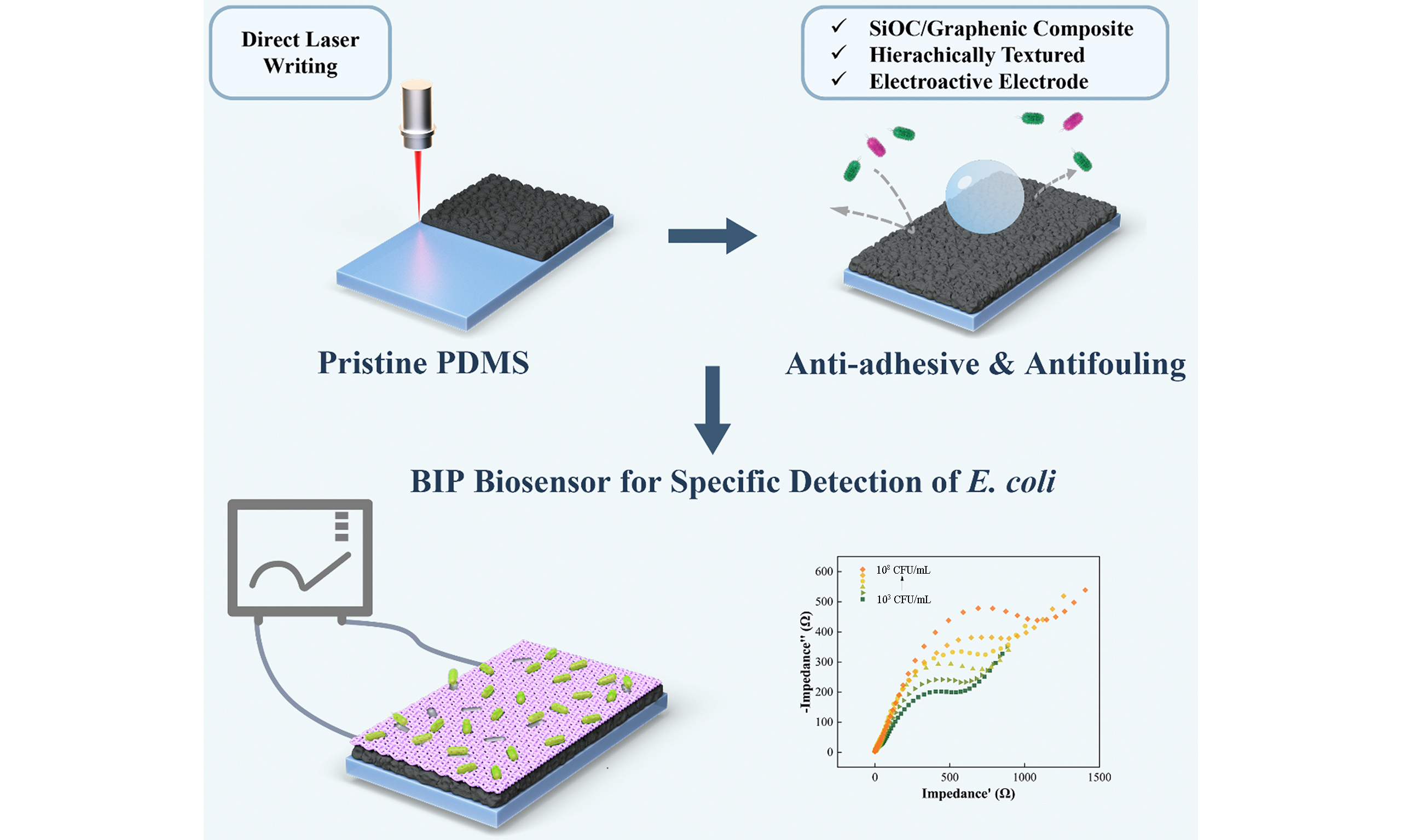
Polydimethylsiloxane (PDMS) is a foundational elastomer for flexible microdevices. Yet, a long-standing materials dichotomy fundamentally constrains its utility: surfaces engineered to resist biofouling typically lack the electrical conductivity and wettability required for electrochemical sensing. Here, we resolve this paradox through a one-step, ambient-air direct laser writing (DLW) strategy that monolithically transforms pristine PDMS into a superhydrophobic hierarchical silicon oxycarbide (SiOC)/graphenic composite (water contact angle = 157°, sliding angle ≈ 3°). Guided by the Young-Dupré equation, the tailored apparent work of adhesion of this architecture provides a thermodynamic barrier to biofouling, achieving a 94.47% reduction in Escherichia coli (E. coli) attachment under high-load conditions (108 CFU/mL). However, under applied electrochemical potentials, the hierarchical domains undergo localized electrowetting, enabling rapid interfacial charge transfer (k0 = 0.00708 cm/s) without compromising macroscopic antifouling properties. Leveraging this switchable interface, we demonstrate the in-situ fabrication of a bacterial-imprinted polymer (BIP) biosensor directly onto the antifouling substrate. Achieving a limit of detection of 348 CFU/mL, this work offers a feasible paradigm for monolithic, self-cleaning bioelectronics, eliminating the need for complex multi-material integration.
Keywords


 Download PDF
INTRODUCTION
Polydimethylsiloxane (PDMS) remains the dominant material in flexible microdevices, valued for its optical transparency, biocompatibility, and facile fabrication[1]. While the inherent insulating nature and susceptibility to hydrophobic biofouling of PDMS limit its utility in complex bioelectronics, researchers have historically managed this tradeoff through the intelligent engineering of composite materials. Strategies incorporating polyurethane copolymers[2], amphiphilic ionic polymers[3], metallic nanomaterials[4,5], and graphene-based materials[6] have successfully demonstrated dual electroactive and antifouling capabilities. However, translating these heterogeneous solutions to PDMS platforms introduces structural and operational compromises.
Surface-grafted hydrophilic polymers and drop-casted conductive inks suffer from thermodynamic incompatibility with the low-surface-energy PDMS substrate, inevitably leading to delamination under sustained microfluidic shear or electrochemical stress. Alternatively, incorporating metallic biocides leads to finite leaching and unintended cytotoxicity, while bulk doping with carbonaceous networks severely embrittles the elastomer. Consequently, a critical gap remains: achieving stable, high-performance electrochemical and antifouling properties in PDMS without relying on unstable heterogeneous coatings or mechanically destructive bulk additives.
While laser-based surface engineering presents a compelling pathway for spatially resolved material modification, its direct application to PDMS is fundamentally limited by the elastomer’s low intrinsic carbon yield. Common laser photothermal treatment fails to directly convert pristine PDMS into high-quality laser-induced graphene (LIG), despite the excellent electrochemical properties that render LIG highly desirable for electrochemical biosensing[7,8]. To circumvent this limitation, current methodologies rely on heterogeneous workarounds, such as doping the matrix with external carbon precursors[9] or employing cumbersome transfer protocols. Specifically, LIG is pre-fabricated on substrates such as polyimide (PI) and subsequently transferred onto PDMS surfaces[10]. Exogenous carbon doping alters the inherent physicochemical and mechanical properties of PDMS, whereas LIG transfer methods are hampered by procedural complexity and limited interfacial affinity. Also, ambient-fabricated LIG is inherently hydrophilic, which accelerates bacterial adhesion and biofouling, failing to meet rigorous antifouling and durability demands for long-term in-situ microbial monitoring.
Fabricating monolithic conductive PDMS requires harsh laser protocols, such as femtosecond-laser irradiation or oxygen-free pyrolysis, to yield conductive SiC[11,12]. Ambient CO2 laser treatment produces more insulating SiO2 that is primarily engineered to create superhydrophobic micro/nanostructures for wettability tuning[13-15]; a few reports functionalize them with Au nanoparticles for heavy-metal surface-enhanced Raman spectroscopy (SERS) detection[16], yet studies on conductive SiC-carbon phases remain largely confined to mechanical strain sensing[17]. Relevant literature rarely includes systematic characterizations of their electrochemical properties or evaluations of their electrochemical biosensing potential, and synergistic integration of inner conductive networks and outer antifouling textures for pathogen detection remains underexplored. The widespread belief that the outer superhydrophobic silica film blocks solid–liquid contact and hinders Faradaic charge transfer further explains why such materials have been largely overlooked as all-in-one biosensing platforms[18,19].
This study resolves this limitation by introducing a monolithic material transformation. Rather than applying an external functional layer, we utilize a one-step, ambient-air direct laser writing (DLW) strategy to photothermally transmute pristine PDMS into a hierarchical silicon oxycarbide (SiOC)/graphenic composite. Critically, we demonstrate that repeated laser passes are essential to allow thermal relaxation while building the hierarchical roughness necessary for both superhydrophobicity and enhanced electroactive surface area. The central innovation lies in the synergistic integration of robust antifouling properties and high-performance electrochemistry onto a single monolithic substrate. By systematically investigating the physicochemical and electrochemical properties of the DLW-modified domains, we demonstrate the in-situ fabrication of a bacterial-imprinted polymer (BIP) sensor directly onto the electroactive surface. The resulting platform [Scheme 1] presents a highly selective Escherichia coli (E. coli) biosensor that maintains mechanical and operational stability, advancing the rational design of multifunctional bio-analytical microdevices.
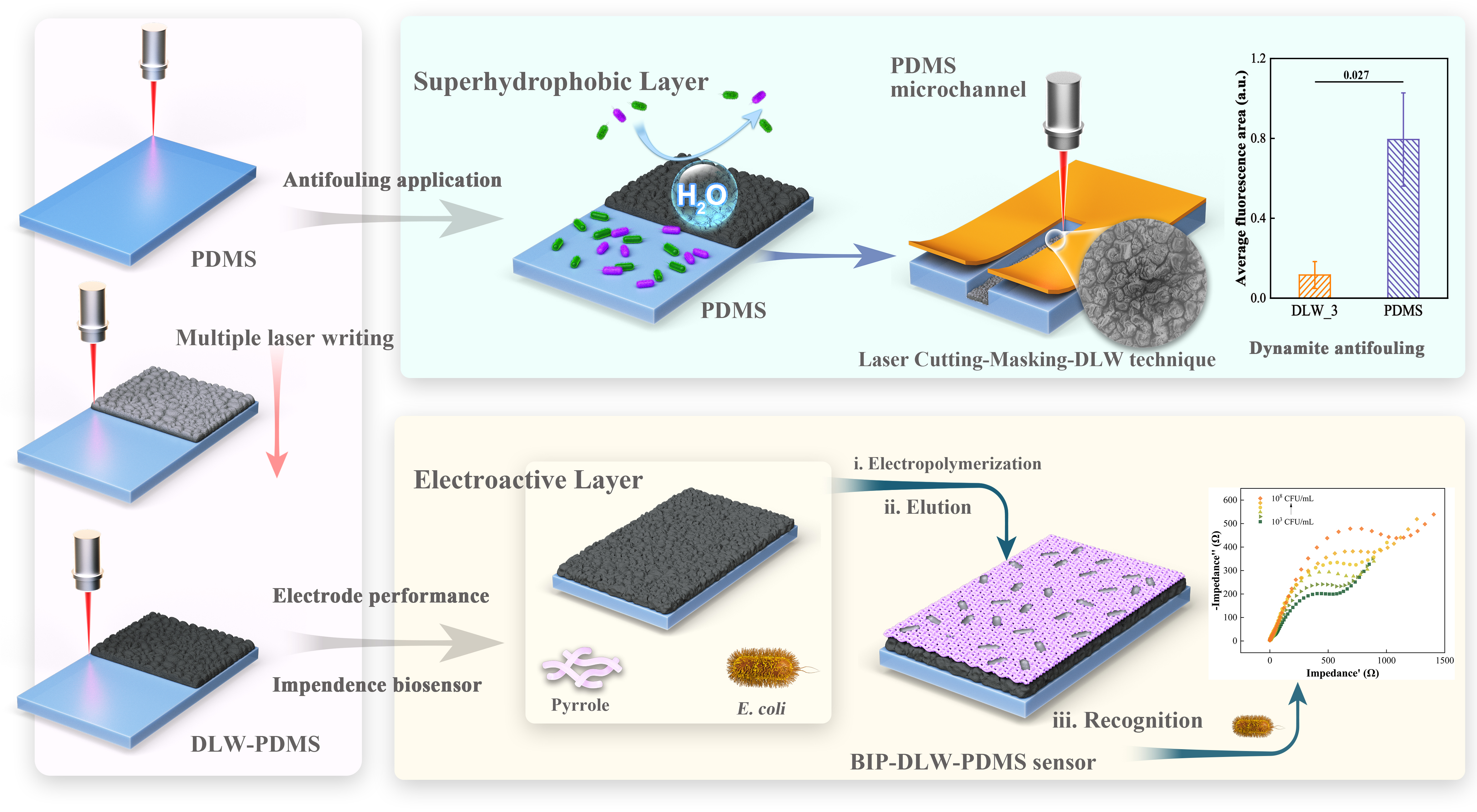
Scheme 1. Fabrication of a superhydrophobic and electroactive layer on PDMS via CO2 laser-based DLW and its bi-applications in antifouling and as functional electrodes and biosensors for BIP technology. PDMS: Polydimethylsiloxane; DLW: direct laser writing; BIP: bacterial-imprinted polymer; E. coli: Escherichia coli.
EXPERIMENTAL
Materials
The details of the chemicals and instruments used are provided in the Supplementary Text 1.
DLW method
The PDMS slices were positioned on a metal table and treated with a commercial 10.6 μm CO2 laser (MSDB-FM60, Minsheng Laser, China, 60 W) in ambient air. The parameters were set at a speed of 1,000 mm/s, a power of 1.8 W, a frequency of 10 kHz, and a pulse density of 5 pulses per dot, henceforth referred to as DLW_1. DLW_n denotes n repetitions of a laser scan under the DLW_1 condition. After treatment, the samples were sectioned into 1 cm × 1 cm pieces for further experiments.
Characterization
The chemical bonding in DLW-PDMS was investigated using attenuated total reflectance-Fourier transform infrared spectroscopy (ATR-FTIR) (Bruker, Germany) over the range 500-3,200 cm-1. A Raman microscope (inVia, Renishaw, UK) equipped with a 532 nm excitation was also employed. The exposure conditions were set to 10% power for 10 s under a 20× lens. The X-ray diffraction (XRD) pattern was obtained using a Rigaku instrument (SmartLab, Japan) with Cu Kα radiation. Each sample was scanned within the 10°-60° range at a rate of 2°/min. X-ray photoelectron spectroscopy (XPS) spectra were collected using a Thermo Scientific K-Alpha spectrometer (USA) operated at an accelerating voltage of 12 kV. Monochromatic Al Kα radiation was employed as the X-ray source. The surface structure of DLW-PDMS was visualized using a Hitachi Regulus 8100 scanning electron microscopy (SEM, Japan). Hydrophobicity was quantified using a DataPhysics OCA 15EC instrument (Germany). A 5 μL deionized-water droplet was deposited onto the sample surface to measure the water contact angle (WCA) and sliding angle (SA). For the SA measurement, the stage was conducted at a tilt speed of 1.0 °/s. Data represent average values from three independent samples per group, with three spots measured on each sample.
Antifouling capabilities assessment
E. coli (ATCC 25922) was prepared in LB broth, resuspended in 1× phosphate-buffered saline (PBS) solution, and adjusted to 108 colony-forming units per mL (CFU/mL). The DLW-PDMS samples were submerged in the bacterial solution at 37 °C for 24 h. The slices were then removed, rinsed gently with 1× PBS, and transferred to centrifuge tubes containing 5 mL of buffer for a sequential vortex-sonicate-vortex treatment (2-4-2 min). The bacteria were then counted using the plate counting method.
Bacterial viability was evaluated using a live/dead kit (Thermo Fisher, USA) and a microreader (SpectraMax iD3, Molecular Devices, USA). The image was captured with a 40× confocal fluorescence microscope (CLSM, Nikon, Japan). For long-term antifouling assessments, a working solution of E. coli expressing green fluorescent protein (E. coli-GFP, ATCC 25922-GFP) was cultured with the DLW-PDMS sheets at 37 °C for 7 days. Following rinsing, surface biofilm formation was captured using CLSM. The excitation wavelength was 488 nm, with a Z-stacking interval of 2.5 μm. The 3D visualizations were generated using the 3D viewer plugin in FIJI[20].
DLW-PDMS microchannel preparation and dynamic antifouling performance evaluation
The preparation of the DLW-PDMS microchannel is detailed in Supplementary Text 3. The treated chips underwent a triple ethanol-deionized (DI) water cleaning before evaluation. The chips were positioned on a heating table at 37 °C. A 50 mL syringe containing a bacterial solution of E. coli-GFP at 108 CFU/mL was circulated through the DLW-PDMS chip at 5 μL/min, with the solution renewed every 48 h to preserve bacterial activity. Following a week-long circulation of the bacterial solution, unattached bacteria were cleared with a PBS rinse, and the chips were then observed at 20× magnification using CLSM.
Electrochemical properties
Electrochemical impedance spectroscopy (EIS) and cyclic voltammetry (CV) experiments were utilized to examine the electrochemical properties of DLW-PDMS as a working electrode. EIS experiments were conducted on a Zahner electrochemical workstation (Zennium XC, Germany) using a 100 mM solution of [Fe(CN)6]3-/4- in 0.1 M KCl. The experimental conditions were set to an amplitude of 10 mV and a frequency range of 0.1-100,000 Hz. The CV experiments were performed in a 5 mM [Fe(CN)6]3-/4- containing 0.1 M KCl. Routine CV characterization was conducted at a scan rate of 50 mV/s over the range of -0.6 to 1.2 V. For parameter-specific measurements, CV at 30 mV/s from -0.1 to 0.5 V was used to calculate the electron transfer rate constant (k0) and CVs at varied scan rates were recorded to determine electrochemically active surface area (A). For double-layer capacitance (Cdl), CVs at varied scan rates were acquired in PBS from 0.2 to 0.4 V. Independent scans from -1.2 to 2.0 V were used to evaluate the electrochemical window (ECW). Detailed calculations are available in the Supplementary Materials.
Electropolymerization fabrication procedure for bacterial (non)-imprinted polymer sensors with DLW-PDMS
The electrochemical polymerization was carried out in a 10 mL solution containing 0.1 M KCl, 0.05 M pyrrole, and E. coli at 108 CFU/mL. The process was performed within an ECW of -0.3 to 0.7 V at a scan rate of 50 mV/s. After eight electrochemical cycles, the sample was transferred to an elution solution containing 5% w/v sodium dodecyl sulfate (SDS) in 5% v/v HAc solution (pH = 2.47) and immersed for 1 h to remove the bacteria. Following the elution step, the electrode was gently rinsed with sterilized DI water. The resulting electrode was identified as the BIP-DLW-PDMS sensor. A non-imprinted polymer (NIP) electrode was prepared using the same steps without adding bacteria during electropolymerization and served as a control. SEM was employed to study the morphology of each step in the process, while E. coli-GFP was used for in-situ confocal fluorescence imaging (60× lens).
Impedimetric detection of E. coli based on BIP-DLW-PDMS
The prepared BIP-DLW-PDMS was incubated with various concentrations of E. coli from 103 to 108 CFU/mL for 2.5 h. EIS measurement was conducted in a 5 mM [Fe(CN)6]3-/4- solution containing 0.1 M KCl over a frequency range of 0.1 to 100,000 Hz. The impedimetric changes were processed using: ΔRct = |Rct1 - Rct0|, where Rct1 is the value of the Rct of the BIP-DLW-PDMS sensor with bacteria, Rct0 is the value without bacteria.
Performance evaluation of BIP-DLW-PDMS sensor
S. aureus (ATCC 25923), S. enterica (ATCC 13076), P. aeruginosa (ATCC 27853), and E. faecalis (ATCC 6569) were selected as interferents [Supplementary Table 1]. Individual measurements for all tested bacteria, including the target E. coli, were performed at 108 CFU/mL. For mixed-culture competitive selectivity tests, E. coli (107 CFU/mL) was mixed with pooled non-target bacteria (total 108 CFU/mL), corresponding to a target-to-nontarget concentration ratio of 1:10.
The real samples, including tap water, seawater, and milk, were collected from local sources. Milk samples were pretreated by a two-fold dilution before measurement. The real samples were spiked with 106 CFU/mL E. coli and tested in triplicate.
RESULTS AND DISCUSSION
Formation and structural evolution of the SiOC composite
Initial single-pass DLW treatments under varied parameters [Supplementary Table 2 and Supplementary Movie 1] produced surfaces with minor visual differences [Supplementary Figure 1]. Still, they induced significant compositional changes in the Si-O, Si-C, and carbon [Supplementary Figure 2]. Owing to PDMS’s substantial infrared absorption at 10.6 μm, a CO2 laser at this wavelength enables efficient photothermal modification under ambient-air conditions[21]. Notably, increased carbon content and reduced SiO2 correlated with improved antifouling efficacy [Supplementary Figure 3]. This observation prompted a strategy of increased laser-overlap exposures, hypothesized to promote a more ordered, graphitic carbon structure[22], which is known to exhibit superior interfacial properties compared to amorphous carbon[23,24].
To investigate this hypothesis, the PDMS substrate was subjected to multiple laser scans under the optimal antifouling parameter condition identified by principal component analysis (PCA), denoted F10k [Supplementary Figure 4]. This repetitive process induced a distinct visual transformation from white to black [Supplementary Figure 5], indicative of substantial photothermal modification. FTIR spectra revealed a surface chemistry dominated by Si–O and Si–C bonds [Figure 1A]. A progressive narrowing of the Si–O peak [Supplementary Table 3] suggests the selective ablation of the amorphous SiO2 layer with repeated laser exposure[25]. Concurrently, Raman spectroscopy demonstrated a pronounced increase in the D and G band intensities [Figure 1B]. The intensity ratio (ID/IG) decreased with successive scans, reaching 1.13 for the DLW_3 condition (DLW_n denotes n laser-scan repetitions), consistent with the formation of an ordered, pyrolyzed carbon structure[26]. Extended scanning further weakened both carbon-related bands and raised the ID/IG ratio to 1.23, reflecting structural evolution and partial degradation of the carbon phase.
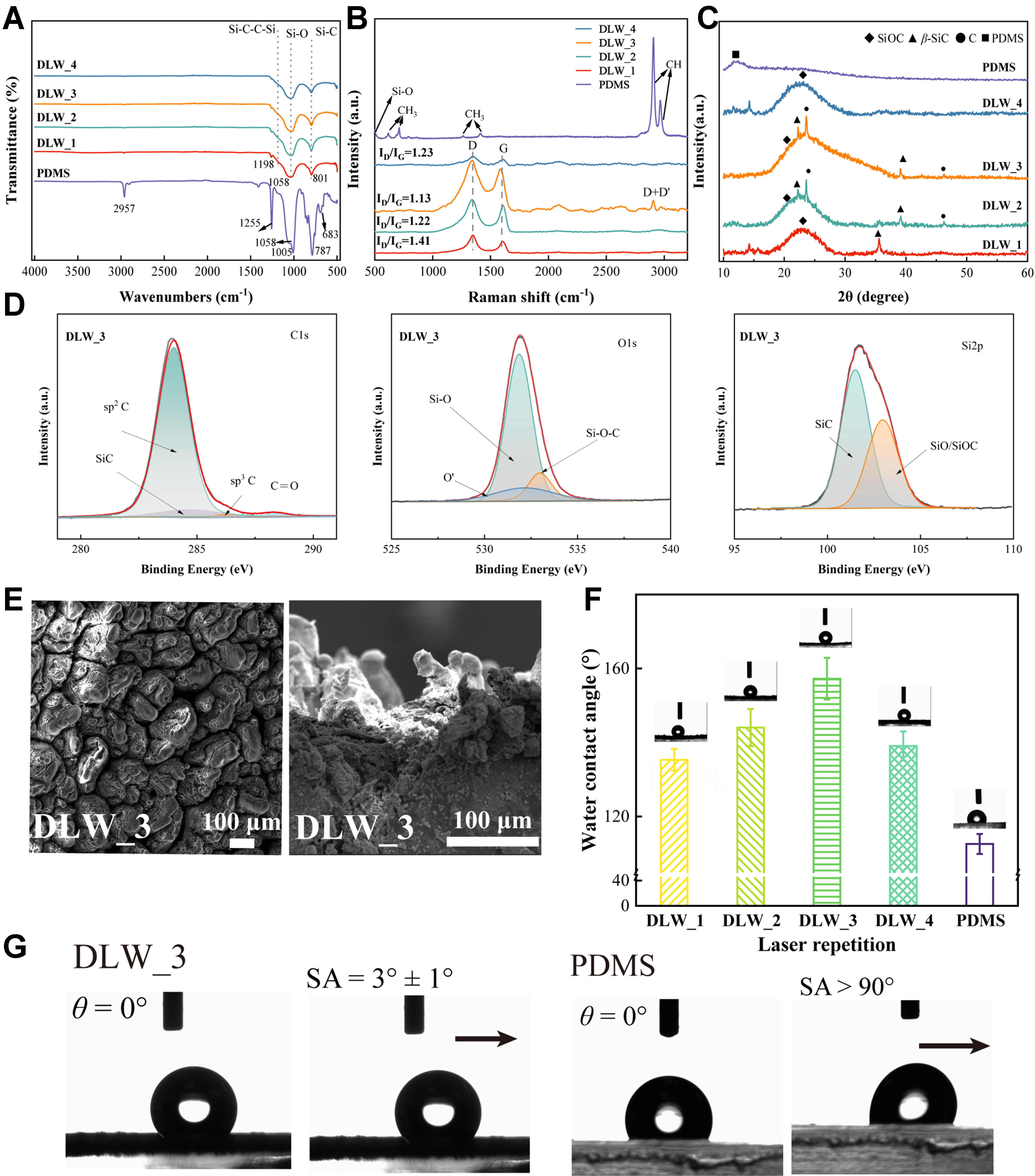
Figure 1. Characterization of the DLW-PDMS under different laser repetitions. DLW_n denotes n repetitions of a laser scan (1,000 mm/s speed, 1.8 W power, 10 kHz frequency, 5 pulses/dot). PDMS refers to the untreated substrate. (A) ATR-FTIR spectra; (B) Raman spectra showing the evolution of D and G bands; the ID/IG ratio is compared; (C) XRD patterns illustrating the emergence of crystalline phases with increased repetitions; (D) XPS spectra of the C 1s, O 1s, and Si 2p regions for the DLW_3 surface. The XPS spectra from other conditions are presented in Supplementary Figure 6; (E) SEM images of the hierarchical morphology of the DLW_3 surface; the scale bar is 100 μm. Other conditions are displayed in Supplementary Figure 7; (F) WCA measurements for untreated PDMS and DLW-treated surfaces; (G) SA measurements of DLW_3 and PDMS. The SA measurements from other conditions are presented in Supplementary Figure 8. Data for WCA and SA are presented as mean ± SD (n = 3, three independent samples per group). DLW: Direct laser writing; PDMS: polydimethylsiloxane; ATR-FTIR: attenuated total reflectance-Fourier transform infrared spectroscopy; XRD: X-ray diffraction; XPS: X-ray photoelectron spectroscopy; SEM: scanning electron microscopy; WCA: water contact angle; SA: sliding angle; SD: standard deviation; SiOC: silicon oxycarbide.
XRD analysis provided further insight into the crystalline evolution of the surface [Figure 1C]. The untreated PDMS exhibited broad amorphous peaks (2θ = 12° and 22.6°). However, the laser-treated surfaces initially showed one broad peak near 23°, characteristic of amorphous SiO2 or SiOC[27,28]. With increasing numbers of laser repetitions, distinct diffraction peaks corresponding to crystalline SiC and graphitic carbon emerged[29,30]. These signals imply phase segregation during the formation of crystalline domains, consistent with previously reported mechanistic analyses[31,32]. Notably, the absence of characteristic SiC diffraction peaks in DLW_4 agrees with the trends observed in Raman measurements and with earlier reports that excessive thermal exposure drives carbon vaporization and suppresses SiC formation[28].
To elucidate the precise surface chemistry, XPS was performed on the DLW samples [Figure 1D and Supplementary Figure 6]. Deconvolution of the high-resolution C 1s spectrum resolved peaks at sp2-C (284 eV), Si-C (284.7 eV), sp3-C (286 eV), and C=O (288.3 eV), while the Si 2p region indicated the presence of Si-C (101.5 eV) and a combined Si-O/Si-OC phase (102.9 eV)[33,34]. The measured elemental composition for the DLW_3 surface (Si = 26.81 at%, O = 34.44 at%, C = 38.75 at%; Supplementary Table 4) is in close agreement with values reported for pure SiOC[34]. The reduction in carbon content for DLW_4 (33.71 at%, Supplementary Table 4) coincides with structural changes detected by Raman and XRD measurements, suggesting carbon depletion under excessive laser irradiation. These results revealed that repetitive laser pyrolysis in ambient air transforms the PDMS surface into a composite material comprising an amorphous SiOC matrix embedded with crystalline SiC and graphene-like carbon nanoparticles[17,35].
The laser-induced physicochemical transformation culminated in a hierarchical surface morphology, characterized by a wrinkled, multi-scale structure, as observed by SEM [Figure 1E and Supplementary Figure 7]. The resulting micro- and nanoscale topography, combined with the altered surface chemistry, drove a transition to superhydrophobicity. The WCA increased from 112° on pristine PDMS to 157° for the DLW_3 condition [Figure 1F], accompanied by a dramatic reduction in the SA to 3° [Figure 1G and Supplementary Figure 8], indicating a robust Cassie-Baxter wetting state consistent with a previous report[15]. Further increases in the number of laser scans produced heavily wrinkled surface topography for DLW_4. These morphological alterations, together with the compositional degradation identified above by Raman, XPS, and XRD, coincided with degraded wetting performance: the WCA decreased to 139°, whereas the SA increased to 11°. The combined set of observations points to partial volatilization of surface-enriched SiOC/C and disruption of the beneficial hierarchical micro-nanotexture, counteracting the advantages achieved under moderate repeated laser exposure.
Thermodynamic antifouling performance
The antifouling efficacy of the DLW-PDMS surfaces was systematically evaluated via CFU assays. The DLW_3 surface exhibited optimal performance, achieving a 94.47% reduction in E. coli attachment after 24 h compared to the untreated PDMS control (P = 0.0019, Figure 2A). Consistently, confocal images revealed synchronous reductions in both live and dead bacteria [Supplementary Figure 9]. This efficiency is highly competitive with state-of-the-art LIG filtration membranes loaded with 0.5 mg/cm2 graphene oxide (GO, 95%)[36] and PDMS doped with gallium-based liquid metal nanodroplets (90%)[4].
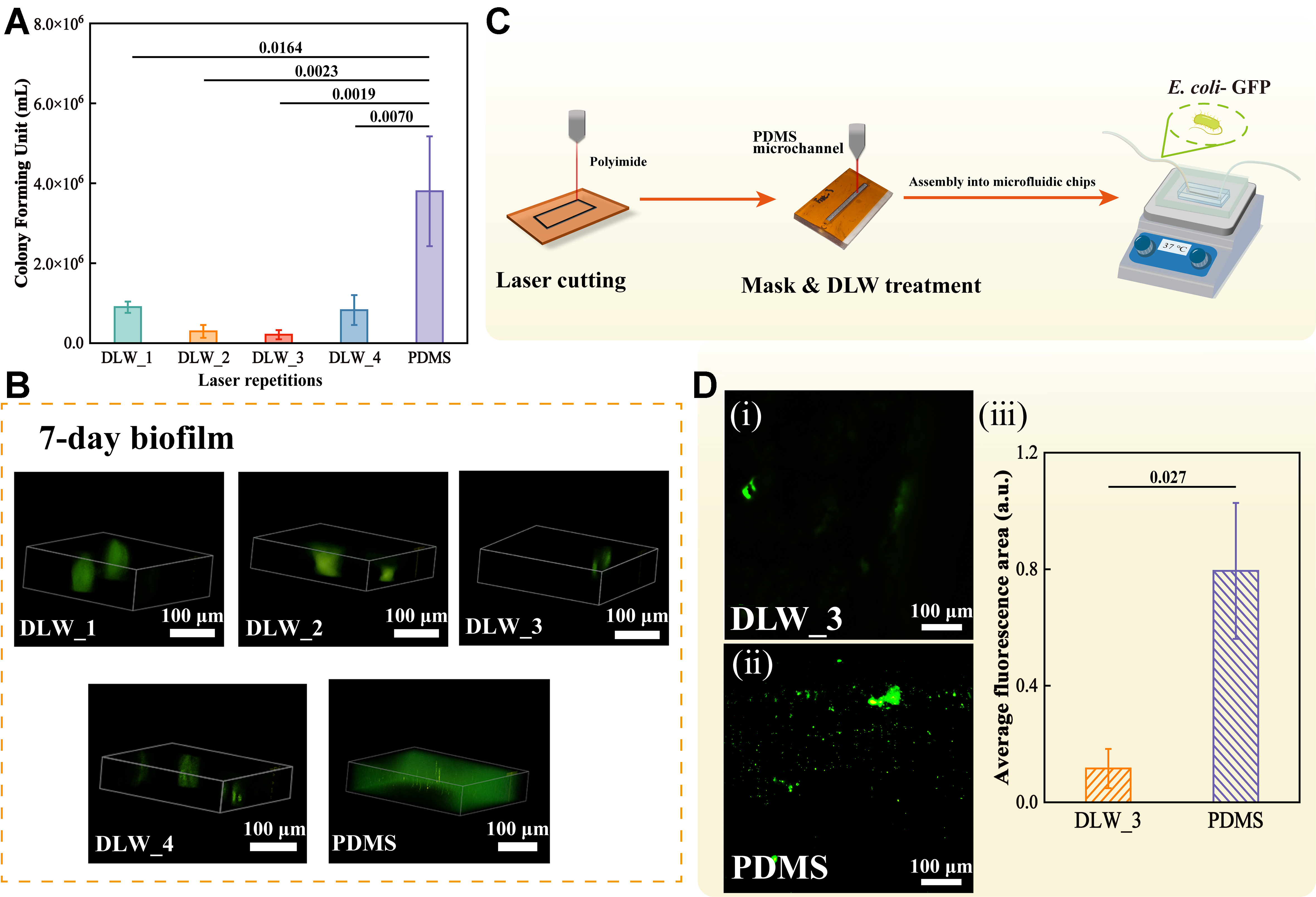
Figure 2. Antifouling performance of DLW-PDMS surfaces. DLW_n denotes n repetitions of the laser treatment, and PDMS refers to the untreated substrate. (A) Quantification of bacterial (E. coli) adhesion via CFU counts after 24 h of incubation. Data are presented as mean ± SD (n = 3, independent substrates per group). Statistical significance was determined using ANOVA with Tukey’s test, with pure PDMS as the control group; (B) Representative 3D CLSM reconstructions of E. coli-GFP biofilm formation after 7 days of static culture on DLW_n surface and an untreated PDMS control. Scale bars: 100 μm; (C) Schematic of the fabrication process for integrating DLW-modified surfaces into a PDMS microfluidic device; (D) CLSM images showing bacterial adhesion in a DLW_3-treated channel (i) and an untreated PDMS control channel (ii) after 7 days of dynamic flow. Scale bars: 100 μm. (iii) The quantification of the fluorescent area was analyzed using an unpaired t-test. Each group replicates (n = 3), with 5 sites per sample as technical replicates, at 20× magnification. Data are presented as mean ± SD. DLW: Direct laser writing; PDMS: polydimethylsiloxane; E. coli: Escherichia coli; CFU: colony-forming unit; SD: standard deviation; ANOVA: analysis of variance; CLSM: confocal fluorescence microscope; GFP: green fluorescent protein.
When moving to the DLW_4 condition, antifouling performance declined. Despite observing a higher ratio of dead attached bacteria on DLW_4 [Supplementary Figure 9F], concurrent increases in both live and dead bacterial and CFU counts do not support a bactericidal effect. The elevated dead-cell proportion likely arises from prolonged residence of bacteria trapped in surface crevices after adhesion, rather than from direct membrane rupture induced by surface topography[37].
Long-term stability assessments under static culture revealed extensive biofilm formation on the pristine PDMS surface. In contrast, the DLW_3 surface supported only sparse, vertically oriented microcolonies [Figure 2B]. This profound resistance to biofilm development is driven by the hierarchical topography, which stabilizes an interfacial air layer, thereby minimizing the solid-liquid contact area available for bacterial anchoring[38].
Recent research highlights laser-engineered topographical patterning as an effective strategy for constructing cell-repellent biointerfaces for implantable bioelectronics, in which surface structuring suppresses cell adhesion without relying on biocidal components[39]. Moreover, prior literature indicates that SiOC particles exhibit favorable biocompatibility and exert no inhibitory effect on bacterial proliferation[40-42]. These findings support our inference of a predominantly anti-adhesive mechanism.
To demonstrate the practical utility of this technology for mitigating biofouling, a critical challenge in PDMS-based microfluidics, the DLW process was applied to the interior of microscale channels [Figure 2C]. The modified channels were assembled into microfluidic devices and subjected to 7 days of continuous circulation with a high-load bacterial suspension (7 days with the wall shear stress only at 0.25 Pa, Supplementary Text 3). Post-experiment analysis revealed substantial bacterial accumulation and biofilm formation within the untreated PDMS control chip, particularly along the channel edges [Figure 2D]. Conversely, the DLW_3-treated channel exhibited only minimal bacterial adhesion, representing a statistically significant reduction in surface fouling (P = 0.027). These results validate the mechanical robustness and operational stability of the superhydrophobic SiOC layer under dynamic flow conditions.
Due to the hierarchical, superhydrophobic nature of the DLW_3 composite (WCA = 157°), the thermodynamic barrier to biofouling was evaluated via the apparent work of adhesion (Wa). Applying the Young-Dupré equation: Wa = γw(1 + cosθw), the Wa of the optimized SiOC matrix was calculated to be exceptionally low (≈ 5.8 mN/m). This thermodynamic barrier to aqueous wetting disfavors the stable anchoring of E. coli extracellular polymeric substances, which could somewhat explain the observed reduction in bacterial adhesion. Nevertheless, it should be noted that the adhesion derived from WCA measurements serves only as an effective surface-wetting descriptor. Bacterial adhesion is governed by multiple additional factors beyond surface wettability, including hydration state, extracellular substances, and surface charge[37].
Interfacial electrochemical kinetics
The formation of graphene-like carbon structures within the SiOC matrix[43], combined with the established biocompatibility and chemical stability of polymer-derived SiOC ceramics as electrode materials[44], motivated a rigorous investigation of the platform’s electrochemical properties. As shown in Figure 3A, the EIS plot revealed a dramatic decrease in Rct, from 44.4 Ω for a single pass (DLW_1) to < 1 Ω (DLW_3). This value significantly outperforms comparable SiOC/C materials (Rct = 82.78 Ω)[45]. These findings, which position the DLW_3 surface as a highly conductive electrode, were corroborated by CV measurements [Figure 3B]. The CV response and redox currents rose with laser repetitions, peaking at DLW_3 and decreasing at DLW_4. This trend aligns with our materials characterization, which indicates that three repetitions yield an optimal balance of high graphitic carbon content and low structural defects [Figure 1 and Supplementary Table 4]. In contrast, excessive exposure may lead to the degradation or vaporization of the conductive carbon phase[46]. This observation further supports the conclusion that DLW-PDMS forms a composite material consisting of SiOC/C. While pure SiO2 is intrinsically insulating, the generated interconnected carbon network provides pathways for efficient electron transport across the substrate[19,47].
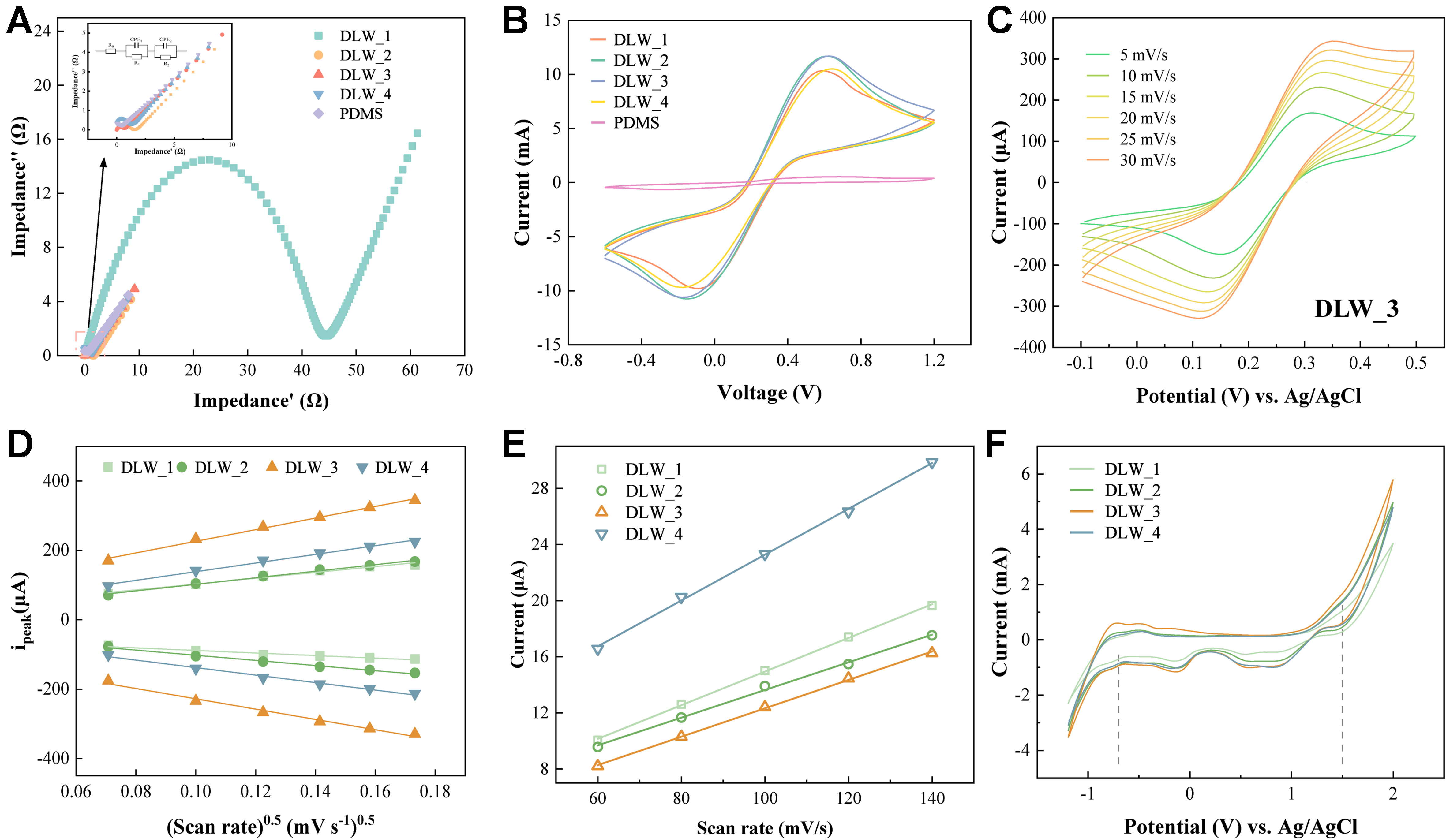
Figure 3. Electrochemical properties of DLW-PDMS electrodes. DLW_n denotes n repetitions of the laser treatment, and PDMS refers to the untreated substrate. (A) Nyquist plots from EIS analysis in 100 mM [Fe(CN)6]3-/4- for surfaces with different laser repetitions. Inset: equivalent circuit model used for fitting; (B) Cyclic voltammograms in 5 mM [Fe(CN)6]3-/4- at 50 mV/s; (C) CV curves for DLW_3 with a redox probe and varying scan rates; (D) Randles-Sevcik plots showing the linear relationship between peak current (ipeak) and the square root of the scan rate (v0.5) for electrodes with different laser repetitions; (E) Estimation of double-layer capacitance (Cdl) of DLW-PDMS electrodes; (F) Comparison of ECWs of DLW-PDMS electrodes. DLW: Direct laser writing; PDMS: polydimethylsiloxane; EIS: electrochemical impedance spectroscopy; CV: cyclic voltammetry; ECWs: electrochemical windows.
Under time-varying electric fields, transient Maxwell electrostatic stress precludes instantaneous steady-state force balance, as illustrated by AC-electrowetting observations where droplet inertia hinders cycle-resolved interfacial relaxation[48]. In the CV measurements, a continuously swept potential generates a transient Maxwell stress concentrated at the liquid-air-solid three-phase contact lines. This stress may locally overcome the Laplace capillary barrier, inducing spatially confined plastron collapse without a global Cassie-to-Wenzel transition or degradation of hierarchical micro/nanostructures[49,50]. Unlike static-DC electrowetting, no steady-state Maxwell-Laplace equilibrium is established during potential scanning. Benefiting from a hierarchical microstructure, wetting is confined to isolated domains, and macroscopic superhydrophobicity is preserved[51].
Quantitative analysis of the CV data [Table 1] revealed a heterogeneous electron transfer rate constant (k0) of 0.00708 cm/s for DLW_3, rivaling standard electrode materials such as glassy carbon (k0 = 0.00782 cm/s) and pyrolytic carbon (k0 = 0.0054 cm/s)[52,53]. A good linear relationship between peak current and the square root of sweep rate demonstrates that the electrochemical reaction is governed by semi-infinite diffusion of redox-active species toward the electrode surface [Figure 3C and Supplementary Figure 10]. As shown in Figure 3D, the electrochemically active surface area (A) reached its maximum of 1.674 cm2 for DLW_3, which is attributed to the laser-pyrolyzed hierarchical roughness.
Comparison of calculated electrochemical parameters based on CV
| Parameters for electrochemical performance evaluation | DLW_1 | DLW_2 | DLW_3 | DLW_4 |
| K 0 (cm/s) | 0.00268 | 0.00416 | 0.00708 | 0.00533 |
| A (cm2) | 0.841 | 0.943 | 1.674 | 1.247 |
| Cdl (μF/cm2) | 142.66 | 104.62 | 60.50 | 131.05 |
Analysis of the non-Faradaic potential regions revealed quasi-rectangular CV curves, indicative of double-layer capacitive behavior [Supplementary Figure 11]. The DLW_3 electrode maintained this ideal rectangular shape even at high scan rates, suggesting excellent charge propagation and electrochemical stability. DLW_3 electrode’s double-layer capacitance (Cdl) reached a minimum of 60.50 μF/cm2 [Figure 3E and Table 1], minimizing non-Faradaic background currents, thereby improving the signal-to-noise ratio. This value is comparable to conventional noble metal electrodes (platinum: 64 μF/cm2, silver: 53 μF/cm2)[52], and superior to other laser-modified materials, such as carbon black-infused polylactic acid (PLA, 96.2 μF/cm2)[54]. The stable ECW of the DLW-PDMS electrode spanned -0.7 to +1.5 V [Figure 3F], a wide operational range similar to that of standard graphite (-0.5 to +1.6 V) and pyrolytic carbon (-0.9 to +1.6 V) electrodes[52], confirming the platform’s suitability for diverse electroanalytical applications.
A defining feature of the DLW_3 platform is its ability to reconcile superhydrophobicity with high-performance electrochemistry. Traditional electrochemical theory dictates that a Cassie-Baxter state (WCA = 157°) should severely impede Faradaic processes due to the insulating air plastron. However, the DLW_3 electrode exhibits interfacial electron transfer kinetics (k0 = 0.00708 cm/s). We attribute this unique capability to a potential-driven electrowetting transition. While the hierarchical SiOC matrix thermodynamically repels bacterial suspensions under open-circuit conditions, preventing the initial formation of focal adhesion complexes, applying sweeping potentials during CV locally reduces the liquid-solid interfacial tension. This transient, potential-induced wetting allows the aqueous electrolyte to penetrate the graphitic nano-domains, dynamically facilitating rapid charge transfer. Crucially, upon removal of the applied potential, the robust hierarchical topography prevents total Wenzel wetting, allowing the macroscopic antifouling properties to persist under dynamic microfluidic flow. This dynamic interfacial behavior distinguishes the DLW_3 composite from conventional LIG electrodes, which become rapidly fouled, and standard superhydrophobic polymers, which remain electrochemically inert. The reproducibility of this favorable electrochemical performance was further verified by impedance measurements, which revealed negligible intra-batch variation and excellent batch-to-batch consistency for DLW_3 [Supplementary Table 5].
Repeated laser scanning produces a suite of competing effects that dictate final surface properties: progressive laser-driven carbonization generates electroactive graphitic domains embedded within the SiOC matrix; evolving multiscale surface roughness builds the hierarchical micro-nano topography essential for Cassie-Baxter superhydrophobic antifouling; cumulative laser input induces heat accumulation, thermal stress, and solid-state phase transformations originating from thermal-relaxation processes; and excessive laser fluence causes material ablation. These effects respond differently as the number of repetitions increases. Moderate repeated scanning from DLW_1 to DLW_3 yields favorable carbon enrichment and well-defined hierarchical morphology without severe ablation or thermal damage, delivering balanced antifouling and electrochemical performance. Further increasing scan passes to DLW_4 breaks this optimal trade-off: carbon content decreases, and electrochemical activity weakens owing to ablation-driven loss of graphitic carbon; meanwhile, excess heat generates thermal-stress-mediated structural defects, and ablation degrades the Cassie-Baxter-supporting hierarchical topography. These combined factors explain the superior overall performance observed for the DLW_3 condition.
In-situ fabrication and evaluation of the BIP biosensor
While microfluidic devices offer an attractive platform for rapid bacterial diagnostics[55,56], integrating biorecognition elements onto PDMS surfaces remains a significant bottleneck, often requiring complex, time-consuming protocols[57,58]. To address this, we integrated BIP technology, a robust approach for creating synthetic receptors using whole cells as templates[59], directly onto the DLW_3 electrode.
The in-situ electropolymerization of pyrrole in the presence of E. coli templates [Figure 4A] resulted in a high-impedance state (Rct = 1.5 kΩ in Figure 4B) due to bacterial encapsulation [Figure 4B-D and Supplementary Figure 12]. SEM and confocal images showed the uniform entrapment of intact bacterial cells within the reticulated 3D Ppy network [Figure 4C(I) and D(I)]. Subsequent template elution generated highly specific, three-dimensional recognition cavities, as evidenced by a sharp decrease in impedance to
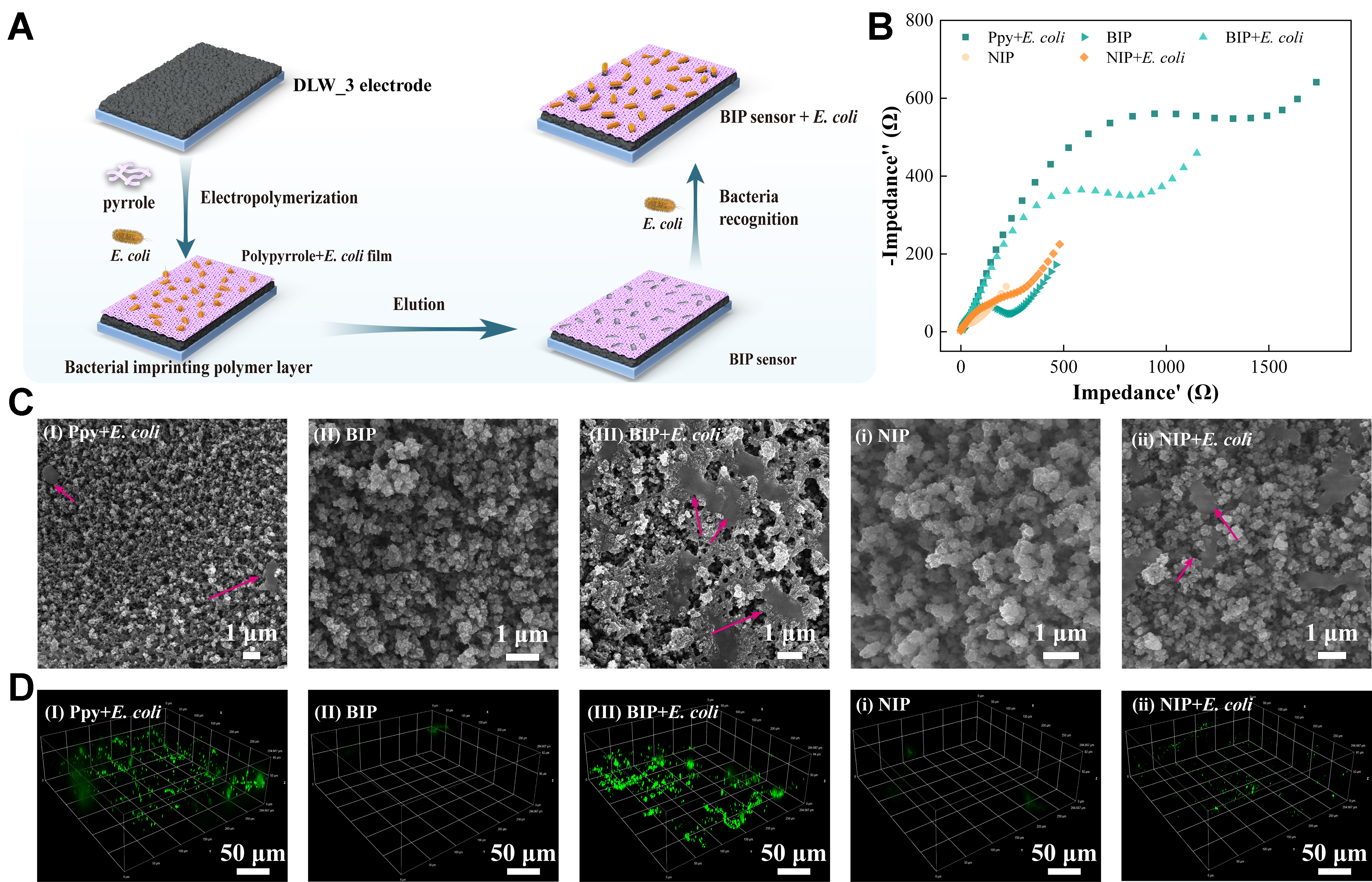
Figure 4. Fabrication and characterization of a BIP sensor on a DLW-PDMS electrode. The DLW_3 condition was used as the base electrode material. (A) Schematic illustrating the fabrication of the BIP sensor via electropolymerization of pyrrole around E. coli templates, followed by template removal and specific rebinding; (B) Nyquist plots from EIS analysis tracking the impedance changes at each stage of sensor fabrication and operation; (C) SEM micrographs and (D) corresponding in-situ confocal fluorescence maps (60× lens) of the electrode surface after: (I) initial polymerization of the polypyrrole layer with entrapped E. coli (Ppy + E. coli); (II) template elution to form the BIP; and (III) rebinding of target bacteria (BIP + E. coli). Control experiments with a (i) NIP for (ii) recognition of E. coli are shown for comparison. The scale bars for SEM and confocal are 1 and 50 μm, respectively. BIP: Bacterial-imprinted polymer; DLW: direct laser writing; PDMS: polydimethylsiloxane; E. coli: Escherichia coli; EIS: electrochemical impedance spectroscopy; SEM: scanning electron microscopy; NIP: non-imprinted polymer.
Under the optimal condition [Supplementary Figure 13], the BIP-DLW_3 sensor demonstrated a wide dynamic range for E. coli (103 to 108 CFU/mL), with an increased Rct in a concentration-dependent manner [Figure 5A], yielding a limit of detection (LOD) of 348 CFU/mL (adjusted R2 = 0.9882, Figure 5B). The fabrication protocol proved highly reproducible, with negligible response variation observed across five independent batches (3 replicate sensors per batch) when challenged by 108 CFU/mL E. coli [Figure 5C].
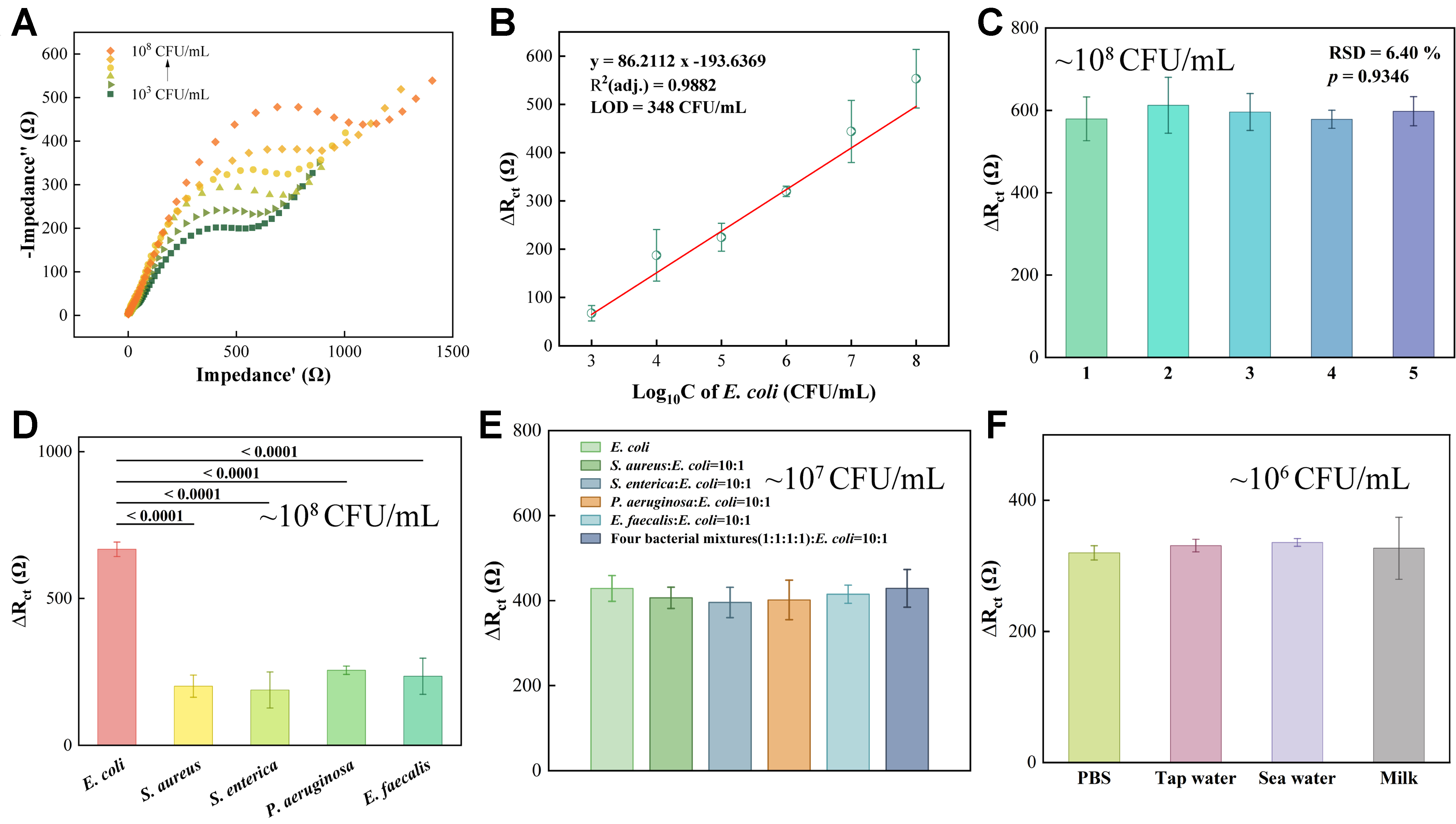
Figure 5. Quantitative and performance evaluations of BIP-DLW-PDMS sensors for E. coli detection. The DLW_3 electrode was used for all experiments. (A) Nyquist plots showing the sensor’s response to E. coli concentrations from 103 to 108 CFU/mL; (B) Calibration curve plotting the change in charge transfer resistance (ΔRct) against the logarithm of bacterial concentration. The linear regression equation is ΔRct = 86.2112Log10CE.coli - 193.6369; (C) Reproducibility assessment across five independent batches, with three technical replicate sensors (n = 3) per batch for detection of E. coli at 108 CFU/mL; (D) Selectivity evaluation against four non-target bacterial species and the target E. coli (all at 108 CFU/mL); (E) Competitive selectivity test in a mixed solution containing target E. coli (107 CFU/mL) and non-target bacteria (108 CFU/mL), with a target E. coli to non-target bacteria ratio of 1:10; (F) Sensor response to E. coli (106 CFU/mL) spiked into real-world samples (tap water, seawater, milk). All experiments were performed in triplicate (n = 3). Data are presented as mean ± SD. Statistical significance was determined using ANOVA with Tukey’s test. BIP: Bacterial-imprinted polymer; DLW: direct laser writing; PDMS: polydimethylsiloxane; E. coli: Escherichia coli; CFU: colony-forming unit; SD: standard deviation; ANOVA: analysis of variance; LOD: limit of detection; RSD: relative standard deviation; PBS: phosphate-buffered saline.
While the BIP-DLW_3 sensor was exposed to four different non-target bacterial species at a high concentration (108 CFU/mL), the impedance response was minimal. In contrast, the response to the target E. coli was 2.6- to 3.5-fold greater (P ≤ 0.0001), confirming the sensor’s high specificity [Figure 5D]. This selectivity was further validated in a competitive assay, where the sensor preferentially detected target E. coli that constituted approximately 9.1% of a mixed bacterial population (Figure 5E, concentration of target E. coli is 107 CFU/mL). This high degree of selectivity is directly attributed to the specific recognition cavities, complementary in size, shape, and chemical functionality to the E. coli template, engineered into the polymer matrix during the imprinting process[60].
Practical utility was validated in complex matrices (tap water, seawater, milk). The sensor’s response to E. coli spiked into tap water and seawater (final concentration = 106 CFU/mL) closely matched the response observed in the clean PBS buffer control. While greater response variability was observed in minimally diluted milk samples, the results demonstrate the sensor’s robust performance and potential for direct application to real-world samples with minimal pretreatment [Figure 5F and Supplementary Table 6].
Compared to conventional bacterial methods such as agar plating and enzyme-linked immunosorbent assay (ELISA)[61,62], this unified DLW-PDMS platform enables rapid (< 4 h), highly sensitive, and label-free bacterial identification. Notably, our BIP-functionalized DLW-PDMS electrode, without additional nanomaterials pre-modification, achieves a LOD comparable to that of gold and carbon electrodes that employ such nanostructured pre-treatment to enhance sensing performance when functionalized with diverse recognition moieties [Table 2].
Comparison of bacterial detection methods
| Bacteria | Modified materials and electrode | Detection methods | Concentration range (CFU/mL) | LOD (CFU/mL) | Ref. |
| E. coli OP50 | MIP-stainless steel microwires | EIS | 102-107 | 2 × 10² | [63] |
| E. coli O157:H7 | Y6NPs@Au@antibody | Colorimetry and photothermometry | 2.5 × 10² - 5 × 10⁶ | CL: 1,725 PT: 311 | [64] |
| E. coli K12 | Lectin/SWCNT/gold electrode | LSV | 2 × 10³ - 2 × 10⁸ | 4.7 × 10³ | [65] |
| E. coli O157:H7 | Ag@PD-GCE | Amperometry and non-faradaic EIS | - | 103 | [66] |
| E. coli | Laser-treated GO/antibody | Amperometry | 917 - 2.1 × 10⁷ | 283 | [67] |
| E. coli | ZnO/CdS/Ag2CO3/graphite | CV | 102-107 | 1 × 10² | [68] |
| E. coli | Ppy/DLW-PDMS | EIS | 103-108 | 348 | This work |
Taking advantage of the template-replaceable nature of molecularly imprinted technology, our BIP sensor shows promise for extending detection from single-target detection of E. coli to a broader spectrum of analytes, including other bacterial pathogens as well as small-molecule targets such as antibiotics and glucose[69]. This platform shows great promise for on-site rapid screening and long-term in-situ monitoring applications.
CONCLUSION
In this study, we have demonstrated a monolithic materials platform that successfully bridges the gap between thermodynamic bio-repulsion and rapid electrochemical kinetics. By utilizing a single-step DLW process to convert native PDMS into a hierarchical SiOC/carbon composite, we bypass the complex integration protocols that have bottlenecked microfluidic biosensor development. The uniqueness of this system lies in its dynamic interfacial behavior: it acts as a robust, Cassie-Baxter antifouling shield at rest while permitting localized electrowetting for high-sensitivity in-situ BIP biosensing under applied potentials. This conceptual advancement not only provides a highly selective diagnostic tool for E. coli but also establishes a foundational design principle for the next generation of continuous-monitoring, self-cleaning bioelectronics.
Several limitations of the present work remain to be addressed. Long-term cycling operational stability has not been validated within fully assembled microfluidic chips. Biosensing performance was assessed only against E. coli as a single target. Future work will expand this strategy by fabricating multiplexed biosensors integrated into one microfluidic chip that can detect various bacterial targets. Additionally, BIP electrode-interface properties will be optimized to enhance sensing performance. Ultimately, this work provides a rational design route for high-performance microdevices that operate with high fidelity within complex environments.
DECLARATIONS
Authors’ contributions
Conceptualization, data curation, methodology, investigation, visualization, validation, formal analysis, software, project administration, writing - original draft, writing - review and editing: Dai, W.
Conceptualization, resources, supervision, project administration, funding acquisition, writing - original draft, writing - review and editing: Chen, J. L.
Resources, supervision, writing - review, and editing: Ye, R.
Methodology, visualization, validation, software: Cheng, L.
Methodology, investigation, visualization, validation, formal analysis, software: Sun, J.
Methodology, investigation, formal analysis: Guo, Q.
Investigation, visualization, formal analysis, software: Wu, X.
Resources, supervision, funding acquisition: Lee, F. W. F.; Tam N. F. Y.
Resources: Peng, Y. K.
Availability of data and materials
All data needed to evaluate the conclusions in the paper are present in the paper and/or the Supplementary Materials.
AI and AI-assisted tools statement
Generative AI tools, ChatGPT (GPT-5.5, released 2026-04-23; OpenAI) and Gemini (Gemini 3.5-Flash, released 2026-05-20; Google DeepMind), were used for language editing and polishing. They were not used to generate the original robot image or any scientific data. These AI-assisted tools did not influence the study design, data collection, analysis, interpretation, or the scientific content of the work. All AI-assisted content was carefully reviewed and verified by the authors.
Financial support and sponsorship
The work described in this paper was supported by a grant from the Research Grants Council of the Hong Kong Special Administrative Region, China (UGC/FDS16/P05/25) and partially by a grant from Hong Kong Metropolitan University Seed Funding (SZSF/2025/1.4) and a grant from the State Key Laboratory of Marine Environmental Health (SKLMEH) Seed Collaborative Research Fund (SKLMP/SCRF/0066).
Conflicts of interest
All authors declared that there are no conflicts of interest.
Ethical approval and consent to participate
Not applicable.
Consent for publication
Not applicable.
Copyright
© The Author(s) 2026.
Supplementary Materials
REFERENCES
1. Li, Y.; Xiao, Z.; Yan, Z.; et al. Polymer substrates for flexible sensors: advances and contributions to smart agriculture. Adv. Funct. Mater. 2026, 36, e77444.
2. Ji, J.; Liu, N.; Tian, Y.; et al. Transparent polyurethane coating with synergistically enhanced antibacterial mechanism composed of low surface free energy and biocide. Chem. Eng. J. 2022, 445, 136716.
3. Shen, N.; Cheng, E.; Whitley, J. W.; et al. Photograftable zwitterionic coatings prevent Staphylococcus aureus and Staphylococcus epidermidis adhesion to PDMS surfaces. ACS. Appl. Bio. Mater. 2021, 4, 1283-93.
4. He, B.; Du, Y.; Wang, B.; et al. Self-healing polydimethylsiloxane antifouling coatings based on zwitterionic polyethylenimine-functionalized gallium nanodroplets. Chem. Eng. J. 2022, 427, 131019.
5. Dai, W.; Sun, J.; Gao, Y.; et al. Substrate selection in antifouling and antimicrobial performances of nanomaterial coatings: a comparison of PDMS, PMMA, and glass. ACS. Appl. Nano. Mater. 2025, 8, 9781-91.
6. Oliveira, I. M.; Gomes, M.; Gomes, L. C.; Pereira, M. F. R.; Soares, O. S. G. P.; Mergulhão, F. J. Performance of graphene/polydimethylsiloxane surfaces against S. aureus and P. aeruginosa single- and dual-species biofilms. Nanomaterials 2022, 12, 355.
7. Huang, L.; Xu, S.; Wang, Z.; et al. Self-reporting and photothermally enhanced rapid bacterial killing on a laser-induced graphene mask. ACS. Nano. 2020, 14, 12045-53.
8. Misra, U.; Jashrapuria, K.; Chouhan, Y.; Kumar, A.; Singh, S. P. Fabrication of electroconductive PVDF membranes: hierarchical architectures for synergistic biofouling suppression and electrochemical regeneration. ACS. Appl. Mater. Interfaces. 2026, 18, 18356-67.
9. Zaccagnini, P.; Ballin, C.; Fontana, M.; et al. Laser‐induced graphenization of PDMS as flexible electrode for microsupercapacitors. Adv. Mater. Interfaces. 2021, 8, 2101046.
10. Li, Z.; Song, Y.; Shao, W.; et al. High-fidelity phase-change transfer platform for laser-induced graphene toward multifunctional flexible electronics. Chem. Eng. J. 2026, 544, 179177.
11. Hayashi, S.; Terakawa, M. Direct writing of fluorescent semiconducting nanoparticles on polydimethylsiloxane by ultrashort-pulsed laser processing: implications for electronic and photonic device fabrication. ACS. Appl. Nano. Mater. 2023, 6, 2125-32.
12. Hayashi, S.; Morosawa, F.; Terakawa, M. Synthesis of silicon carbide nanocrystals and multilayer graphitic carbon by femtosecond laser irradiation of polydimethylsiloxane. Nanoscale. Adv. 2020, 2, 1886-93.
13. Wang, W.; Liu, Y.; Liu, Y.; et al. Direct laser writing of superhydrophobic PDMS elastomers for controllable manipulation via marangoni effect. Adv. Funct. Mater. 2017, 27, 1702946.
14. Zhang, J.; Ren, C.; Liu, L.; Li, Z.; Zhao, H.; Jin, H. Ultrafast laser direct writing superhydrophobic/superoleophilic microfluidic chips for uniform water-in-oil droplet generation. Opt. Laser. Technol. 2026, 195, 114589.
15. Zhang, W.; Yan, W.; Zheng, H.; Zhao, C.; Liu, D. Laser-engineered superhydrophobic polydimethylsiloxane for highly efficient water manipulation. ACS. Appl. Mater. Interfaces. 2021, 13, 48163-70.
16. Gao, R.; Guan, Z.; Yu, Y.; et al. Femtosecond laser-aided fabrication of graphitic carbon based micro-nano hierarchical interfaces for SERS detection on microfluidic sensor. Sens. Actuators. B. Chem. 2026, 466, 140238.
17. Nair, V.; Yi, J.; Isheim, D.; et al. Laser writing of nitrogen-doped silicon carbide for biological modulation. Sci. Adv. 2020, 6, eaaz2743.
18. Zhao, L.; Peng, Y.; Dou, P.; Li, Y.; He, T.; Ran, F. Surface chemistry of electrode materials toward improving electrolyte‐wettability: a method review. InfoMat 2024, 6, e12597.
19. Shao, Y.; Starikova, T.; Xi, M.; et al. Sensitive and spatially‐resolved electrochemiluminescence via micropatterning. Adv. Mater. Interfaces. 2026, 13, e70560.
20. Cao, H.; Habimana, O.; Safari, A.; Heffernan, R.; Dai, Y.; Casey, E. Revealing region-specific biofilm viscoelastic properties by means of a micro-rheological approach. NPJ. Biofilms. Microbiomes. 2016, 2, 5.
21. Zhang, Q.; Wang, Y.; Zhang, H.; Lei, J. High‐stability flexible patterned perovskite optoelectronic structures fabricated by a laser pyrolysis assisted process. Adv. Mater. Technol. 2023, 8, 2300741.
22. Chyan, Y.; Ye, R.; Li, Y.; Singh, S. P.; Arnusch, C. J.; Tour, J. M. Laser-induced graphene by multiple lasing: toward electronics on cloth, paper, and food. ACS. Nano. 2018, 12, 2176-83.
23. Liu, S.; Zeng, T. H.; Hofmann, M.; et al. Antibacterial activity of graphite, graphite oxide, graphene oxide, and reduced graphene oxide: membrane and oxidative stress. ACS. Nano. 2011, 5, 6971-80.
24. Xu, X.; Cao, D.; Wei, Y.; et al. Impact of graphitization degree on the electrochemical and thermal properties of coal. ACS. Omega. 2024, 9, 2443-56.
25. Wang, H.; Kong, J.; Xu, M.; et al. Laser ablation behavior of SiO2-Nd2O3/Si-SiC-MoSi2 coated C/C composites repaired by laser cladding. Corros. Sci. 2022, 198, 110132.
26. Harada, D.; Tunmee, S.; Euaruksakul, C.; et al. Investigation of the structure and tribological properties of laser-irradiated hydrogenated amorphous carbon films. Diamond. Relat. Mater. 2023, 131, 109573.
27. Kuo, C.; Faber, K. Permeable carbon nanotube-reinforced silicon oxycarbide via freeze casting with enhanced mechanical stability. J. Eur. Ceram. Soc. 2020, 40, 2470-9.
28. Kumar S, R.; Prasad, R. M. Yttrium-modified polymer-derived SiOC ceramics with enhanced high-temperature thermal stability. Ceram. Int. 2026, 52, 5567-76.
29. Wang, X.; Qian, J.; He, C.; Shui, A.; Du, B. The structural evolutions and enhanced thermal stability of Al cation-modified silicon oxycarbide ceramics. J. Sol. Gel. Sci. Technol. 2023, 106, 616-25.
30. Feng, X.; Chen, H.; Zhang, J.; et al. Constructing a conductive nest to improve the electrochemical properties of SiOC anodes through CNT additives. J. Electron. Mater. 2024, 53, 1074-82.
31. Luo, Y.; Li, L.; Su, Z.; et al. High-strength and superior thermal insulating SiOC ceramic aerogels derived from thermostable silicone aerogels. Ceram. Int. 2025, 51, 53181-91.
32. Du, B.; Hong, C.; Wang, A.; et al. Preparation and structural evolution of SiOC preceramic aerogel during high-temperature treatment. Ceram. Int. 2018, 44, 563-70.
33. Scharin‐Mehlmann, M.; Saarmann, S.; Gobert, C.; Köhler, R.; Berwian, P. Passivated SiC surfaces for photonic and quantum applications: balancing chemical stability and surface luminescence. Adv. Mater. Interfaces. 2026, 13, e70525.
34. David, L.; Bhandavat, R.; Barrera, U.; Singh, G. Silicon oxycarbide glass-graphene composite paper electrode for long-cycle lithium-ion batteries. Nat. Commun. 2016, 7, 10998.
35. Shin, J.; Ko, J.; Jeong, S.; et al. Monolithic digital patterning of polydimethylsiloxane with successive laser pyrolysis. Nat. Mater. 2021, 20, 100-7.
36. Thakur, A. K.; Singh, S. P.; Thamaraiselvan, C.; Kleinberg, M. N.; Arnusch, C. J. Graphene oxide on laser-induced graphene filters for antifouling, electrically conductive ultrafiltration membranes. J. Membr. Sci. 2019, 591, 117322.
37. Ashok, D.; Cheeseman, S.; Wang, Y.; et al. Superhydrophobic surfaces to combat bacterial surface colonization. Adv. Mater. Interfaces. 2023, 10, 2300324.
38. Ellinas, K.; Kefallinou, D.; Stamatakis, K.; Gogolides, E.; Tserepi, A. Is there a threshold in the antibacterial action of superhydrophobic surfaces? ACS. Appl. Mater. Interfaces. 2017, 9, 39781-9.
39. Seo, H.; Ko, G. J.; Song, S.; et al. Topographical patterning of cell-repellent interfaces for immune-stealth implantable electronics via multiphoton ablation lithography. Adv. Sci. 2025, 12, e06482.
40. Vakifahmetoglu, C.; Zeydanli, D.; Ozalp, V. C.; Borsa, B. A.; Soraru, G. D. Hierarchically porous polymer derived ceramics: a promising platform for multidrug delivery systems. Mater. Design. 2018, 140, 37-44.
41. Chen, H.; Yu, S.; Li, Y. Bioactivity of SiOC and SiCaMgOC coatings prepared by sol-gel method. Ceram. Int. 2023, 49, 34248-54.
42. Rauchenwald, K.; Shirvani, R.; Edtmaier, T.; Steiger, M.; Konegger, T. Freeze-cast SiOC ceramics supporting the growth of industrially relevant microorganisms. PLoS. One. 2025, 20, e0325311.
43. Niu, M.; Jayanthi, K.; Gao, H.; et al. Structural and thermodynamic evolution of an amorphous SiOC ceramic after swift heavy ion irradiation. Acta. Mater. 2023, 242, 118475.
44. Jang, J.; Warriam Sasikumar, P. V.; Navaee, F.; Hagelüken, L.; Blugan, G.; Brugger, J. Electrochemical performance of polymer-derived SiOC and SiTiOC ceramic electrodes for artificial cardiac pacemaker applications. Ceram. Int. 2021, 47, 7593-601.
45. Ma, M.; Wang, H.; Li, X.; Peng, K.; Xiong, L.; Du, X. Free-standing SiOC/nitrogen-doped carbon fibers with highly capacitive Li storage. J. Eur. Ceram. Soc. 2020, 40, 5238-46.
46. Amato, L.; Schulte, L.; Heiskanen, A.; Keller, S. S.; Ndoni, S.; Emnéus, J. Novel nanostructured electrodes obtained by pyrolysis of composite polymeric materials. Electroanalysis 2015, 27, 1544-9.
47. Ricohermoso, E.; Solano-Arana, S.; Fasel, C.; Riedel, R.; Ionescu, E. SiOC-based strain gauge with ultrahigh piezoresistivity at high temperatures. ACS. Appl. Eng. Mater. 2023, 1, 1093-105.
48. Tan, J.; Du, J.; Lv, D.; et al. Orbital electrowetting: from continuous droplet transport to programmable microfluidics. Adv. Mater. 2026, 38, e73401.
49. Oh, J. M.; Manukyan, G.; Ende, D. V. D.; Mugele, F. Electric-field–driven instabilities on superhydrophobic surfaces. EPL 2011, 93, 56001.
50. Manukyan, G.; Oh, J. M.; van den Ende, D.; Lammertink, R. G.; Mugele, F. Electrical switching of wetting states on superhydrophobic surfaces: a route towards reversible Cassie-to-Wenzel transitions. Phys. Rev. Lett. 2011, 106, 014501.
51. Zhong, X.; Xie, S.; Guo, Z. The challenge of superhydrophobicity: environmentally facilitated cassie-wenzel transitions and structural design. Adv. Sci. 2024, 11, e2305961.
52. Park, J. H.; Song, Z.; Bong, J. H.; et al. Electrochemical one-step immunoassay based on switching peptides and pyrolyzed carbon electrodes. ACS. Sens. 2022, 7, 215-24.
53. Li, X.; Triskova, I.; Trnkova, L. A new experimental approach to understanding microscopic details of the electrode/electrolyte interface. Electrochim. Acta. 2023, 442, 141921.
54. Carvalho, M. S.; Rocha, R. G.; Nascimento, A. B.; et al. Enhanced electrochemical performance of 3D-printed electrodes via blue-laser irradiation and (electro)chemical treatment. Electrochim. Acta. 2024, 506, 144995.
55. Dai, W.; Inumbra, B.; Wong, P. Y.; et al. A dye-assisted paper-based assay to rapidly differentiate the stress of chlorophenols and heavy metals on Enterococcus faecalis and Escherichia coli. Biosensors 2023, 13, 523.
56. Zhu, Z.; Lv, Z.; Wang, L.; et al. A pump-free paper/PDMS hybrid microfluidic chip for bacteria enrichment and fast detection. Talanta 2024, 275, 126155.
57. Jiang, T.; He, X.; Du, X.; et al. Simultaneous detection of Staphylococcus aureus and Escherichia coli in blood by the integrated multifunctional microfluidic SERS chip. Microchem. J. 2025, 213, 113833.
58. Agnihotri, S. N.; Fatsis-Kavalopoulos, N.; Windhager, J.; Tenje, M.; Andersson, D. I. Droplet microfluidics-based detection of rare antibiotic-resistant subpopulations in Escherichia coli from bloodstream infections. Sci. Adv. 2025, 11, eadv4558.
59. Xu, X.; Lin, X.; Wang, L.; Ma, Y.; Sun, T.; Bian, X. A novel dual bacteria-imprinted polymer sensor for highly selective and rapid detection of pathogenic bacteria. Biosensors 2023, 13, 868.
60. Tokonami, S.; Nakadoi, Y.; Takahashi, M.; et al. Label-free and selective bacteria detection using a film with transferred bacterial configuration. Anal. Chem. 2013, 85, 4925-9.
61. Koçak, E.; Ozkul, C.; Bozal‐Palabiyik, B.; Süslü, İ.; Uslu, B. Electrochemical biosensors for rapid diagnosis of bacterial infections: design, targets and applications in clinical setting. Electroanalysis 2023, 35, e202300106.
62. Subjakova, V.; Oravczova, V.; Tatarko, M.; Hianik, T. Advances in electrochemical aptasensors and immunosensors for detection of bacterial pathogens in food. Electrochim. Acta. 2021, 389, 138724.
63. Akhtarian, S.; Kaur Brar, S.; Rezai, P. Electrochemical impedance spectroscopy-based microfluidic biosensor using cell-imprinted polymers for bacteria detection. Biosensors 2024, 14, 445.
64. Zheng, Z.; Zhang, G.; Xu, W.; et al. Organic-inorganic core-shell satellite nanostructures for dual-readout detection of Escherichia coli O157:H7. Anal. Chem. 2026, 98, 10072-82.
65. Saucedo, N. M.; Gao, Y.; Pham, T.; Mulchandani, A. Lectin- and saccharide-functionalized nano-chemiresistor arrays for detection and identification of pathogenic bacteria infection. Biosensors 2018, 8, 63.
66. Patel, R.; Mandal, N.; Pramanick, B. Biosynthesized AgNP modified glassy carbon electrode as a bacteria sensor based on amperometry and impedance-based detection. IEEE. Trans. Nanotechnol. 2024, 23, 257-64.
67. Zhao, L.; Rosati, G.; Piper, A.; et al. Laser reduced graphene oxide electrode for pathogenic Escherichia coli detection. ACS. Appl. Mater. Interfaces. 2023, 15, 9024-33.
68. Heram, A. Y.; Mengesha, E. T.; Mengesha, A. T.; et al. E. coli imprinted CdS/ZnO/Ag2CO3 nanocomposites for rapid, sensitive and selective electrochemical E. Coli sensing. Electrochim. Acta. 2026, 554, 148335.
Cite This Article
 Open Access
How to Cite
Dai, W.; Cheng, L.; Sun, J.; Guo, Q.; Wu, X.; Lee, F.F.; Tam, N.Y.; Peng, Y.K.; Ye, R.; Chen, J. L. A unified antifouling and electroactive platform from laser-pyrolyzed PDMS. Soft Sci. 2026, 6, 92. https://dx.doi.org/10.20517/ss.2026.178
Download Citation
If you have the appropriate software installed, you can download article citation data to the citation manager of your choice. Simply select your manager software from the list below and click on download.
Export Citation File
Type of Import
Tips on Downloading Citation
Citation Manager File Format
Type of Import
Direct Import: When the Direct Import option is selected (the default state), a dialogue box will give you the option to Save or Open the downloaded citation data. Choosing Open will either launch your citation manager or give you a choice of applications with which to use the metadata. The Save option saves the file locally for later use.
Indirect Import: When the Indirect Import option is selected, the metadata is displayed and may be copied and pasted as needed.
About This Article
Special Topic
Copyright

Data & Comments
Data
0












Comments
Comments must be written in English. Spam, offensive content, impersonation, and private information will not be permitted. If any comment is reported and identified as inappropriate content by OAE staff, the comment will be removed without notice. If you have any queries or need any help, please contact us at [email protected].