

Advances in early human embryonic development and organogenesis from spatiotemporal multi-omics
 0

Graphical Abstract
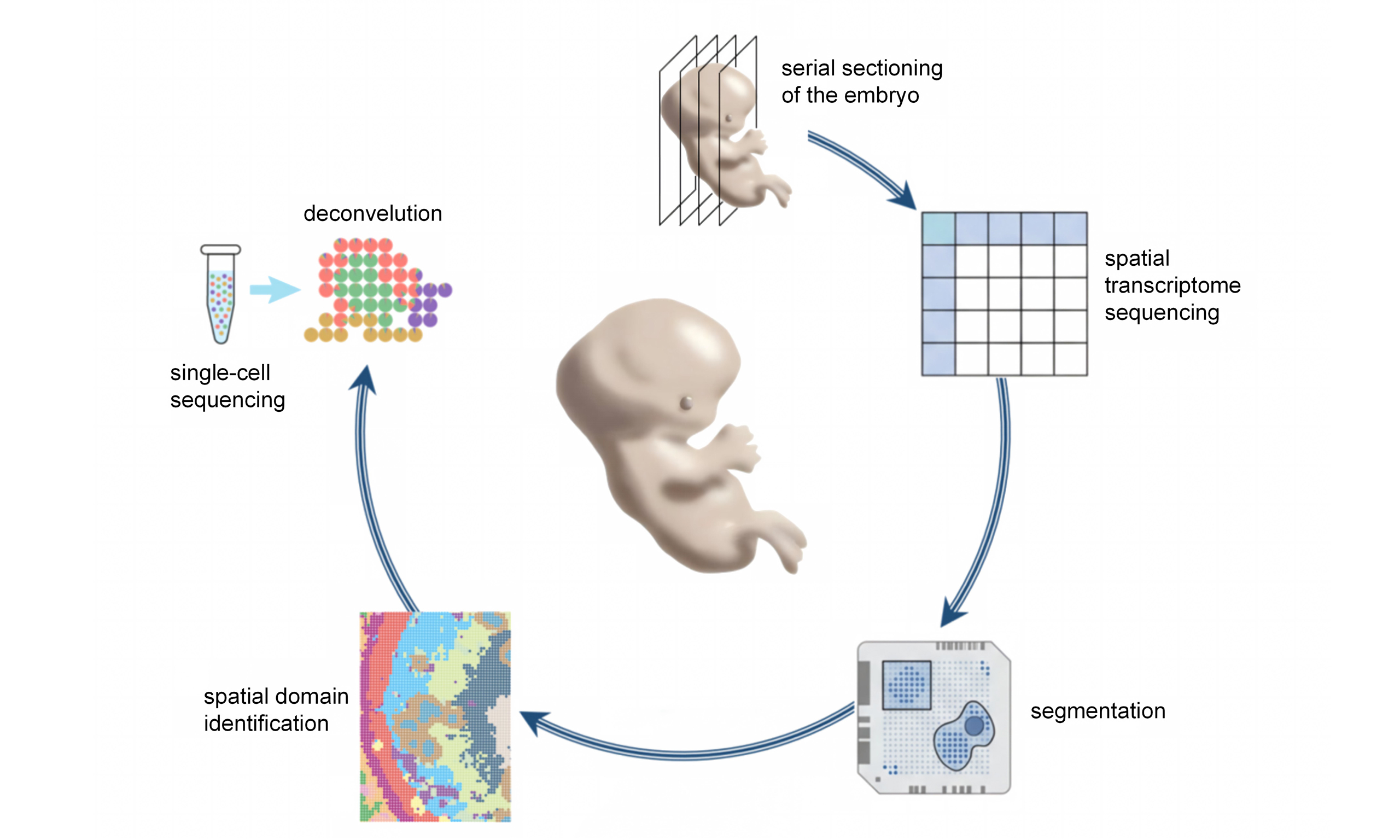
Abstract
Deciphering early human embryonic development and organogenesis is constrained by limited access to in vivo specimens and the intrinsic complexity of spatiotemporal cell fate dynamics. Recent advances in spatiotemporal multi-omics have begun to overcome these barriers by enabling the reconstruction of in silico human embryos and developmental trajectories with spatial and temporal resolution. These approaches have revealed fundamental principles of lineage specification, cell migration, tissue morphogenesis, and axial patterning in early human development. In this Review, we summarize how single-cell and spatial multi-omics technologies, particularly spatial transcriptomics, have transformed the study of human early embryogenesis, organogenesis, and stem-cell-derived embryoid systems. We highlight analytical frameworks that integrate spatial context, temporal progression, and multimodal datasets, and discuss how biological discovery and algorithmic innovation have co-evolved. Finally, we outline how artificial intelligence, foundation models, and large-scale multimodal learning may eventually enable predictive and causal modeling of human development, while addressing current technical, interpretability, and ethical challenges.
Keywords


 Download PDF
INTRODUCTION
Understanding how spatiotemporal regulatory programs govern early human embryonic development and organogenesis remains a central question in developmental biology and regenerative medicine. It is also directly relevant to the mechanistic interpretation of congenital disorders and to the long-term goal of precise regenerative intervention. Prior to implantation and between 5-6 days post-fertilization, the fertilized human egg evolves into a blastocyst. At this time, the blastocyst is surrounded by the trophectoderm (TE), and the inner cell mass (ICM) in the blastocoel consists of both epiblast and hypoblast cells. Approximately 6-7 days after fertilization, the blastocyst implants into the uterus and continues to develop; this stage is defined as Carnegie stage 4 (CS4)[1]. During the post-implantation to peri-gastrulation period (7-28 days after fertilization), the embryo first undergoes gastrulation initiation, trilaminar formation, axial specialization, and rapid and intense cellular differentiation and spatial rearrangement at CS7-CS8 (15-19 days)[2]. Subsequently, during the CS9 (19-21 days) stage, neural tube development begins, and simultaneously, the primitive heart, eyelids, otic vesicle, and other organs also begin to develop[3]. Organ growth displays unique spatial traits, underscoring the need for molecular characterization that preserves spatial information.
The period around 14 days post-fertilization coincides with the formation of the three germ layers and rapid cellular rearrangement, yet it is also when precisely staged human embryonic samples are most scarce. Ethical and regulatory constraints compound this scarcity, although the limits placed on the in vitro culture of intact human embryos differ across jurisdictions; many countries adopt the widely referenced "14-day rule"[4,5]. Systematic, high-resolution molecular and cellular research in real human embryos across this window therefore remains consistently challenging.
Addressing this limitation, numerous teams have created a range of in vitro research methodologies, such as the derivation of pluripotent human embryonic stem cell (hESC) lines and the establishment of human pluripotent stem cell (hPSC)-based embryo models (embryo-like structures), together with organoids representing diverse organs, enabling systematic in vitro interrogation of early human development and organogenesis. The development of technologies such as single-cell RNA sequencing (scRNA-seq) has enabled researchers to systematically study the cellular heterogeneity and developmental trajectories of human embryos and multiple organs from gastrulation to even later stages of organogenesis at single-cell resolution. However, these research frameworks share a fundamental limitation: once tissues are dissociated or reconstructed in vitro, the original spatial relationships among cells are only partially retained and are often lost altogether[6]. Early human embryos undergo rapid axis specification, germ-layer organization and the induction of organ primordia, processes in which cellular position, neighborhood interactions and local mechanical and molecular cue fields are tightly coupled to fate decisions, lineage trajectories and morphogenetic programs. Although in vivo and in vitro human embryo systems profiled by single-cell transcriptomics have catalogued diverse cell types and states, dissociative single-cell readouts are intrinsically limited in resolving core spatial questions such as where cells reside within the embryo and how multicellular communities interact in 3D space to assemble coherent tissue architecture. This may sound like a technical detail, but in embryology it often changes the biological question itself. Spatial transcriptomics (ST), as the leading component of spatiotemporal multi-omics, has emerged to address this gap by quantifying gene expression while preserving tissue architecture.
Spatial transcriptomic technology did not emerge overnight. Initial versions of spatial transcriptomic platforms frequently encountered obstacles including insufficient sequencing depth, inadequate spatial resolution, probes failing to encompass the entire genome, and elevated expenses[7]. This spurred the development of algorithms such as single-cell reference-based deconvolution to infer potential cellular composition and states at low spatial resolution bins/spots[8,9]. With advances in probe density and detection sensitivity, ST has progressed toward subcellular resolution, enabling finer-grained analyses including spatial domain delineation, cell segmentation, 3D (and pseudo-3D) reconstruction—where pseudo-3D refers to the simple stacking of aligned 2D sections without volumetric interpolation, whereas true 3D involves computational inference of continuous spatial relationships across sections, inference of migratory trajectories, and the reconstruction of cell-cell communication networks[10]. With the rapid maturation of spatial epigenomics and spatial proteomics, ST is now being integrated into multimodal spatiotemporal omics frameworks spanning transcriptional, epigenetic, proteomic, and metabolic layers[8,11], and their computational integration with in vivo imaging, graph representations, deep learning, and generative modeling is reshaping our understanding of the spatiotemporal and hierarchical regulatory mechanisms underlying early human embryogenesis and organogenesis. For scarce human embryonic specimens, such multimodal strategies offer a uniquely information-dense route to extract maximal molecular and spatial insight from minimal material, while reducing the experimental and ethical costs associated with repeated sampling.
In this Review, we argue that the integration of spatiotemporal multi-omics with computational modeling is fundamentally reshaping our understanding of early human development. This integration has made spatial context an intrinsic component of developmental inference, rather than a supplementary annotation. This Review is organized around six central biological questions that define the major challenges in studying early human development: (1) How faithfully do stem-cell-based models reproduce in vivo development? (2) Which cell states arise during early embryogenesis? (3) Where are these cells located within the developing embryo? (4) How do cells migrate and change fate over time? (5) How do local signaling centers organize tissue development? and (6) How can descriptive spatial maps become predictive developmental models? Each of the following sections restates the question it addresses before turning to the relevant technologies and analytical strategies [Figure 1]. We first return to embryo models, organoids and single-cell studies, which defined the current framework of the field while exposing the limits of dissociation-based or in vitro systems in resolving spatial organization and in vivo developmental complexity. We then move to the major spatial transcriptomic platforms, with emphasis not only on their technical principles but also on when they are most useful in practice. For the same reason, we include a summary table that links representative embryonic sample types and research questions to suitable sequencing and analytical strategies. We next discuss how a series of downstream analytical approaches, including binning and segmentation, deconvolution, spatial domain analysis, lineage and migration inference, cell-cell communication analysis, and 3D reconstruction, have gradually pushed the field beyond descriptive mapping. In the final part, we extend this discussion to multimodal integration and artificial intelligence, which may further help organize scattered observations into more connected, and perhaps eventually predictive, models of human embryogenesis and organogenesis. This Review is structured as a narrative review with systematic elements. The literature covers approximately 2000-2026, with emphasis on major advances from 2020 onward, prioritizing methodologically rigorous and technically innovative contributions relevant to embryonic development.
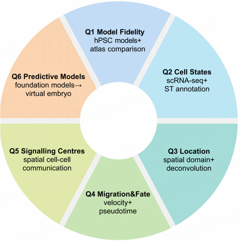
Figure 1. Conceptual roadmap linking the six guiding biological questions of this Review to the spatiotemporal multi-omics technologies and analytical approaches that address them. Q1, model fidelity: benchmarking the fidelity of stem-cell-based embryo models against in vivo human atlases through integrated multi-omics comparisons. Q2, cell states: identifying the cell types and states that arise during early embryogenesis by single-cell and multimodal profiling, with spatial annotation where applicable. Q3, tissue architecture: resolving spatial organization through ST, cell-type deconvolution and spatial-domain identification. Q4, directional transitions: inferring how cells migrate and change fate over time by trajectory inference, RNA velocity and three-dimensional reconstruction. Q5, signalling centres: dissecting local signalling centres through ligand-receptor inference and spatial signalling analysis. Q6, predictive models: moving from descriptive maps toward predictive models through multimodal integration, foundation models and perturbation prediction.
EMBRYO-LIKE SYSTEMS AND SINGLE-CELL TRANSCRIPTOME-SUPPORTED EMBRYO RESEARCH ETHICAL DILEMMAS AND THE "14-DAY RULE" IN HUMAN EMBRYO RESEARCH
The first question is how faithfully stem-cell-based embryo models reproduce in vivo human development. Answering it requires comparing in vitro models with single-cell transcriptomic atlases of early human embryos, which provide the current reference for cellular composition and lineage relationships; this section therefore examines embryo models and single-cell studies together, emphasizing where such systems remain limited in resolving spatial organization and in vivo developmental complexity. The fundamental dilemma in early human embryonic development research is that the most critical stage (from post-implantation to gastrulation and early organogenesis) is precisely the least visible stage. On the one hand, according to the latest guidelines issued by the International Society for Stem Cell Research in 2021, which are still widely regarded as the benchmark, human embryo research must be conducted within a strict ethical framework, with in vitro culture generally not exceeding 14 days after fertilization or the appearance of the primitive streak[5]. On the other hand, during the critical period of 14 to 28 days in vivo, when gastrulation and organogenesis are in the early stages, human embryo samples are extremely scarce and can often only be obtained in a few special clinical situations, and they cannot be reproduced or systematically collected[4,12,13]. Therefore, the decisive interval spanning lineage reconfiguration, axis establishment, and the emergence of organ primordia is effectively compressed in time, bounded by ethical constraints and limited by experimental tractability, creating a gap with both institutional and technological dimensions. Notably, within the 0-14 days window adopted by many jurisdictions for in vitro culture, human embryo research has only recently shifted from being largely inaccessible to becoming partially observable. Over the past decade, extended embryo culture platforms have enabled human blastocysts to develop to approximately 13 days in vitro, recapitulating key hallmarks of early post-implantation embryos[14-16]. However, this research paradigm still faces two major structural limitations: First, culture experiments are required to be terminated around 14 days in jurisdictions that adhere to this limit [Table 1], precluding analysis of gastrulation and organogenesis—the very processes that lie within the E14-E28 “black box” of human development [Figure 2]; second, the limited sampling capacity of single embryos prevents multi-timepoint, multi-omics profiling and robust spatiotemporal mapping. Under these dual constraints, relying solely on in vitro embryo culture or occasional in vivo samples is no longer sufficient for systematic mechanistic analysis of early human development.
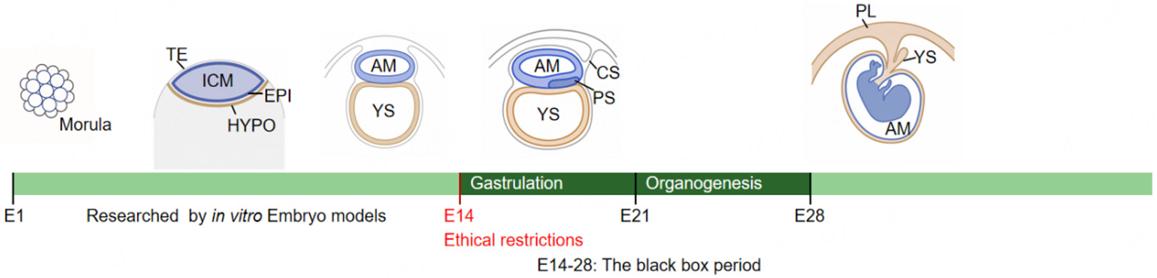
Figure 2. E14 serves as a widely recognized ethical boundary for in vitro embryo culture in many jurisdictions, aligning with the onset of gastrulation. E14-E21 represents the gastrulation period; E21-E28 represents the early organogenesis period. Together, E14-E28 constitutes the "black box" period of early human development, for which our current understanding remains limited. TE: Trophectoderm; ICM: inner cell mass; EPI: epiblast; HYPO: hypoblast; AM: amnion; YS: yolk sac; CS: connecting stalk; PS: primitive streak; PL: placenta.
Regulatory approaches to human embryo research and embryo models across representative countries/regions
| Country/Region | 14-day rule status | Embryo research policy | Embryo model regulation | Primary legal/policy basis |
| United Kingdom | Legally binding | Permitted up to 14 days | Not explicitly covered by statute; 2024 voluntary Code of Practice issued | Human Fertilisation and Embryology Act 1990[17]; SCBEM Code of Practice (2024)[18] |
| United States | No federal statute | Varies by state; federal funding restricted by Dickey-Wicker Amendment (1996) | No federal-specific regulation; subject to IRB, state laws, and NIH funding policy | Dickey-Wicker Amendment (annual federal appropriations rider, first enacted 1996)[19] |
| China | National guidelines | Permitted for 14 days; institutional ethics review required | Governed by the 2003 Ethical Guidelines through institutional review; Human Genetic Resources Regulation applies to cross-border data/sample transfer | Ministry of Science and Technology & Ministry of Health, Ethical Guiding Principles for Human Embryonic Stem Cell Research, 2003[20]; Regulation on the Administration of Human Genetic Resources, 2019[21] |
| Canada | Legally binding | Surplus IVF embryos permitted; creation for research prohibited | Not explicitly covered | Assisted Human Reproduction Act, S.C. 2004[22]; Tri-Council Policy Statement: Ethical Conduct for Research Involving Humans, 2022[23] |
| Germany | Not applicable | Human embryo research not permitted | hESC research: stem cell act (StZG) | Embryo protection act (ESchG), 1990[24]; Stem cell act (StZG), 2002[25] |
| Netherlands | Legally binding | Permitted up to 14 days, using surplus IVF embryos only | Pending; the national Health Council recommended a 28-day limit in 2023 | Embryo act, 2002[26]; Health Council of the Netherlands, advice on extending the 14-day rule (2023)[27] |
| Brazil | No day-limit provision in statute | Permitted on unviable IVF embryos or IVF embryos frozen ≥ 3 years | Regulatory status not yet clearly defined | Biosafety law (2005)[28] |
The research paradigm has thus undergone a profound shift: on the one hand, embryonic models and organoid systems based on hPSCs have developed rapidly. On the other, spatiotemporal multimodal omics, building on single-cell profiling and increasingly integrating spatial transcriptomic, epigenomic, and proteomic layers, is enabling unified computational reconstruction across both in vitro models and scarce in vivo human embryo specimens. Together, stem cell-based systems and algorithmic innovation are more than an adjunct to classical embryology; they define an ethically bounded route to knowledge production in which controllable, repeatable, and scalable experimental platforms generate high-dimensional measurements that computational frameworks can integrate into continuous developmental trajectories and computable spatiotemporal representations. Trajectory inference, spatial reconstruction, graph-based modeling of cellular neighborhoods and communication, and generative approaches collectively assemble discrete observations into coherent developmental narratives and predictive maps of human embryogenesis and organogenesis.
In vitro embryo-like models serve as a new platform for studying early human development
To overcome the ethical and technological bottlenecks in human embryo research, scientists have developed a series of in vitro models based on hPSCs, collectively known as "embryo-like/embryo models". These models aim to simulate the structure and developmental process of real human embryos, providing unprecedented tools for studying early development[29]. At the same time, they do not fully reproduce the biological complexity of in vivo human embryos, especially with respect to native tissue architecture, physical environment, and endogenous cell-cell interactions[30]. Based on their cellular composition and developmental potential, these models are generally divided into two main categories: non-integrated embryo models and integrated embryo models.
Non-integrative embryonic models typically contain only intraembryonic cell types (such as epiblast cells) and contain little or no extraembryonic tissue. They do not have the potential to develop into a complete fetus and therefore face less ethical controversy, primarily being used to study specific tissues or developmental events. Their development can be traced back to 2000, when Itskovitz-Eldor et al. from Benvenisty's team at Rambam Medical Center discovered that suspended cultured hESCs spontaneously assembled into embryoid body (EB) structures, but these structures were highly disordered and randomly differentiated, quite different from the ordered developmental pattern of real embryos[31]. In 2014,
Integrative embryonic models incorporate both intraembryonic and extraembryonic cell types, enabling a more complete simulation of the structure of a real embryo. For example, blastoids mimic the pre-implantation blastocyst structure, while post-implantation embryonic models simulate the post-implantation developmental stages. In 2021, Liu et al. and Yu et al. at Monash University in Australia reported methods for generating human blastoids using naive-state hPSC induction and somatic cell reprogramming, respectively[41,42]. Liu's team's work is particularly noteworthy; they successfully constructed iBlastoids by reprogramming fibroblasts into induced pluripotent stem cells (iPSCs), providing an important platform for studying genetic diseases using patient-specific iPSCs[41]. In 2023, Weatherbee et al. overexpressed key regulatory factors of trophoblast and primitive endoderm cells in hPSCs via transgenic methods, inducing trophoblast-like and primitive endoderm-like cells to spontaneously assemble into structures containing three lineage-like cell types, reproducing structures similar to amniotic sacs, extraembryonic mesoderm cells, and primordial germ cells (PGCs)[43]. In the same year, Oldak et al. at the Weizmann Institute of Science in Israel systematically optimized the induction system of human hPSCs, constructing an embryo-like structure in vitro that simulates a real human embryo, known as a synthetic complete embryo model (SEM) based on stem cell synthesis. This model relatively completely simulates the key developmental events of a human embryo after implantation in the uterus until the onset of gastrulation[44]. Even in these more integrated systems, increased structural resemblance does not mean full developmental equivalence to in vivo embryos, and careful benchmarking against real embryonic data remains necessary.
After the aforementioned technologies enabled embryo culture to reach the post-implantation stage and simulate the development of key primitive organs such as the neural tube, heart, and aorta-gonad-mesonephros region hematopoietic region[45-47], recent embryo-like studies have expanded beyond embryo-like structures to include joint modeling of the maternal-fetal interface. In December 2025, Molè et al. from Peter J. Rugg-Gunn's team developed the Cell-Enhanced Receptivity Endometrial Scaffold (CREST) technology. This system successfully simulated the tissue receiving the implanted embryo (superficial endometrium) and cultured both real embryos and embryo-like structures to day 14[48]. Following this, in January 2026, the laboratories of Li et al. not only constructed a small-scale microarray of the maternal-fetal interface system but also developed a relatively complete drug screening process based on it, enhancing the clinical trial value of this system[49].
The main contributions of embryo-like technologies lie in their scalability, reproducibility, and operability. Compared to scarce human embryo samples, hPSCs (including hESCs and iPSCs) can be expanded indefinitely, enabling large-scale experiments. The introduction of iPSCs is particularly important because they can be obtained by reprogramming adult cells, avoiding the ethical controversies associated with using embryonic stem cells, while also enabling the creation of patient-specific disease models. Furthermore, these models are easily genetically manipulated, allowing researchers to precisely analyze the function of specific genes during development. However, these models have also raised complex ethical debates, particularly integrative models, which structurally resemble real embryos, making their developmental potential and ethical standing a focus of attention for the scientific community and the public[50]. With the rapid accumulation of single-cell reference atlases, it has become increasingly common to compare in vitro embryo models with real embryonic datasets in order to identify both shared features and model-specific differences.
A key question in the aforementioned embryo-like studies is the extent of their similarity to real embryos. Can they accurately replicate the development of an in vivo embryo? To answer this question, conventional experimental methods require collecting real human embryos at the corresponding stage or embryos for immunofluorescence staining verification to ensure that the key expression characteristics of the cell types obtained from embryo-like studies are similar to those of real embryos. However, this verification requires precious in vivo embryo samples and has limited throughput. Bulk RNA-seq has been used to compare the transcriptomic differences between induced amniotic membranes and hPSCs as a control group to demonstrate that the induced cells possess the expression characteristics of the amniotic membrane[38] [Figure 3].
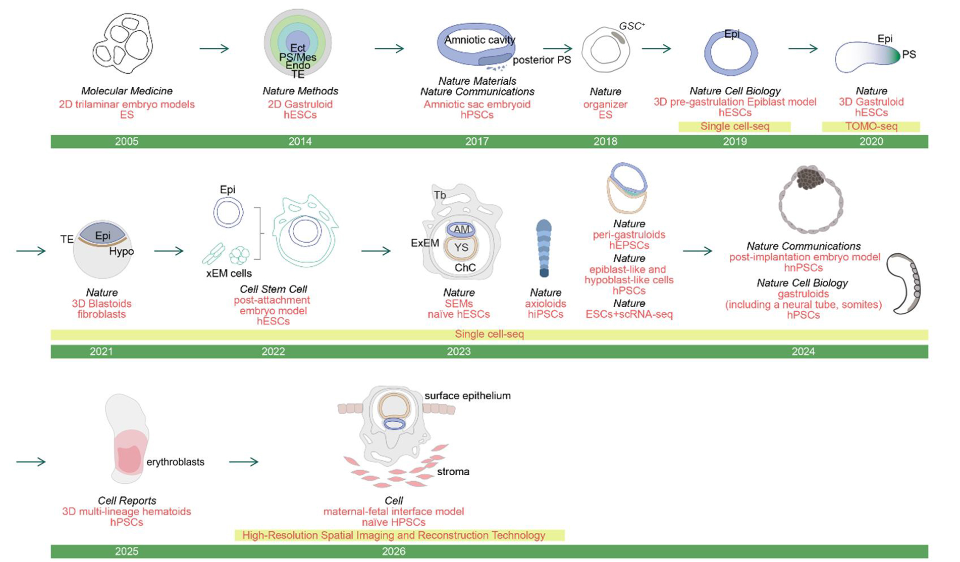
Figure 3. Timeline of Embryo Model Technology Development. Publications utilizing single-cell and spatial omics technologies are highlighted. AM: Amnion; ChC: chorionic cavity; EB: Embryoid body; Ect: ectoderm; Endo: endoderm; Epi: epiblast; ES: embryonic stem cell; ExEM: extraembryonic mesoderm; hESC: human embryonic stem cell; hiPSC: human induced pluripotent stem cell; hnPSC: human naive pluripotent stem cell; hPSC: human pluripotent stem cell; Hypo: hypoblast; Mes: mesoderm; PL: placenta; PS: primitive streak; SEM: synthetic complete embryo model; Tb: trophoblast; TE: trophectoderm; xEM cells: extraembryonic mesoderm cells; YS: yolk sac.
scRNA-seq technology has opened the door to comprehensive mapping studies of embryonic development mechanisms
The rise of scRNA-seq substantially changed the study of early human embryonic development. For the first time, researchers could systematically resolve developmental heterogeneity at cellular resolution and begin to assemble transcriptomic atlases across successive embryonic stages. This shift was important not only because it expanded cell-type discovery, but also because it provided a practical reference framework for comparing embryo-like models with real embryos in vivo. At the same time, scRNA-seq still profiles development in a dissociated manner. It can describe cell states with increasing precision, but it cannot by itself explain how those states are positioned within intact embryonic structures or how spatially adjacent tissues shape developmental trajectories[39]. Therefore, the development of embryo-like cells and the verification of their accuracy are inseparable from the development of sequencing technology, and even more so from the study of the mechanisms of real embryos in vivo (as prior knowledge for verifying the accuracy of embryo-like cells). In the context of preimplantation development, in 2013, Xue et al. first applied scRNA-seq to human and mouse embryos[51]. By comparing gene expression profiles from the oocyte to morula, they not only identified specific expression patterns of maternal and paternal alleles (such as paternal activation of the CDCA2 gene), but also constructed stage-specific functional modules using weighted gene co-expression network analysis, elucidating the sequential activation process of cell cycle, transcriptional regulation, and metabolic pathways in embryonic development[51]. This work not only laid the foundation for cross-species comparative studies of embryonic data but also highlighted the advantages of scRNA-seq for analyzing low-abundance transcripts and allele expression, directing research focus to deeper lineage-separation mechanisms.
Following the morula, researchers began to focus on the peri-implantation period. In 2016, Xue et al. used scRNA-seq to analyze human preimplantation embryos (3-7 days post-fertilization), revealing for the first time at the single-cell level the transcriptomic differences in TE, ICM, epiblast (EPI), and primitive endoderm (PE)[51]. The study found that the expression of lineage-specific genes (such as GATA3, SOX2, and GATA4) begins to differentiate at the day 5 stage, and that X chromosome dose compensation is achieved through biallelic downregulation rather than complete inactivation, challenging the traditional view of X chromosome inactivation in mouse models[51]. Furthermore, in 2021, Meistermann et al. reconstructed the developmental trajectories of human and mouse preimplantation embryos through integrated pseudotime analysis, finding that transcriptome segregation of human TE and ICM occurs during the blastocyst expansion stage (B2-B3), later than the mouse morula stage, indicating significant differences in developmental timing between species[52]. In 2020, Xiang et al. developed a 3D culture system that successfully simulated human embryonic development from implantation to pre-gastrulation[14]. scRNA-seq analysis showed that 3D cultured embryos could form structures such as the EPI, amnion, and yolk sac, and revealed the molecular pathways by which trophoblast cells differentiate into the cytotrophoblast, syncytiotrophoblast, and extravillous trophoblast[14]. In the same year, Tyser et al. performed the first scRNA-seq analysis on CS7 gastrulating embryos and constructed a cellular atlas of germ layer differentiation and organ primordia formation, including cell groups from the ectoderm, mesoderm, and definitive endoderm, providing a valuable resource for the study of human gastrulation[53]. With the accumulation of data, a reliable reference dataset is urgently needed for the identification of real embryonic stages and cells, or for the reliability assessment of embryonic-like systems. Therefore, recent research has focused on constructing a single-cell atlas of embryos across a longer developmental period. In 2023,
ST brings a new paradigm to embryo research
The second question is which cell states arise during early human embryogenesis and how transcriptomic data can resolve them. We address it by first introducing the major spatial profiling platforms and then showing how each has been applied to embryonic tissues. ST became important in this context because it began to reconnect molecular identity with physical location. This did not simply add spatial coordinates to existing datasets. More importantly, it allowed developmental time, tissue architecture, and cell-state information to be interpreted in relation to one another, making it possible to revisit questions that dissociation-based profiling could only approach indirectly. In practice, however, no single spatial strategy is equally suitable for all embryonic samples and research aims. Choices about assay design and downstream analysis depend on sample scarcity, developmental stage, spatial scale, transcriptome coverage, and the type of biological inference required. To make these considerations more explicit, we summarize below a comparative framework linking representative embryonic sample types and research aims to commonly used spatial assays and analytical strategies in current embryonic research [Table 2].
Representative spatial assay and analytical choices across embryonic sample types and research aims
| Embryonic sample scenario | Research aim | Commonly used spatial assay | Representative analytical strategy | Practical note | Supporting references |
| Very scarce peri-gastrulation human embryos | Maximize spatial and molecular information from minimal material | High-resolution barcode-based ST | Square-bin optimization, spatial domain analysis, selective 3D reconstruction | High information density, but replication is often limited | Xiao et al., 2024[2]; Yuan et al., 2025[3]; Cui et al., 2025[56] |
| Samples with matched scRNA-seq references | Improve annotation and resolve mixed spots or bins | Moderate- to high-resolution ST | Reference-based deconvolution, label transfer, atlas integration | Strong interpretation, but depends on reference quality | Xu et al., 2023[54]; Zeng et al., 2023[57]; Zhang et al., 2024[58] |
| Samples without matched references | Explore spatial organization in poorly characterized stages | Higher-resolution ST preferred | Reference-free domain identification, unsupervised analysis | Useful for rare stages, but annotations are less certain | Hara et al., 2025[59]; Pan et al., 2026[60] |
| Embryo-like models, gastruloids or blastoids | Benchmark model fidelity against in vivo embryos | ST combined with reference atlas comparison | Cross-system mapping, trajectory alignment, spatial comparison | Identifies shared and model-specific features, but not full in vivo equivalence | Oldak et al., 2023[44]; Weatherbee et al., 2023[43]; Zhao et al., 2025[55]; Van Den Brink et al., 2020[61] |
| Later-stage organs or organ primordia | Map tissue architecture and signaling niches | Large-area barcode-based ST | Domain analysis, communication inference, histology integration | Good tissue coverage, but fine boundaries may be blurred | Xu et al., 2023[54]; Zhang et al., 2024[58]; Farah et al., 2024[62] |
| Serial sections or whole-embryo reconstruction studies | Infer three-dimensional organization | Serial ST or 3D-compatible assays | Registration, alignment, 3D reconstruction | Extends section-based data, but alignment errors matter | Xiao et al., 2024[2]; Yuan et al., 2025[3]; Qiu et al., 2024[63] |
| Targeted signaling or marker-localization studies | Precisely localize predefined pathways or cell states | Imaging-based assays | Segmentation, neighborhood analysis, constrained communication analysis | High spatial fidelity, but limited gene breadth | Chen et al., 2024[64]; Farah et al., 2024[62]; Qian et al.,[65] |
| Budget-limited comparative studies | Balance throughput and interpretability across stages or sections | Lower-cost or moderate-resolution ST | Deconvolution, smoothing, domain analysis | Practical for larger comparisons, but more dependent on computation | Jiang et al., 2023[66]; Zhu et al., 2024[67] |
The following sections build on this overview and examine spatial profiling technologies and analytical strategies in greater detail, with emphasis on the embryological questions they help address [Box 1].
Summary of embryo-like systems and single-cell transcriptomics
| Main biological advance | The "14-day rule" confines in vitro culture to approximately the first 14 days in jurisdictions that apply it[5,13]. The E14-E28 interval, which spans gastrulation and early organogenesis, can therefore be studied mainly through scarce in vivo specimens. Embryo models have evolved from non-integrated (EB[31], gastruloids[39]) to integrated systems (blastoids[41], SEM[44], CREST[48]). Meanwhile, scRNA-seq has constructed transcriptomic reference frameworks spanning preimplantation to CS16 for model benchmarking and the interpretation of developmental events[14,51-55] |
| Preferred methods | hPSC-based differentiation protocols for embryo models[41-44]; scRNA-seq for molecular characterization[51-55]; integrated computational frameworks for aligning model data with in vivo reference atlases[54,55] |
| Current limitation | No model fully recapitulates in vivo development, most lack extraembryonic tissues or native tissue architecture[39,43,44]; scRNA-seq cannot resolve spatial organization; the 14-day rule prevents cultured embryos from progressing beyond the gastrulation and organogenesis[5,13]; ethical and regulatory frameworks remain heterogeneous across jurisdictions[4,50] |
| Next experimental priority | Evaluate embryo-model fidelity against in vivo human reference atlases across consecutive developmental stages[54,55]; integrate multi-omics datasets into stage-resolved spatiotemporal maps[2,3] |
DECODING THE DIGITAL BLUEPRINT OF EARLY HUMAN EMBRYONIC DEVELOPMENT: EVOLUTION AND PARADIGM SHIFT OF SPATIAL MULTI-OMICS TECHNOLOGIES
The diversity of spatial multi-omics platforms matters in embryonic research not simply because the technologies differ in principle, but because those differences determine which developmental questions can be addressed with sufficient spatial resolution, molecular depth, and anatomical coverage. Based on their technical principles, spatial transcriptome sequencing technologies can be mainly subdivided into the following categories: whole-transcriptome sequencing based on spatial barcodes, fluorescence in situ hybridization (FISH) imaging technology, and in situ sequencing (ISS) technology[68], Complementary approaches also include microdissection-based and microfluidic methods for specialized applications[69]. They can also be classified according to the characteristics of their sequencing results, such as low-resolution, subcellular resolution, single-cell (nuclear) sequencing technology, and low-cost sequencing technology.
Spatial barcode-based transcriptomic profiling is widely used to generate digital and spatially resolved maps of embryonic development. Early human embryos or organs are cryosectioned into 5-10 µm-thick sections and mounted on a barcoded capture slide. The tissue is then permeabilized, and the released transcripts are captured by carriers with spatial barcodes (such as magnetic beads or nanospheres). After reverse transcription, library construction, and next-generation sequencing (NGS), the RNA sequence is obtained and compared with all genes in a reference genome. The maximum resolution of this technology depends on the unit size and density of the capture points (called spots or bins). Early platforms like 10X Visium offered a spot resolution of 55 μm[70], which exceeds the 10-15 μm diameter of a typical mammalian cell[71]. Consequently, each spot in such low-resolution spatial transcriptomic data typically contains transcripts from multiple cells, precluding precise cellular boundary delineation. The idea to overcome this limitation and improve resolution is to reduce the size of individual capture regions. In 2024, 10X Genomics launched the 10X Visium HD sequencing technology[72], which improved the maximum resolution to 2 micrometers. Slide-seq uses magnetic beads carrying spatial information with a center-to-center spacing of 10 micrometers, approaching that of a single cell[72]. BGI's Stereo-seq[73] uses DNA nanospheres (DNB) to shrink the spot to 220 nm (with a two-point spacing of 500-750 nm), achieving a maximum capture area of 13 cm × 13 cm. This provides both subcellular resolution and a large sequencing field of view. This class of methods offers three principal advantages that have made it particularly well suited for embryonic applications: subcellular spatial resolution, near-transcriptome-wide coverage in principle, and a large capture area that accommodates whole sections. In addition, high-resolution barcode sequencing technologies such as Stereo-seq and 10X Visium HD achieve subcellular-level segmentation and can generate Cell Bins through various cell segmentation methods, such as morphology-based Watershed[74], deep learning-based Cellpose[75-77], and graph theory-based Baysor[78], which aim to accurately segment individual cells from subcellular data. However, due to the limitation that this type of sequencing technology cannot capture absolutely accurate cell boundaries, some researchers have considered transferring the spatial barcode on the chip directly through the cell membrane into the cell nucleus, and then reconstructing the position after sequencing the single-cell nucleus to achieve true single-cell resolution. Examples include sci-Space[79] and SeekSpace[80]. The former showed a high degree of matching with sci-RNA-seq data annotation on mouse E14.0 embryos and accurately represented the boundaries between major organs, such as the boundary between the forebrain and midbrain. The latter successfully and accurately identified 11 major cell types on mouse E13.5 liver samples, which were mutually validated by single-cell data.
Complementary to spatial barcode-based sequencing approaches, which rely on the release and sequencing of spatial barcodes using conventional next-generation sequencing platforms, image-based in situ molecular decoding technologies preserve transcripts within their native tissue context. These approaches include FISH based imaging methods and ISS based strategies. FISH-based methods, such as MERFISH or SeqFISH, use pools of gene-specific probes labeled with fluorescent barcodes. Through multiple rounds of hybridization and imaging, they decode the identity and location of thousands of RNA species directly in situ[68]. For example, MERFISH, due to its extremely high capture efficiency, can reveal the polar distribution of mRNA within cells, which is critical for understanding the asymmetric allocation of certain transcription factors in early human embryos. Although this reduces gene throughput compared to barcode-based technologies, these methods provide reliable molecular-level resolution, laying the foundation for studying the spatial expression patterns of genes in tissues and cells. ISS technologies such as STARmap[81] and 10X Xenium In situ[82] attempt to improve depth on the basis of molecular-level resolution, and directly perform sequencing reactions on cDNA with spatial information inside tissues.
Microdissection-based methods sacrifice cellular throughput to achieve high sequencing depth, facilitating the integration of spatial information with conventional single-cell transcriptomic profiling. LCM-seq[83] utilizes microdissection technology to extract cells (1-3 cells) from labeled regions in frozen tissue for single-cell sequencing (e.g., smart-seq2[84]), thus maximizing sequencing depth. GEO-seq[85] and Tomo-seq[86] can further enhance this by dissecting thicker frozen sections and constructing 3D maps. While providing exceptional depth, these methods inherently compromise spatial continuity because they profile only a sparse, discrete subset of cells within the tissue architecture.
Microfluidic platforms offer a route to lower-cost spatial multi-omics profiling, though often with a trade-off in spatial resolution. For example, Jiang et al. applied MISAR-seq to profile the mouse brain across four developmental time points (E11.0, E13.5, E15.5, E18.5) in duplicate, performing joint ATAC-RNA sequencing. This identified 16 cell types and revealed complex gene regulatory networks underlying brain development[66]. The MAGIC-seq platform employs a microfluidic mesh chip to lower cost and increase throughput. Using MAGIC-seq for spatial transcriptomic profiling, Zhu et al. mapped gene expression in an E17.5 whole mouse embryo and in postnatal brain sections (days 0 and 4), enabling precise organ localization and fine-grained brain region annotation[67]. Simultaneously, by integrating 94 serial sections from an E18.5 mouse brain, they reconstructed a 3D transcriptomic atlas and delineated 11 distinct 3D brain regions. Further investigation revealed significant differences in the distribution of GABAergic neurons across brain regions, and variations in gene expression among neurons of the same type.
Because cells reside and interact in three dimensions, 3D ST is essential to model intact embryonic structure and dynamics. Recently, several true 3D spatial profiling methods have emerged. The 3D MERFISH[87] developed by Fang et al., by increasing the slice thickness (60-200 μm) and combining it with computational imaging algorithms, can quantitatively analyze hundreds of genes in 3D space, marking a leap from 2D slices to 3D digital modeling of entities in spatial omics[87]. In a demonstration, Zhuang's team quantified 242 genes in a 100 μm-thick mouse cortex section and mapped gene expression in a 200 μm-thick hypothalamus, enabling robust 3D clustering of neuronal cell types. This work significantly pushed the multiplexing capacity of 3D spatial imaging towards practical, genome-scale application. The Open-ST[88] developed by Nikolaus Rajewsky et al. converts Illumina flow cells into spatial capture regions, achieving low-cost ST sequencing at a maximum resolution of 0.6 micrometers. Unlike other ST data that mostly rely solely on ST information for alignment and point cloud generation, Open-ST integrates its proprietary OpenST algorithm end-to-end to align HE staining images with ST data, and further combines it with the STIM algorithm for multimodal 3D reconstruction and generation of a 3D virtual tissue block. Deep-STARmap and Deep-RIBOmap (for detecting ribosome-bound mRNA)[89] extend the thickness of slices to 60-200 micrometers and support the quantification of thousands of genes [Table 3].
ST sequencing technologies applied in embryonic development
| Platform category | Representative platforms | Effective cellular resolution | Gene throughput | Best suited for |
| Capture-array barcode sequencing | Stereo-seq[73], 10X Visium HD[72], 10X Visium[70], Slide-seq[90] | Subcellular capture units (Stereo-seq, Visium HD); near-cellular bead spacing (Slide-seq); multi-cell spots analyzed by deconvolution (Visium) | Whole transcriptome (capture-based) | Large-area/whole-embryo sections; transcriptome-wide discovery with scRNA-seq deconvolution |
| Imaging-based (FISH/ISS) | MERFISH[91], seqFISH+[92], STARmap[81], 10X Xenium[93] | Individual-cell imaging resolution after cell segmentation | Targeted panels—from hundreds of genes up to ~10,000 genes (seqFISH+) | Precise in situ localization of predefined markers; high spatial fidelity |
| Microdissection/regional profiling | LCM-seq[83], Geo-seq[85], Tomo-seq[86] | Single captured cells to small cell groups (region-level, user-selected) | Whole transcriptome at high sequencing depth | Scarce samples; anatomically defined micro-regions needing maximal transcript depth |
| Microfluidic indexing/joint profiling | MAGIC-seq[67], MISAR-seq[66] | Near-single-cell to | Whole transcriptome (MISAR-seq additionally profiles chromatin accessibility) | Budget-constrained studies, large comparative cohorts, multi-region screening |
| Volume/thick-tissue 3D imaging | 3D MERFISH[87], Deep-STARmap[89], RT&T-AMP-MERFISH[94]; Open-ST[88] | Individual-cell 3D segmentation in thick blocks (3D MERFISH, Deep-STARmap); subcellular capture with segmentation (Open-ST) | Targeted (hundreds-thousands) or transcriptome-scale imaging | Intact-volume 3D reconstruction; moving from 2D sections to digital 3D models |
In practice, the choice of spatial technology in human embryo studies is dictated by the varying requirements of throughput, spatial resolution, and the specific biological scientific questions and scenarios. When the study involves a large sample size across multiple developmental stages and has matching single-cell transcriptomes as reference data, researchers tend to choose sequencing methods like 10X Visium and Magic-seq, which are lower in cost and resolution. They compensate for these resolution deficiencies by using deconvolution to computationally increase annotation accuracy and expression matrix information. By contrast, for scarce early-stage human embryos (such as peri-gastrulation embryos) with extremely low cell numbers and intricate structures, which are difficult to obtain in large quantities, capturing as much transcript information as possible at the highest resolution is paramount. Therefore, subcellular resolution technologies such as 10X Visium HD, Stereo-seq, or Slide-seqv2 are primarily used. During mid-to-late organogenesis, when embryos are larger and anatomically more elaborate, cost and field of view become dominant considerations; microfluidic barcoding strategies can be attractive under budget constraints, whereas large-field platforms such as Stereo-seq offer a practical balance between tissue coverage and sequencing depth. Finally, when the goal is precise in situ localization of predefined markers or pathways, image-based approaches, including FISH-based multiplex imaging or ISS, provide direct localization with high spatial fidelity. For anatomically well-defined microregions with extreme requirements for sensitivity to rare transcripts, targeted region enrichment coupled to high-depth profiling, including laser capture microdissection followed by sequencing, can further complement whole-section spatial assays[95].
In studies of early human development and organogenesis, spatial multi-omics has advanced substantially in throughput and measurement fidelity. Yet the central challenge has not disappeared; rather, it has shifted from simply obtaining spatial data to integrating heterogeneous measurements into biologically coherent and scalable models of development. However, a persistent bottleneck remains the inability to simultaneously achieve four desiderata: in situ single-cell or subcellular resolution, robust multimodal coupling across molecular layers, high detection sensitivity, and low marginal cost at scale. Overcoming this constraint will require not only continued innovation in assay chemistry and instrumentation, but also a deeper integration of artificial intelligence (AI) as a driving force for developmental inference. Foundation models for spatial biology, including transformer-based and diffusion-based generative architectures, could learn from large collections of spatiotemporally resolved differentiation trajectories to build generalizable representations of human development. Such models may support principled alignment across individuals, stages, and modalities, and enable in silico perturbation studies that predict how genetic or regulatory interventions reshape organogenetic programs. In parallel, algorithmic advances are beginning to move the field beyond descriptive spatial correlations toward spatiotemporal causal inference. Graph-based learning frameworks, including graph neural networks, are well suited to capture nonlinear cell-cell communication and neighborhood effects, offering a route to mechanistically dissect how local signaling microenvironments program cell fate decisions. Together, the tight coupling of spatiotemporal omics with modern AI has the potential to shift developmental biology from phenomenological mapping to a predictive, computable, and experimentally testable quantitative science. We will systematically review the progress in this field in the final chapter of this article [Box 2].
Summary of spatial transcriptomics technologies
| Main biological advance | Spatial transcriptomics anchors molecular profiles to anatomical coordinates. It enables reconstruction of gene expression gradients, signaling centers, and tissue architecture across embryonic space[2,3,56,57] |
| Preferred methods | For scarce early-stage embryos: high-resolution barcode-based platforms (Stereo-seq[73], 10X Visium HD[72]) or imaging-based methods (MERFISH[91], STARmap[81]). For later-stage larger embryos: large-area barcode platforms (10X Visium[70], Slide-seq[90]) or microfluidic approaches (MAGIC-seq[67]) for cost-effective coverage. For 3D reconstruction: serial-section ST with registration algorithms[96], or true 3D platforms (3D MERFISH[87], Deep-STARmap[89], Open-ST[88]). For targeted marker localization: imaging-based assays (MERFISH[91], 10X Xenium[93], STARmap[81]). For multi-omics profiling: microfluidic platforms (MISAR-seq for ATAC-RNA)[67] |
| Current limitation | No platform simultaneously achieves single-cell resolution, genome-wide coverage, high sensitivity, and low cost; imaging methods have high spatial fidelity but limited gene throughput[81,91-93]. Barcode methods offer transcriptome-wide coverage but lower resolution[70,72,73,90]; 3D reconstruction is computationally intensive and often requires manual curation[2,3,63] |
| Next experimental priority | Develop platforms that balance resolution, throughput, and cost; reduce manual intervention in 3D reconstruction; profile transcriptomic, epigenomic, and proteomic layers from the same section |
A SPATIALLY-RESOLVED SINGLE-CELL MULTI-OMICS ATLAS OF EARLY HUMAN EMBRYONIC DEVELOPMENT
Knowing which cell states arise is not enough; we must also know where they are located within the embryo, how they migrate and change fate over time, and how local signaling centers organize tissue development. These questions shape the analytical strategies discussed below, from bin selection, segmentation and spatial domain identification to migration inference, communication analysis and 3D reconstruction. In early human embryos, where sample scarcity, structural complexity and developmental heterogeneity often coexist, analytical choices can substantially influence what kinds of cell states, tissue domains and lineage relationships become visible. For this reason, the following sections focus on how key computational frameworks have evolved alongside spatial profiling technologies, and how they have progressively improved the resolution and interpretability of embryonic atlases.
Bin selection and cell segmentation: a trade-off between sequencing depth and spatial precision
One of the first analytical challenges in high-resolution ST is that increased spatial precision often comes at the cost of increased data sparsity. In embryonic research, this trade-off is especially consequential because rare cell states and fine anatomical boundaries may be lost if analysis units are defined too coarsely, whereas excessively small units may lack sufficient transcript depth for robust interpretation. A common response to this problem is to aggregate neighboring capture spots into more information-rich analytical units. In practice, this is usually done in two ways: by merging neighboring spots into square bins or by using image- or transcript-informed segmentation to generate near-single-cell units, here referred to as Cell Bins[97] in this paper.
Square bin remains a widely used method for embryonic atlas construction due to its computational efficiency and its ability to preserve transcriptomic information at the bin level. However, it inherently involves a trade-off between bin size and spatial resolution, and larger bins may reduce single-cell resolution and obscure rare cell populations. In the field of human embryos, the number of detected genes per unit is typically controlled between 400 and 1,300 [Table 2] to achieve a balance between capture quality and single-cell resolution. If the number of genes or the sequencing depth (total unique molecular identifier counts) is too low, the clustering algorithm will lack sufficient information; conversely, if spatial bin size is too large, it will lose its near-single-cell resolution, hindering the identification of valuable cell types. To address this issue, for example, 10X Genomics recommends starting with an 8 µm square bin to identify cell types through clustering when using data generated by Visium HD sequencing technology, and gradually increasing the square bin range to 16 µm to empirically determine an optimal balance between quality and resolution. Other technologies also employ similar strategies to select appropriate square bins; for example, in Stereo-seq with a spot spacing of 500nm, bins from 20 (10 µm) to 50 (25 µm) can be selected. In practice, Square Bins remain particularly useful when the priority is to preserve transcript depth and analytical stability across large embryonic sections, rather than to approximate single-cell boundaries as closely as possible [Table 4].
Samples, resolution, and depth of embryonic ST studies across species
| Species | Sample | Technology platform | Resolution | Median gene number | Publication date | PMID |
| Mouse | E9.5-E16.5 embryos | Stereo-seq | Cellbin/bin50 (25 µm) | 529-792/1,770-3,900 | 2022 | 35512705[73] |
| Drosophila | Late-stage embryos and all stages of larvae | Stereo-seq | bin20 (10 µm)/bin50 (25 µm) | > 400/> 400 | 2022 | 35512700[98] |
| Zebrafish | 3.3hpf-24hpf embryos | Stereo-seq | bin15 (10 µm) | 786 | 2022 | 35512701[99] |
| Marmoset | CS5-CS7 embryos | STEP (LCM + Smart-seq2) | 1-3 cells | > 8,000 | 2022 | 35709828[95] |
| Human | PCW4 embryo | 10X Visium | 55 µm | NA | 2023 | 37192616[57] |
| Human | CS13 | 10X Visium | 55 µm | ~8,000 | 2023 | 36928764[100] |
| Mouse | E13.5 embryo | 10X Visium | 55 µm | 5,668 | 2023 | 37524711[101] |
| Mouse | E8.5, E9.0, E9.5 embryos | Slide-seq | 10 µm | 1,224 | 2023 | 37414952[102] |
| Mouse | E9.5, E11.5 embryos | MERFISH | Single-cell level | < 295 | 2024 | 38348657[64] |
| Human | CS8 embryo | Stereo-seq | bin50 (25 µm) | 1,233 | 2024 | 38657603[2] |
| Human | PCW6.7, 5.7, 7.3 embryos/PCW9 | ISS/10X Visium | Cellbin/55 µm | < 150/x | 2024 | 39567793[103] |
| Mouse | E9.5, E11.5 embryos | Stereo-seq | Cellbin | ~600 (hearts) | 2024 | 39532097[63] |
| Human | PCW 5.6, 6.2, 7.0, 8.1 Human posterior limb | 10X Visium | 55 µm | ~2,500 | 2024 | 38057666[58] |
| Human | GW8, GW10, GW18 Dorsal Root Ganglion | TF-seqFISH | Single-cell level | NA | 2024 | 39536745[104] |
| Human | CS7 embryo | Stereo-seq | bin50 (25 µm) | 1,603 | 2025 | 39794460[56] |
| Human | CS9 embryo | Stereo-seq | bin50 (25 µm) | 1,607 | 2025 | 40345192[3] |
| Mouse | E12.5, E13.5, E15.5 embryos | SeqFISH | Single-cell level | < 94 | 2025 | 40355462[105] |
Ideally, each analytical unit should be a real, bounded cell rather than a bulk. This is crucial for preserving heterogeneity in dense and small tissues like the embryo. Cell segmentation delineates cell (nuclei) boundaries by identifying structures in DAPI- or antibody-stained images, and subsequently aggregates the spatial transcripts (spots) falling within each delineated region into a Cell Bin, which approximates a single cell. Cell Bin can greatly reduce erroneous transcript allocation caused by square bin. As sequencing depth and segmentation algorithms improve in ST platforms, Cell Bin segmentation is becoming increasingly feasible[106]. The evolution of cell segmentation algorithms has progressed from image processing techniques to deep learning-based paradigms. Early methods, such as the Watershed algorithm, are mathematical morphology-based segmentation algorithms grounded in topological principles. They infer cell boundaries by iteratively analyzing pixel intensity gradients in the stained image, treating them analogously to a topographic landscape to identify catchment basins (corresponding to cells) separated by Watershed lines[107]. However, this algorithm struggles to accurately delineate cell boundaries in high-density scenarios, often leading to over-segmentation or under-segmentation[108], and is now primarily used as a baseline for benchmarking more advanced methods. Cell segmentation is a long-standing challenge in computer vision and image analysis, and deep learning-based algorithms are currently the mainstream approach. For instance, CellPose employs a U-Net-based neural network to predict spatial flow fields (gradients) for each pixel. By learning to predict these vector fields, the algorithm directs pixels toward the centroid of their respective cells, effectively grouping them to reconstruct cell morphology[77]. Through subsequent iterations, CellPose 2.0 incorporated denoising modules[75,76], significantly enhancing its robustness and generalizability when processing low-quality images. In contrast, StarDist utilizes a U-Net to predict parameters defining star-convex polygons[109]. This representation, which models objects as convex when viewed from a central point, performs better at segmenting densely packed nuclei and accommodating non-rectangular cell morphologies. However, the above methods rely on high-quality images that match the transcripts. When high-quality staining images are lacking, methods are needed to parse cell boundaries from transcript data and spatial information. This technical approach explores segmentation strategies that do not rely on traditional imaging information, such as EM + BP, gOTSU, Moran's I, VI + BP, and Baysor methods[63,78]. These methods rely solely on spatial representation data features and employ various mathematical methods to segment cell boundaries. Notably, Baysor's Markov random fields (MRFs) framework is not a transcript-based segmentation method; rather, it is an algorithm that directly identifies cell affiliation of transcripts through clustering without requiring segmentation. A conceptually similar approach is embodied in FICTURE. It learns a latent representation with a probabilistic generative model and, at each spatial location (pixel/spot), infers a probability distribution over “factors” or states. This enables direct derivation of cluster or annotation maps without performing explicit cell segmentation[110].
Although cell segmentation technology has not yet been widely used in human embryonic research, it has been demonstrated in other mammalian embryology research to accurately cluster cells, identify rare cell types, and enable co-visualization with microscopic images, thereby enhancing the interpretability of spatial gene expression patterns. Xie et al. used Cellpose to process three mouse embryos from the E7.5-E8.0 stage and performed cell segmentation based on ssDNA images[111]. Following integration with scRNA-seq-derived cell annotations, they achieved highly concordant results, reconstructed the spatial expression gradient of HOX genes along the anteroposterior (AP) axis, and delineated the precise molecular boundary between the embryonic and extraembryonic tissues[111]. Similarly, Stereo-seq's Cell Bin was applied to mouse E9.5-E11.5 embryos[112] and salamander embryos[113] to reveal the asynchronous development of the left and right lungs and the neurodevelopmental patterns of brain regeneration. In zebrafish, research spanning gastrulation to organogenesis has employed weMERFISH-based Cell Bin segmentation to reveal dynamic changes in gene expression across multiple cell types. The weMERFISH technique enhances gene detection efficiency on three-dimensional tissue sections[114].
In early human embryonic research, the choice between Square Bins and Cell Bins is not merely a preprocessing step, but a decision that directly affects the balance between anatomical precision and transcriptomic robustness. Due to staining and algorithmic errors, cell segmentation cannot identify cell boundaries with absolute precision, resulting in 40%-60% of transcripts being unable to be assigned to neighboring cells because they are far from the cell boundaries[110]. Therefore, the Cell Bin approach is advisable primarily when sequencing depth is high, as this is a prerequisite for approaching true single-cell resolution and maximizing annotation accuracy. Conversely, Square Bins are recommended when sequencing depth is limited or when the tissue of interest contains multinucleated or anucleate cells (e.g., some blood cells), which are challenging for nucleus-based segmentation algorithms [Figure 4].
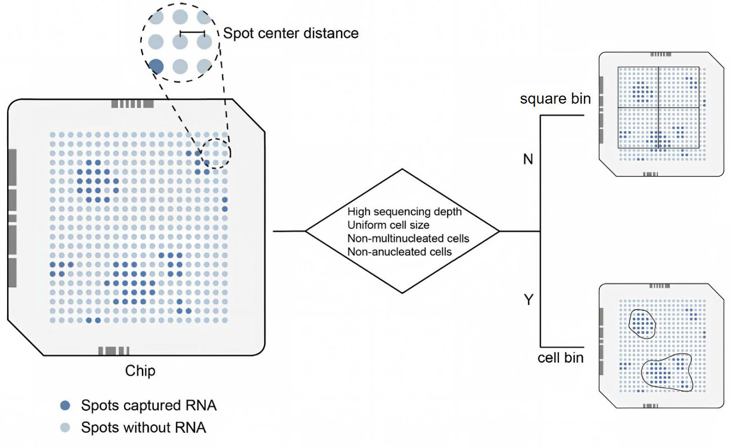
Figure 4. Selection of different analysis unit segmentation strategies based on sample condition and data quality. The square lattice area represents the sequencing chip, and the blue spots indicate the regions where RNA has been captured. The diamond-shaped boxes contain specific criteria for determining the segmentation strategy. The choice between these strategies determines the trade-off between transcriptomic depth and spatial resolution, directly affecting the detection of rare cell populations and fine anatomical structures.
Leveraging deconvolution and read-depth enhancement for embryonic studies
Deconvolution becomes important in embryonic ST when the spatial assay does not directly resolve individual cells, but the biological question still requires cell-type level interpretation. This situation is common in human embryonic studies, where low-resolution ST platforms, limited sample availability, and scarce matched sections often make direct cell-level annotation difficult. In such settings, deconvolution uses scRNA-seq-derived reference profiles to infer the likely cellular composition of each spatial spot or bin, thereby improving the interpretability of spatial maps.
The landscape of deconvolution algorithms encompasses a variety of approaches, including complex probabilistic models and deep learning-based methods[115]. In practice, the main distinction is not simply algorithmic form, but whether the method relies on matched single-cell references, how well it tolerates platform and batch differences, and whether it is intended primarily for annotation transfer, composition inference, or expression enhancement. Commonly used deconvolution methods in embryonic research include RCTD[116] and Cell2location[117]. Both methods employ generalized linear models, utilizing Poisson (RCTD) and negative binomial (Cell2location) distributions, respectively, to infer the cellular composition (i.e., the proportion of each cell type) within each ST spot or bin from scRNA-seq-derived reference profiles. Although these methods were published several years ago, their performance remains robust and has been widely validated across diverse ST platforms[115,118,119]. In 2021, Tangram introduced a deep learning approach to ST deconvolution. Building upon deconvolution, it further integrates single-cell and ST expression matrices to impute or enhance gene expression information in the spatial data, offering unique advantages for gene expression prediction. It continues to be actively used in embryonic research[120]. To improve accuracy, Xiao Yang and Jinjin Xiang developed the Spatialsmooth software package, which integrates multiple deconvolution methods and uses a convolutional autoencoder to smooth different cell-type combinations in space[121]. Beyond continuous improvements in accuracy, optimization of deconvolution algorithms primarily focuses on three directions: (1) enhancing computational scalability to handle ever-increasing cell numbers (e.g., jMF2D[122], NLSDeconv[123]); (2) integrating histological image information to improve reliability (e.g., SpaDecon[124], GraphST[125], FAST[126]); and (3) enabling deconvolution without reliance on scRNA-seq reference data (e.g., STdeconvolve[127], RETROFIT[128], gwSPADE[129], SpatialDeX[130]). For embryonic applications, these three directions correspond to three recurring practical needs: scaling to increasingly large atlases, improving robustness in histologically complex tissues, and reducing dependence on perfectly matched reference datasets. Given the scarcity of early human embryonic samples and the practical challenge of obtaining multiple replicates, reference-free deconvolution methods (those not reliant on scRNA-seq data) hold particular promise for this field.
Deconvolution techniques have been successfully applied to low-resolution ST data to resolve the spatial localization of diverse cell types within human embryonic tissues. Xu et al. (2023) employed the RCTD algorithm to map 239 scRNA-seq-derived cell type annotations onto two CS13 human embryonic tissue sections profiled by 10X Visium (55 µm resolution)[54]. This successfully localized both distinct and subtle anatomical structures visible in H&E staining, such as the optic vesicles, renal epithelium, head muscles, and pancreas[54]. Similarly, Cell2location has been applied in limb development studies to precisely identify key mesenchymal cell subsets, thereby delineating the spatial expression patterns of genes implicated in limb malformations within specific niches. Beyond the probabilistic models discussed, the deep learning-based method Tangram has been applied to human embryonic brain sections at the postconceptional week (PCW) 4 stage. It generated annotations for brain structures, including the neural tube, that align accurately with established anatomical landmarks[57]. Collectively, these studies show that deconvolution is most convincing when supported by anatomically interpretable landmarks and by reference data that are reasonably matched in developmental stage.
Current applications demonstrate that deconvolution excels at identifying spatially dispersed cell populations (e.g., immune cells) and is also highly effective for detecting rare cell types. To mitigate batch effects arising from differences in technology platforms, biological sample variability, and experimental protocols, it is ideal to perform deconvolution using matched, concurrently generated scRNA-seq data from the same embryonic stage as the reference. Therefore, this algorithm is widely used in human embryonic samples from later, relatively easily obtainable organ development stages. This is because the embryonic samples from a relatively later period can be obtained more readily and repeatedly.
Finding the anatomical structures of spatially adjacent tissues: spatial domain identification
Spatial domain identification becomes necessary when the goal is not only to annotate cell states, but also to recover anatomically coherent tissue regions from spatial data[131]. This is particularly important in embryonic research, where neighboring tissues often differ sharply in developmental potential and signaling environment despite sharing partially overlapping transcriptional features. Traditional clustering methods based only on gene expression similarity therefore tend to miss one of the key features of embryo organization: the continuity of local structure in space. To fill this gap, a range of spatial domain identification methods have been developed to integrate gene expression with spatial topology and, in many cases, histological context.
The application of convolutional neural networks (CNNs) and graph neural networks (GNNs) represents a standard computational approach to address this challenge. In practice, these methods mainly differ in how they weight spatial continuity, histological information, computational cost and robustness to spatially dispersed cell populations. Since the recognition task often involves co-registered histological image data, it naturally extends into a multimodal deep learning problem. Statistical model-based methods, such as Hidden Markov Random Field models[132], were the first to adapt to spatial multi-omics[133]. These methods are based on the assumption that neighboring cells have similar gene expression (BayesSpace[134]). Subsequently, deep learning-based methods have rapidly advanced in the field of spatial domain identification. Initial efforts focused on leveraging CNNs to extract features that capture spatial context. stLearn[135] uses a pretrained CNN model, ResNet-50, to extract image features and integrates them with a smoothed representation matrix to obtain a new matrix. Based on the integrated matrix, traditional Louvain/Leiden clustering is performed, and further sub-clustering based on the DBSCAN algorithm (an algorithm based on pure spatial location information clustering) is performed on spatially discrete clusters to further distinguish spatially distant clusters of the same type. SpaGCN[136] integrates gene expression, spatial information, and H&E image information using graph convolutional networks (GCNs). STAGATE significantly improves upon SpaGCN by introducing an adaptive graph attention autoencoder to dynamically learn the similarity between neighboring points[137]. Following this, methods for integrating multimodal information in deep learning to achieve spatial domain dimensionality reduction have proliferated (e.g., DeepST[138], SpaceFlow[139], GraphST[125], SpaSEG[140]).
In the past two years, the introduction of the transformer architecture and large-scale pretrained models has substantially addressed a key limitation of previous methods: incorrectly classifying spatially non-clustered cell types, such as immune cells, into nearby anatomical hierarchical structures (TransformerST)[141]. spaLLM utilizes the foundation model scGPT[142] pretrained on a single-cell dataset to obtain the spatial group expression enhancement matrix, and then fuses multimodal information through graph neural networks and multi-view attention mechanisms, significantly outperforming existing methods on multiple datasets[143]. Similarly, Novae adapts the SwAV visual self-supervised learning framework to ST, enabling accurate spatial domain identification and dimensionality reduction even for complex tissue sections[144]. While the aforementioned neural network-based methods are gradually improving, they often come with substantial computational costs. In contrast, Banksy adopts a distinct strategy. It constructs a feature matrix for each cell by integrating its own gene expression profile with neighborhood information. The latter includes the average expression of neighboring cells and the spatial gradient (magnitude and direction) of expression changes. This composite representation is termed the Banksy matrix. This matrix encapsulates the intrinsic cell expression, the local microenvironment expression profile, and the spatial patterns of expression variation. The weight of the cell's own expression information is flexibly adjusted by the λ parameter. Standard clustering algorithms like Leiden or Louvain, when applied to the Banksy matrix, can identify spatially coherent domains while simultaneously preserving the identity of spatially dispersed cell populations. Compared to neural network methods, it greatly saves computation time while maintaining competitive and stable accuracy[145]. The algorithms mentioned above all emphasize spatial domain identification on a single two-dimensional slice, while SpaDo emphasizes the continuity of three-dimensional spatial domains on continuous slices. This algorithm can accurately transfer scRNA-seq annotations to continuous slices, i.e., supervised identification of spatial domains, and can also accurately identify adjacent spatial domains on three-dimensional continuous slices without supervision[146].
Using spatial domain identification algorithms alone struggles to balance the contradiction between the heterogeneity of single cells and the continuity of their neighborhoods. This means they may overlook small clusters or scattered cells in space and categorize them into neighboring large anatomical cell types. In embryonic analysis, spatial domain identification is most informative when the objective is to delineate contiguous tissue architecture or to locate developmental organizers within complex local neighborhoods. However, because these methods may obscure scattered or rare cell populations, they are best interpreted together with deconvolution, single-cell references, or conventional clustering results when higher annotation fidelity is required [Figure 5].
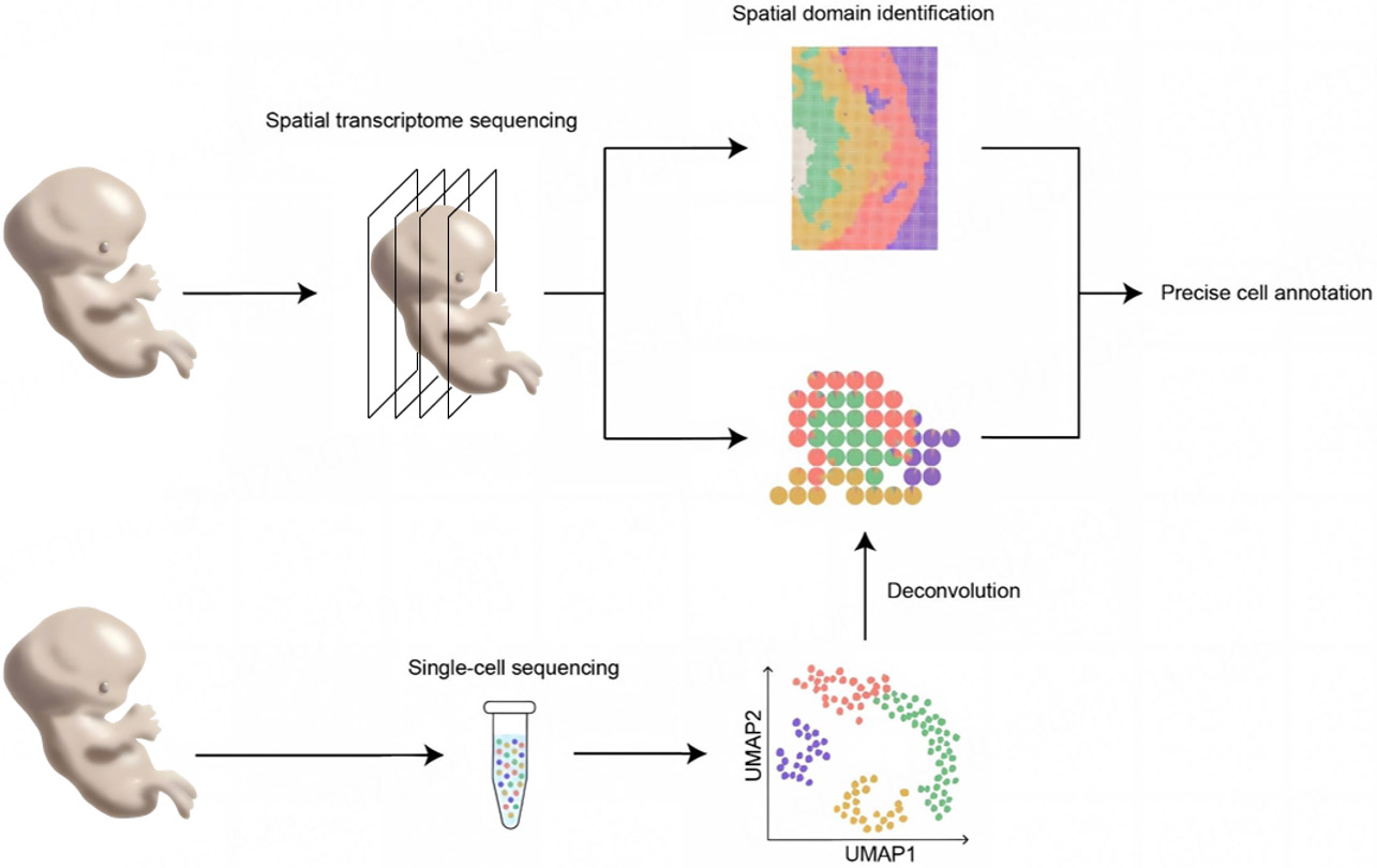
Figure 5. The workflow for cell annotation in human embryonic spatiotemporal omics studies, from raw data processing (bin selection and deconvolution) through spatial domain identification and cell-type assignment, enabling the transition from spatial measurements to biologically interpretable cell-type maps.
Spatial domain identification technology is mainly used in early embryonic research. Chen et al. applied Stereo-seq in mice and proposed spatially constrained clustering[73]. Based on the union of spatial proximity maps and transcriptome similarity maps, they generated a combinatorial map and then used Leiden to perform clustering on the combinatorial map[73]. This relatively simple and computationally efficient spatial domain identification strategy was subsequently extended to study human early embryonic development during the peri-gastrulation period[2,3,56], also utilizing Stereo-seq technology. This application helped researchers locate key structures, such as the anterior visceral endoderm (AVE). This illustrates why spatial domain methods are especially valuable in early embryonic samples, where identifying small but spatially organized signaling centers can be more informative than transcript-only clustering.
Human early embryonic cell migration and cell fate decisions in spatiotemporal dimensions
In embryonic development, lineage progression and cell migration are inseparable from space: cells do not differentiate in isolation, but within changing tissue geometries, signaling gradients, and neighborhood relationships. For this reason, trajectory inference in ST is valuable not only because it estimates developmental order, but because it allows inferred fate transitions to be interpreted in their anatomical context. This is especially important during gastrulation and early organogenesis, when transient cell states and directional movements are tightly coupled to morphogenesis.
In practical analysis, mainstream differentiation methods can be divided into two main categories: graph-based or minimum spanning tree-based methods and velocity-based methods based on mRNA splicing. The main distinction in practice is whether the method infers developmental progression from transcriptional similarity alone or estimates directional change from RNA dynamics. Algorithms such as Monocle2[147], Monocle3[148], and Slingshot[149] work by constructing a cell-to-cell graph within a low-dimensional embedding [e.g., derived from DDRTree or uniform manifold approximation and projection (UMAP)] of the gene expression matrix. This graph is then used to infer a pseudotemporal ordering of cells along putative differentiation trajectories. Trajectory inference methods that have developed alongside spatial omics technologies primarily rely on velocity-based approaches. Traditional velocity methods, such as the steady-state model Velocyto[150], the dynamic model scVelo[151], and deep learning-based CellDancer[152] and DeepVelo[153], work by comparing the abundance of unspliced and spliced mRNA in cells to infer the instantaneous direction of gene expression changes, thereby predicting the future differentiation state of cells. Although these methods do not directly utilize spatial information, the calculated pseudotime, latent time, or vector projection can be projected onto the spatial dimension (or onto a 3D point cloud if reconstruction is involved), allowing visual observation of cell differentiation in space. These methods typically require principal component analysis (PCA) dimensionality reduction, and high-quality spatial PCA results generated by SpatialPCA can be used as preliminary data to further improve performance in ST[154].
The past three years have also seen the proposal of velocity algorithms specifically designed for ST. These methods are particularly attractive for embryonic studies because they attempt to couple dynamic inference with local spatial context, rather than treating developmental progression as independent of tissue position. For high-resolution ST based on NGS, loom files containing splicing information can be directly generated from the aligned BAM files. For instance, Stereopy[97] can directly generate loom files from Stereo-seq data. For other platforms like 10X Visium HD, the BAM files first need to be merged and processed according to the chosen spatial bin size, after which tools like Velocyto[150] can be used to generate the loom files. These upstream files can serve as the basis for various velocity algorithms. Spateo[63] introduced the concept of vector fields from geoinformatics, combined with Dynamo[155] to model the differentiation rate, and determined the spatial differentiation and migration direction of cells in mouse developmental data across time periods. TopoVelo[156] employs a GNN to model interactions between spatially neighboring cells, thereby deriving velocity parameters that are intrinsically coupled to spatial context. It can be further integrated with lineage inference tools like scFate[157] to estimate the direction and rate of cellular migration within the tissue space. spVelo[158] is a generative model designed for ST. Building upon advancements in RNA velocity analysis, it specifically addresses the challenge of batch effect correction when integrating data from multiple developmental time points. For techniques like MERFISH based on fluorescent probes, the velocity calculation results from scRNA-seq need to be deconvolved spatially (Cell2Fate[159], SIRV[160]).
In actual differentiation analysis of embryonic development, stemness genes, the CytoTRACE stemness scoring algorithm[161,162], and the researcher's prior knowledge are often used to comprehensively identify cells in the current differentiation lineage that are suspected to be at the differentiation origin. Next, traditional methods such as minimum spanning trees and the Velocity algorithm are used to determine the differentiation path in a combination of supervised and unsupervised methods. A representative application is the study of human CS7 embryos by Cui et al.[56]. They projected pseudotime inferred by Monocle3[148] onto both UMAP embeddings and the actual 3D embryo model. These pseudotime projections were then integrated with inferences from the dynamic model of scVelo[151] and the partition-based graph abstraction (PAGA) method[163]. These three complementary approaches were used to reconstruct the spatial differentiation trajectory of the mesoderm lineage. In the same study, analysis of PGC lineage differentiation utilized the scVelo dynamic model within the UMAP space. This suggested an epiblast origin for PGCs and pointed to a potential differentiation pathway from amniotic ectoderm towards PGCs. In a study of mouse E9.5 embryos[102], the authors also used Monocle3 to infer pseudotimes of neuromesodermal progenitor cell (NMP) lineages to presomitic mesoderm (PSM), somites, and neural tube, etc., and projected the results into three-dimensional space. They found that the degree of transcriptome change was not a simple linear relationship with spatial distance, revealing a gene expression change pattern along the dorsoventral (DV) axis. The studies mentioned above employed high-resolution ST methods. When using platforms with relatively lower spatial resolution, such as in a study of human embryonic limb development[58], researchers have successfully combined multiple computational approaches. These include Diffusion Maps[164], force-directed graph layout[165], and PAGA to infer differentiation trajectories of cells originating from the lateral plate mesoderm. This revealed a differentiation network that radiated from six core mesenchymal progenitor cells at PCW5, differentiating into various cell types such as cartilage, bone, and tendon.
Traditional methods like Monocle2 and Monocle3 often require extensive prior biological knowledge to determine reliable cell types at the origin of differentiation, which can introduce subjective bias. While RNA velocity provides an unsupervised framework, the accuracy and assumptions underlying its inferences have been subjects of ongoing discussion and refinement[166]. Furthermore, standard ST data often represent transcript counts from spots or bins containing multiple cells, rather than pure single-cell measurements. Consequently, the observed spliced and unspliced mRNA counts are aggregated signals, which may not accurately reflect the kinetics within individual cells. These issues all challenge the accuracy of differentiation analysis with ST data. Therefore, until a completely reliable, unsupervised single differentiation method emerges, complex differentiation events like embryonic cell differentiation require the use of multiple differentiation methods to cross-validate and determine the direction and sequence of differentiation. In embryonic research, trajectory and velocity analyses are most informative when they are supported by recognizable anatomical landmarks, temporally ordered sampling, or independent lineage knowledge. They are especially useful for reconstructing directional processes such as gastrulation movements, germ-layer transitions, and early organ-specific patterning. However, because inferred trajectories remain sensitive to data sparsity, batch effects, and the assumptions of individual models, they should be interpreted as spatially constrained hypotheses rather than direct proof of lineage progression or migration.
Analyzing developmental signals: communication algorithms under spatial constraints
In embryonic development, signaling is not simply a property of cell type, but of spatially organized interactions among neighboring tissues. This is especially important during gastrulation and early organogenesis, when local signaling centers and short-range inductive interactions help establish body axes, tissue boundaries, and organizer function. Under these conditions, spatially constrained communication analysis becomes valuable not only because it reduces false positive interactions caused by tissue dissociation, but also because it helps reconstruct the signaling architecture of developing embryos. The CellChat v2 upgrade introduces spatial constraints by defining spatial neighborhoods, which identify each cell's neighbors based on calculating the Euclidean distance threshold between cells, thereby effectively filtering out false positive signals introduced by tissue dissociation[167]. CellPhoneDB v5, a classic communication analysis tool designed for ST upgrades, introduces the concept of microenvironments. When inferring communication, it only considers the interactions between cell types belonging to the same microenvironment, thereby refining the communication network and making it more consistent with the actual structure of the tissue[168]. Furthermore, new algorithms designed natively for ST data are continuously emerging. SpaTalk uses graph networks and knowledge graph methods to model and score the co-expression of ligand-receptor-target signaling networks between spatially adjacent cells[169]. COMMOT, developed by the same team as CellChat, incorporates optimal transport theory to model ligand diffusion from secretory to receiving cells. This provides a more realistic simulation of signal propagation through tissue space[170]. Signaling regulation operates across multiple hierarchical levels. Building upon COMMOT's output, FlowSig reveals more precisely how ligand-receptor interactions drive downstream signal output by regulating intracellular gene modules through a causal inference graph model that includes signal inflow, gene expression modules, and signal outflow[171]. The limitation of the above methods is that they rely on known, human-summarized communication network databases such as CellChatDB, and cannot discover potential new communication control relationships. To address this limitation, OrgaCCC[172] was developed. This method utilizes two independent variational graph auto-encoders to capture features from the cellular/spot domain and the gene domain, respectively. These two autoencoders are integrated by maximizing the similarity between their respective reconstructions of the gene expression matrix. The resulting intercellular relationship matrix is the inferred communication network. This method was tested on mouse placental ST data and identified receptor-ligand pairs that were not detected by other communication methods. Significant interactions between trophoblast cells and endometrial spiral artery endothelial cells were observed to anchor trophoblast cells within the vascular lumen, thereby ensuring blood perfusion and embryonic health. Real cells live in three-dimensional space and communicate with each other. Spateo[63] developed a complete analytical framework. After rearranging cells using the concept of a spatial field, its communication analysis module integrates spatial location, gene expression, and morphological information to construct a spatially aware regression model to identify receptor-ligand interactions in three-dimensional space and reveal complete cascade signaling pathways.
Computational inference alone does not establish that cell-cell communication is active during embryonic development. We therefore distinguish four levels of evidence for inferred interactions. (1) A predicted interaction is inferred from ligand-receptor co-expression and serves only to formulate hypotheses. (2) A spatially supported interaction is additionally constrained by spatial proximity between ligand- and receptor-expressing cells, which raises confidence without providing functional validation. (3) An experimentally perturbed interaction is supported by perturbation experiments in embryo models. Such experiments provide direct functional evidence that the candidate interaction can influence downstream responses, although they do not by themselves establish a causal developmental mechanism. (4) A causally validated developmental mechanism is demonstrated through perturbation together with rescue and lineage tracing, a standard rarely achievable in human embryos. In the CS7-CS9 examples discussed below, we primarily report evidence at the first two levels, and these interactions should therefore be interpreted as spatially constrained hypotheses requiring experimental validation.
These analytical advances are particularly relevant to embryonic studies, where signaling centers are often small, spatially restricted, and developmentally transient. The evolution of communication algorithms, from single-level to multi-level regulation and from two-dimensional slices to three-dimensional structures, also influences research on embryonic development, particularly in the study of axial signal regulation and the identification and regulation mechanisms of Organizer cells. In the early CS7 stage of gastrulation, researchers, using CellChat v2, discovered that the AVE may serve as a major source of signaling molecules such as DKK1 and CER1, as well as the transcription factor OTX2. These secreted factors inhibit signaling activity associated with the anterior primitive streak, thereby helping to establish the embryo’s anteroposterior polarity[56]. As gastrulation proceeds into the CS8 stage, the body axis undergoes further extension and regional specialization. Spatial communication analysis identified two key signaling centers: the notochord and the connecting stalk. These centers are predicted to jointly regulate the spatial gradients of signaling factors such as WNT, SHH, and BMP within the embryo[2]. In the CS9 stage, the neural tube begins to close, and brain regions begin to specialize. Researchers identified the isthmic organizer at the midbrain-hindbrain boundary as a candidate signaling center. This organizer exhibits high expression of signaling factors like Fgf8 and Wnt1, which are crucial for patterning the anterior (midbrain) and posterior (hindbrain) regions[3].
When selecting specific communication algorithms for embryonic data, it is important to note that not all spatial communication methods are currently compatible with Square Bin-based spatial sequencing units. Therefore, careful consideration should be given to cell segmentation methods when choosing a specific communication tool. Furthermore, it's necessary to consider whether using only known database interactions as a basis is sufficient for discovering new communication relationships. The development of spatial communication inference tools mainly focuses on the following directions: first, ensuring broad applicability across ST platforms with varying sequencing depths and segmentation methods; second, refining spatial distance constraints to reduce false positives while preserving genuine signals; and third, modeling multi-cascade signaling networks and long-range communication.
Embryo reconstruction in serial slices
Embryo reconstruction in serial slices becomes necessary when the biological question extends beyond isolated sections to embryo-scale organization. This is particularly important because body axes, signaling gradients, tissue continuity, and cell migration are inherently three-dimensional, whereas most spatial assays still generate data as two-dimensional slices. The close co-development of reconstruction algorithms and embryonic applications has therefore been driven by several intrinsic features of embryonic samples: their rarity, which makes maximal information recovery from each specimen especially valuable; their relatively small size, which makes serial sectioning technically feasible; and their highly ordered spatial organization, which is best interpreted in a continuous 3D framework. For this reason, embryonic datasets have become both a major application scenario and an important benchmark resource for the development of 3D reconstruction algorithms. For example, the MOSTA[73] embryo database derived from the mouse embryo article published by BGI in 2022, besides being used for 3D reconstruction in that article, has also appeared as a test dataset in many subsequent 3D reconstruction algorithm articles (SLAT[173], SPATEO[63]). Similarly, the STitch3D algorithm was applied to integrate multi-slice spatial transcriptome and scRNA-seq data, achieving the first 3D spatial reconstruction of human heart development[174]. In addition, sequencing technologies such as Magic-seq have directly performed continuous slices and 3D reconstruction of the developing mouse brain. These cases illustrate not only that embryonic ST data are key benchmark resource for reconstruction algorithms, but also that reconstruction is most informative when the aim is to recover spatial continuity across sections rather than to interpret each slice in isolation.
Three-dimensional reconstruction algorithms have been widely used in human embryonic research. ST data of the human CS8 embryo enabled researchers, for the first time, to accurately map gradients of morphogenetic signals and spatial distributions of cell types across a complete intact early human embryo[2]. Specifically, researchers utilized the PASTE algorithm for initial alignment of sequential ST slices. The positions of key, spatially coherent cell populations (e.g., pre-annotated notochord cells) were then manually refined to generate a coherent 3D point cloud. Subsequently, Maya was used to model the outer shell of key tissue anatomy within the point cloud, and the modeling results were imported into Plotly (https://plot.ly) for dynamic visualization. Finally, a visualization webpage was built to publicly display the aforementioned three-dimensional virtual embryo model containing the expressed information [Figure 6].

Figure 6. The workflow for the 3D reconstruction of embryonic spatial transcriptomics data from serial sections. From left to right: serial sectioning and sequencing, algorithmic generation of the 3D point cloud, refinement of the point cloud using prior knowledge, and shell modeling.
This pioneering pipeline was subsequently referenced in several human embryology papers, including CS7[56] and CS9[3], which also constructed high-quality three-dimensional virtual embryos. Leveraging these reconstructions, researchers delineated the spatial distribution of signaling molecules along the AP and DV axes, characterized the 3D morphology of key tissue structures and signaling centers, and uncovered 3D cellular migration patterns during CS7 and CS8[2]. At the CS9 stage, analysis revealed that NMPs are organized into a three-dimensional, bilayered structure along the DV axis. These findings show that reconstruction is particularly powerful when developmental interpretation depends on axial geometry, cross-section continuity, or spatially extended signaling architecture that cannot be inferred reliably from single slices alone.
From an analytical perspective, the evolution of 3D reconstruction algorithms has progressively addressed three recurring challenges in embryonic spatial data: cross-section alignment, non-rigid tissue deformation, and incomplete section continuity. This shift reflects progress toward more efficient, flexible 3D reconstruction of spatial transcriptomic data. Early attempts at 3D reconstruction often adapted traditional computer vision image registration algorithms, such as SIFT[175]. These methods relied solely on pixel-level image features, entirely disregarding the biological information in gene expression similarity, and were incapable of handling the complex, nonlinear deformations inherent to embryonic development. Consequently, their application to biological data faced significant limitations[176]. This was particularly problematic for embryonic samples, in which rapid morphological change and curved body plans make purely image-based alignment unreliable. A significant breakthrough came with PASTE in 2020, which achieved alignment of slices using ST data based on optimal transport theory[96]. However, PASTE employed a rigid alignment strategy, which could not accommodate natural tissue deformations introduced during sectioning. Furthermore, its computational scalability was limited. Substantial progress commenced with the advent of high-resolution ST platforms and concurrent innovations in algorithmic design. The SLAT[173] algorithm, proposed in 2023, breaks through the bottleneck of heterogeneous data integration. Its core innovation involves modeling spatial topology as a graph structure and employing a lightweight graph convolutional network trained with an adversarial strategy. This framework not only enhances compatibility and computational efficiency across diverse technological platforms but also enables cross-timepoint slice alignment, thereby facilitating the inference of differentiation and migration trajectories during mouse kidney development. Also released in 2023, the SPACEL algorithm leverages an adversarial graph convolutional neural network model and a global optimization strategy to perform well in cell type prediction, spatial domain recognition, and 3D alignment tasks[177]. These methods improved cross-platform compatibility and made reconstruction more scalable in heterogeneous datasets. In 2024, the Spateo framework innovatively and effectively solved the problem of non-rigid alignment and morphological correction at the whole embryo scale through a probabilistic model of Gaussian processes and variational inference. Building upon this, it can automatically construct surface meshes from the aligned point clouds. These modules achieved excellent alignment results for both mouse and Drosophila embryos, accurately reconstructing complex morphological features such as the curled body plan of the mouse embryo. To address the common issue of non-consecutive or missing sections in ST datasets—which can disrupt 3D reconstruction—the SpatialZ algorithm was developed. It compensates for missing adjacent slices, ensuring the continuity of the reconstructed volume along the z-axis[178]. Together, these advances moved the field closer to embryo-scale reconstruction under realistic conditions of deformation and incomplete sampling.
The value of 3D reconstruction in embryonic studies extends beyond visualization. Once embryo-scale spatial continuity is established, many downstream analyses become more biologically interpretable in three-dimensional context. For example, Spateo's spatial communication module allows researchers to study modular representations and cascaded communications in 3D space. Metrics such as stemness scores from CytoTRACE[161,162], pseudotime from Monocle3[148], and latent time[151] from RNA velocity can be projected onto the 3D reconstruction, providing spatial context to understand the dynamics of cell differentiation and migration. Gene clustering algorithms developed specifically for ST, such as HotSpot[179], can also more accurately identify co-expressed gene modules in three-dimensional space, helping researchers deepen their understanding of regional tissue functions.
Although 3D reconstruction algorithms have become increasingly sophisticated, assessing the accuracy of a reconstructed embryo remains a considerable challenge. In imaging-based reconstruction tasks, ground truth can sometimes be obtained from an independent modality. This is not possible for human embryonic ST: once a section has been sequenced, the same tissue cannot be re-sequenced for validation. Reconstruction accuracy must therefore be assessed through alternative strategies. Several complementary checks can help evaluate reconstruction accuracy. Cross-validation with histology provides a qualitative but essential check. Overlaying spatial transcriptomic spots on H&E or immunofluorescence images from adjacent sections can expose misalignments that transcriptomic data alone would not reveal. Alignment error can also be quantified, for example as the root mean square deviation (RMSD) between corresponding spots in adjacent sections. Independent anatomical landmarks provide a further check of global consistency. The relative positions of the notochord, the neural tube and the developing cardiac primordium can be compared with the expected anatomy. Such comparisons confirm that biologically meaningful spatial relationships are preserved in the reconstruction. To make 3D reconstructions transparent and interpretable, we recommend that future studies report: (1) the number and thickness of sections used, and the proportion of sections that were missing or discarded; (2) the alignment algorithm employed, any manual refinement steps, and the anatomical criteria used for curation; (3) quantitative alignment metrics, such as RMSD or correlation coefficients between neighboring sections; and (4) a sensitivity analysis relating variation in alignment parameters or bin size to downstream biological interpretation.
Despite these advances, current 3D reconstruction methodologies still face several limitations. First, most current algorithms utilize only sequencing-derived gene expression and spatial coordinate data, largely neglecting the complementary information available from high-resolution histology images. Future multimodal 3D reconstruction software that integrates transcriptomic and imaging data could not only improve alignment accuracy but also produce more anatomically realistic and visually informative models. Second, sequencing of continuous thin sections is too costly and unsuitable for larger samples such as late-stage embryos. A low-cost end-to-end thick-section ST-running sequencing technology needs to be developed to balance the cost of large-sample reconstruction[89]. Finally, despite improvements in alignment accuracy, the process often still requires manual curation. Researchers must rely on prior biological knowledge to select tissue types with clear spatial continuity to guide and validate z-axis alignment. This places certain demands on the researchers' biological literacy and introduces some subjective bias into the alignment process. In embryonic research, serial-slice reconstruction is most informative when the goal is to interpret cell states, signaling gradients, and tissue morphology within a continuous three-dimensional framework. Its main limitation remains that alignment quality, section loss, and manual refinement can still influence the final biological interpretation [Box 3].
Summary of analytical strategies for spatial data
| Main biological advance | Computational pipelines convert raw spatial transcriptomic data into interpretable outputs, including spatial-domain identification, deconvolution, trajectory and RNA-velocity inference, cell-cell communication analysis and 3D reconstruction. Together, they move the field from descriptive mapping toward mechanistic interpretation |
| Preferred methods | Spatial unit selection: Square Bins for transcript depth and stability, Cell Bins for single-cell resolution. Cell-type annotation: reference-based (RCTD[116], Cell2location[117]) or reference-free (STdeconvolve[127], SpatialDeX[130]). Spatial domains: graph-based (SpaGCN[136], STAGATE[137], BANKSY[145]) or foundation models (spaLLM[143], Novae[144]). Trajectory: Monocle3/Slingshot for pseudotime[148,149]; scVelo for spatial RNA velocity[151]. Communication: CellChat v2/CellPhoneDB v5 for standard use[167,168]; COMMOT/Spateo for spatially constrained inference[63,170]. 3D reconstruction: SLAT or Spateo[63,173] |
| Current limitation | Batch effects, data sparsity, and platform differences strongly affect results. Trajectory and velocity analyses rest on assumptions and require cross-validation with multiple approaches. Communication inference depends on curated interaction databases and cannot readily discover novel interactions[167,168]. 3D reconstruction often requires manual curation, and missing sections can compromise continuity[2,3,63] |
| Next experimental priority | Develop embryo-specific tools accounting for rapid morphogenetic changes and sample scarcity; improve uncertainty quantification and reproducibility; develop standardized reference datasets for cross-method benchmarking; further integrate AI and foundation models to improve accuracy and automation |
FUTURE OUTLOOK: EMBRYONIC RESEARCH DRIVEN BY SPATIOTEMPORAL OMICS MODELS
The final question is whether descriptive spatial maps can become predictive models of human development. We address it by considering the current barriers to applying foundation models to embryonic research, the evidence that already exists for each proposed application, and the directions most likely to move these models from description toward prediction. Despite major advances in spatial multi-omics, several central challenges in embryonic research remain unresolved. Current analyses are still limited by sparse spatial measurements, incomplete reference atlases, labor-intensive annotation, weak causal inference, and the difficulty of integrating molecular, spatial, and temporal information into a unified developmental model. It is in this context that large-scale generative models, including Transformer-based and diffusion-based architectures, have begun to attract attention. Their potential value lies not simply in modeling complex data distributions, but in potentially helping convert fragmented embryonic measurements into more coherent, predictive, and biologically interpretable representations[180]. Having revolutionized natural language processing in recent years, Transformer-based models are now rapidly expanding their application to multimodal bioinformatics data analysis. The classic Transformer architecture consists of an encoder and a decoder[181]. Encoder-based models like BERT excel at understanding context and performing tasks like masked language modeling (often likened to a cloze test), whereas decoder-based models like GPT specialize in autoregressive text generation (text continuation). Another generative model that has shone brightly in the field of computer vision (image and video generation) is the diffusion model[182]. These models operate through a progressive denoising process, which allows them to effectively model complex data distributions and thereby generate high-fidelity, high-quality synthetic content. Currently, these architectures have been preliminarily applied to spatiotemporal transcriptomics data analysis and modeling. In the following discussion, we distinguish three levels of inference—data recovery (imputation and denoising), statistical prediction (annotation transfer and perturbation response), and causal biological inference (mechanistic understanding of developmental programs). The first two are already showing practical utility, while the third remains a long-term goal. We then examine how current models address practical challenges in embryonic development analysis across these three levels.
The first opportunity for large-scale foundation models lies in addressing the two bottlenecks that recur throughout the preceding sections: sparse spatial measurements and incomplete or imperfect reference data. A prevailing challenge is that the sequencing depth of current ST platforms is typically significantly lower than that of scRNA-seq[8,183]. This interferes with the accuracy of analyses of early human embryos with scarce samples and limited access to scRNA-seq for deconvolution. A semantic enhancement matrix based on a foundation model can transform the original sparse gene expression matrix into a biologically meaningful low-dimensional representation by integrating prior biological knowledge (such as pathways and regulatory networks) and extracting high-order features using a pre-trained model. This matrix can significantly improve the accuracy of downstream analyses such as dimensionality reduction, clustering of cells, and spatial domain identification in benchmark datasets. Models specifically pretrained on single-cell transcriptomes, such as scGPT[142], scFoundation[184], and GeneFormer[185], can generate information-dense low-dimensional matrices as the basis for subsequent dimensionality reduction and clustering. To explicitly model the relationship between scRNA-seq and ST data, NicheFormer[186] was co-trained on combined datasets from both human and mouse, incorporating both single-cell and spatial transcriptomic profiles. Notably, a significant portion (76.45%) of the spatial data came from MERFISH, a lower-depth imaging-based platform. This training strategy enhances the model's capability for cross-modal and cross-depth-scale analysis. Consequently, NicheFormer acquires the ability for bidirectional annotation transfer between scRNA-seq and ST data and can predict cellular composition within spatial microenvironments. This functionally extends the capabilities of traditional deconvolution algorithms. Beyond Transformer-based approaches, diffusion-based models such as CausalGeD[187] and SpaDiT[188] are designed to integrate scRNA-seq with spatial transcriptomic data. CausalGeD has demonstrated state-of-the-art performance across ten benchmark datasets using metrics including PCC, SSIM, RMSE, and JS divergence, while SpaDiT achieved an 8%-12% improvement over eight baseline methods on ten datasets. Specifically, these models can leverage information from scRNA-seq to impute missing values or denoise low-depth spatial transcriptomic data, thereby generating gene expression patterns that more accurately approximate the true underlying spatial expression.
Imputation can recover biological signal from sparse measurements, but it also risks introducing expression patterns that were not directly measured. This risk is greatest in rare human embryonic samples, where computationally generated signals could be mistaken for genuine biological findings. Diffusion-based models illustrate the risk: when applied to sparse spatial data, they may infer spatially coherent expression in regions where few or no transcripts were captured. The resulting expression gradients could be mistaken for developmental signaling centers. Human embryos offer limited scope for experimental perturbation and replication compared with model organisms, so independent references for distinguishing true biological signal from imputation artifacts are scarce. Imputed values should therefore be treated as hypotheses requiring confirmation by imaging-based methods or experimental perturbation, rather than as direct measurements.
A second recurring bottleneck is annotation itself, especially in embryonic datasets where marker definitions are incomplete, rare cell states are frequent, and spatial bins do not correspond neatly to classical cell-type signatures. Manual annotation requires supervision by a professional embryologist, is complex and time-consuming, and can introduce subjective bias. Traditional annotation methods often rely on markers for various cell types to score cells, which is poorly adapted to the ST data of Square bin segmentation. Moreover, comprehensive, curated marker databases specifically for human embryonic cell types are scarce, further limiting the utility of these methods for discovering novel cell types[189]. Both single-cell and ST-based foundation models have automatic annotation capabilities and do not rely on built-in marker databases. Although their performance depends on the quality and relevance of their pretraining and fine-tuning datasets, these models can be effectively adapted to embryonic datasets through targeted fine-tuning. After targeted fine-tuning, scBERT[190] and scGPT have demonstrated annotation accuracy substantially higher than that of traditional methods on benchmark datasets[191]. On the multiple sclerosis dataset, scGPT achieved an accuracy of approximately 0.85. scBERT has been evaluated on multiple public scRNA-seq datasets, including Zheng68k PBMC and five pancreas datasets, with PEFT methods substantially outperforming conventional methods such as support vector machines and logistic regression. To enhance usability and generalizability, scFoundation expanded the pre-trained dataset and learned as many potential connections between each gene as possible without gene screening during training and inference. This approach enables scFoundation to clearly distinguish rare cell types, such as memory T cells and monocytes, on benchmark datasets like Zheng68K even without task-specific fine-tuning[184]. SCimilarity employs an autoencoder architecture combined with metric learning to perform rapid annotation by searching a massive, integrated human cellular atlas encompassing 23.4 million cells from 412 studies[192].
Current gene-level analyses, such as TF regulatory factor analysis, communication network analysis, and co-expression module analysis, often yield large gene sets. Researchers can use functional enrichment analyses (e.g., Gene Ontology, Kyoto Encyclopedia of Genes and Genomes, Human Phenotype Ontology, HALLMARK[193]) and known gene functions to infer the potential biological roles of these gene sets. However, these approaches are limited in their ability to elucidate the specific interaction networks and causality among the genes. Experimental validation using actual human embryos is severely constrained by sample scarcity. Embryo model preparation and associated experimental workflows are complex, time-consuming, and often exhibit low reproducibility, failing to meet the demand for rapid verification of massive amounts of genes. To address this gap, researchers have developed large-scale omics models that learn potential gene interaction networks and possess virtual knockout capabilities. For example, the scFoundation framework[184] extracts cell-specific gene context embeddings from its pretrained decoder. Each gene embedding encapsulates information about its functional associations within the specific cellular context. These embeddings serve as node features for downstream graph-based models (e.g., GEARS[194]) to construct cell-type-specific gene co-expression networks. GEARS has been evaluated on perturbation prediction benchmarks (Norman et al.[195], Adamson et al.[196], Replogle et al.[197]) using metrics including Pearson correlation between predicted and measured expression changes. When simulating gene knockout in validated cell-line models, the approach has shown promise in predicting differentially expressed genes (DEGs) by masking the gene node and inferring expression changes of downstream genes. During the pretraining of GeneFormer[185], the team led by X. Shirley Liu and Patrick T. Ellinor employed a masked language modeling objective, randomly masking 15% of the input genes. The model learns to infer the masked genes from the context of the remaining genes, thereby capturing gene-gene regulatory relationships in a self-supervised manner. Furthermore, analysis of the model's attention mechanisms revealed that it disproportionately attends to core regulatory network nodes, such as transcription factors, during pretraining. This inherent focus on key regulators likely enhances the model's ability to predict outcomes of gene network perturbations. Similarly, diffusion models, renowned for their high-quality data generation, also exhibit potent predictive capabilities for cellular perturbation responses. He et al.[198] developed Squidiff, which further extends predictive capabilities from gene knockout to cell differentiation, drug perturbation, and changes in the physical environment. In the task of predicting single-cell expression profiles by inputting perturbation vectors, based on data from both ends of the 0-3 days culture period, it demonstrated promising performance in predicting the single-cell state of iPSCs differentiating into the three germ layers at 1-2 days.
AI Agents developed by researchers can intelligently integrate the aforementioned analytical capabilities with traditional bioinformatics workflows. This is especially relevant to embryonic research, where analytical workflows are long, sample numbers are limited, and errors introduced at early stages of interpretation can propagate through multiple downstream inferences. This enables general-purpose language models, which are prone to hallucinations, to acquire domain-specific expertise in bioinformatics analysis or specialized biological knowledge. Scientists can then interact with these agents using natural language to bridge knowledge gaps in bioinformatics or developmental biology, thereby accelerating their analysis and improving its reliability. Agents designed for biological knowledge synthesis assist scientists in rapidly and comprehensively surveying the current state of a research field. The development of such agents does not rely on sequencing or omics data but rather addresses classic challenges in text processing and knowledge base construction (e.g., PubMedGPT[199], Flan-PaLM[200], Microsoft BioGPT[201], GeneGPT[202], AgentMD[203]). While the aforementioned models primarily aid in hypothesis generation, agents that directly execute analytical workflows represent a more transformative shift in productivity. Examples include CellAgent[204] for single-cell transcriptomics and SpatialAgent[205] for ST. These agents integrate a suite of reliable and robust analytical tools with the natural language interface of traditional large language models (LLMs). Researchers can use them to quickly obtain analysis solutions and corresponding code suitable for their data, or directly upload data for automated analysis, fundamentally changing the traditional paradigm of bioinformatics analysis. Two clear developmental trajectories are emerging in this field. The first is the development of multi-omics analysis agents, building upon matured capabilities for individual omics layers. For instance, AutoBA[206] supports the integrative analysis of transcriptomic, chromatin accessibility (ATAC-seq), and proteomic data. The second trajectory involves deeply integrating large-scale foundational omics models with LLMs, enabling researchers to query these complex models directly through natural language dialogue. This approach relies on rigorous and reliable massive expression matrix data. For instance, CellWhisperer[207] allows researchers to use natural language to access large-scale omics models and directly retrieve novel markers for various key cell types during the CS8 stage of human embryos, which was validated on three-dimensional ST data. The research goal for this direction in the coming years will be to develop super agents proficient in multi-omics and biological knowledge, potentially capable of providing reliable analytical approaches and code support based on the data collected by researchers. Ultimately, the integration of sequencing hardware control and scheduling intelligence into these large-scale foundation models points toward a future of fully automated, intelligent laboratories. In this vision, both sequencing platforms and analytical workflows would be comprehensively automated and orchestrated.
To avoid overstating their readiness for embryonic applications, we summarize in Table 5 the level of evidence currently available for each AI application. Because several of these methods have been used in only a limited number of embryonic or embryo-model contexts, we distinguish benchmark-level maturity from validated performance on human embryonic data.
Current evidence level of AI applications in embryonic research
| AI application | Evidence level | Supporting evidence/status |
| Data imputation and denoising (e.g., CausalGeD[187], SpaDiT[188]) | Emerging | Reported on public spatial transcriptomic benchmarks; evaluation on human embryonic ST data is currently sparse and lacks independent benchmarking |
| Cross-modal annotation transfer (e.g., scGPT[142], scFoundation[184], NicheFormer[186]) | Emerging | Benchmark-validated on single-cell and spatial datasets; human embryonic applications remain case-level demonstrations that require fine-tuning and independent confirmation |
| Cell type annotation (e.g., scBERT[190], SCimilarity[192]) | Emerging | Reliable on public single-cell benchmarks; performance on early human embryonic cell states has not yet been systematically benchmarked against embryo-specific references |
| Virtual gene knockout/perturbation prediction (e.g., scFoundation[184], GEARS[194]) | Emerging | Validated on perturbation screens in cell lines and model organisms; embryo and embryo-model validation is still limited |
| Differentiation trajectory prediction (e.g., Squidiff[198]) | Emerging | Demonstrated in iPSC differentiation toward three germ layers; requires further validation in embryonic contexts |
| Predictive virtual embryo reconstruction | Speculative | Conceptually promising; requires substantial data accumulation and model development |
| Causal developmental modeling | Speculative | Currently at conceptual stage; major challenges remain in causal inference from observational data |
| Fully automated intelligent laboratories | Speculative | Envisioned future direction; current AI agents are early-stage prototypes envisioned |
The development of large-scale foundational models for bioinformatics and multi-omics integration is still in its nascent stages and faces significant challenges. First, there is a pronounced lack of high-quality, embryo-specific datasets for targeted pretraining or fine-tuning of these models. The common training strategy for foundation models is to collect diverse datasets, train the model jointly on them, and then fine-tune it for a specific domain. However, transcriptomic data from embryos, especially human embryos, is extremely scarce and varies across sequencing platforms, diluting their effectiveness during pre-training and making it difficult to demonstrate good generalization ability after fine-tuning. In addition to data scarcity, several other barriers limit the application of foundation models to embryonic research: biological validation is particularly challenging due to the lack of ground truth for rare developmental events; ethical constraints restrict data sharing and model development; and the scarcity of standardized training datasets with consistent formats and annotations further complicates large-scale pretraining. Many AI models described above were developed and validated on specific datasets, and it is not yet established whether they generalize to embryonic contexts. Embryonic data differ from these training data in tissue architecture, cell-state diversity, and data sparsity. Technical variation across sequencing platforms and batches adds a further source of uncertainty. Cross-species transfer raises additional concerns when models trained largely on mouse data are applied to human embryos. Performance metrics reported in the original publications may therefore not translate to embryonic datasets. Independent validation remains limited, and most evaluations have been carried out by the teams that developed the methods. Systematic benchmarking on human embryonic data, coordinated across independent groups, will be needed to establish the reliability of these tools. Second, deploying and running these foundation models demands substantial graphics processing unit (GPU) computational resources, a requirement that contrasts sharply with traditional spatial omics analyses, which typically have minimal GPU demands. This may require researchers to purchase additional graphics cards for local deployment of large-scale foundation models. In addition, deploying and fine-tuning large-scale foundation models places more stringent demands on the interdisciplinary knowledge and professional skills of bioinformatics technicians. Furthermore, real cells are not a single-omics study, but involve the entire process of the central dogma and cell interactions. Current large-scale omics models are still limited to single-omics studies and cannot form multimodal, virtual cells similar to real cells. This limitation is particularly acute in ST, where current models fail to simultaneously and effectively integrate spatial, temporal, and high-resolution histological image information. In summary, the development of large-scale omics models and virtual cells faces a multifaceted set of challenges. Addressing these challenges requires a concerted, interdisciplinary effort: professional biologists to define the core biological questions and validate models, bioinformaticians to process complex multimodal data and serve as crucial translators between domains, and AI/computer scientists to design innovative model architectures and optimization strategies.
Returning to the domain of embryonic development, the fundamental prerequisite for developing large-scale foundation models is the accumulation of a substantial and diverse data foundation. As of 2025, large-scale foundation models trained on a single biological domain have already emerged. For example, BrainBeacon, trained on 133 million spatial transcriptomic units from human, mouse, and monkey brains, has learned interpretable brain feature representations and integrates functional modules for tasks like gene perturbation prediction and cross-species conserved region analysis[208]. Data on mouse embryos throughout all stages have been largely analyzed and compiled into high-quality databases such as MOSTA. With the release of 3D reconstructed virtual embryos from CS7, CS8, and CS9, a corner of the mystery of early human embryos has been unveiled. In the foreseeable future, virtual 3D human spatial transcriptomic models encompassing various developmental stages may become available, paving the way for a comprehensive, cross-stage human embryo ST database. Such a comprehensive database will constitute the essential foundation for training next-generation, embryo-specific large-scale foundation models. In this sense, the long-term significance of embryo-specific large-scale foundation models is not merely computational. They may ultimately provide the conceptual and technical foundation for moving from descriptive embryonic atlases toward predictive virtual embryos, although substantial technical and biological challenges remain [Box 4].
Summary of future outlook
| Main biological advance | Large-scale foundation models (scGPT[142], scFoundation[184], NicheFormer[186], GeneFormer[185]) address bottlenecks including sparse measurements, incomplete atlases, and labor-intensive annotation; AI agents (CellAgent[204], SpatialAgent[205]) provide natural language interfaces that democratize access to complex analytical workflows, collectively offering a pathway toward predictive, experimentally testable models of human development |
| Preferred methods | Annotation/integration: scGPT[142] or NicheFormer[186] for cross-modal tasks; SCimilarity[192] for rapid cell search. Perturbation prediction: GEARS[194] or scFoundation[184]; Squidiff[198] for diffusion-based response. Automated analysis: CellAgent[204] for single-cell; SpatialAgent[205] for ST |
| Current limitation | High-quality embryo-specific datasets remain scarce for model pretraining; foundation models require substantial computational resources; most models are limited to single-omics analysis; data imputation risks generating artificial patterns that may be mistaken for genuine biological findings in rare human embryonic samples |
| Next experimental priority | Accumulate and standardize multi-stage human embryo spatial datasets for embryo-specific foundation models; develop multimodal models integrating spatial, temporal, and histological information; strengthen experimental validation using embryo models and organoids |
CONCLUSION
Spatiotemporal multi-omics, especially ST, has begun to reshape early human embryonic research. It unites developmental time, tissue architecture, and molecular identity within an integrated analytical framework. This shift has already expanded what can be inferred about lineage specification, signaling organization, morphogenesis and embryo-model fidelity. Biological discovery and algorithmic innovation have co-evolved, allowing spatial maps, single-cell references and three-dimensional reconstructions to serve as linked lines of evidence. The field is moving from descriptive atlases toward mechanistic and predictive models, although this transition remains incomplete.
At the same time, major challenges remain in technology, computation, ethics, and data sharing, and these limitations continue to constrain the construction of truly predictive and clinically meaningful models of human embryogenesis. Although ST platforms are constantly progressing towards higher resolutions that approach those of real single cells, current technology still cannot achieve a balance between resolution, cost, and sequencing depth. Constructing a three-dimensional virtual embryo requires high sequencing costs, which is the first major technological hurdle. Significant challenges also exist on the analytical front. With the release of virtual embryo models for individual developmental stages, there is a pressing need to study cellular dynamics across multiple, consecutive stages. For example, while tools like SLAT and Spateo include modules for inferring differentiation and migration across stages, they often fail to reconstruct complete cellular migration trajectories and face limitations when applied to data spanning more than two developmental time points. Most current trajectory inference methods are better suited for analyzing differentiation sequences within a single developmental stage. Batch effects further compromise trajectory inference from integrated multi-stage data. Therefore, there is an urgent need for robust computational methods that can reliably correct for batch effects, predict differentiation trajectories across multiple stages and samples, and identify key driver genes for specific cell fates. Breakthroughs leveraging large-scale foundational models are anticipated to address these analytical gaps. Transferring developmental annotations across species or platforms poses a further challenge. Staging systems differ fundamentally, because mouse embryos are staged by embryonic days (E), whereas human embryos are staged by CS based on morphological landmarks. Core developmental programs are broadly conserved, but their timing, spatial domains, and target gene repertoires diverge across species. Some cell types and anatomical structures are species-specific or lack direct homologs, which complicates annotation transfer. Technical differences between platforms and protocols add batch effects that can obscure genuine biological differences or create spurious similarities. Direct transfer of developmental annotations is therefore inadvisable without species-specific validation. Wherever possible, predictions derived from model organisms should be benchmarked against available human embryonic data. However, a separate and related challenge is the lag in applying even existing advanced algorithms specifically to human embryonic studies. Analyzing human embryonic data demands high scientific literacy, advanced programming skills, and the ability to keep pace with evolving algorithms from bioinformaticians. This disparity is primarily due to the extreme scarcity and precious nature of human embryonic samples, which limits opportunities for methodological benchmarking and reduces the incentive for rapid tool adoption compared to studies on more accessible model organisms. Novel algorithms for spatial domain identification and trajectory inference are also typically benchmarked using mouse embryonic data. However, the application of these cutting-edge algorithms to human embryo data lags behind.
Realizing the full potential of spatiotemporal multi-omics in human embryogenesis will require coordinated progress on data resources, computational models, and governance. Standardized reference datasets spanning consecutive developmental stages should be assembled first. Harmonized formats, metadata, and quality control would enable cross-platform integration and benchmarking, and provide the embryo-specific training data that foundation models currently lack. Such models could then advance cross-modal integration, multi-stage trajectory inference and predictive modeling of developmental outcomes. Virtual embryo reconstructions should be validated experimentally against stem-cell-derived embryo models and, where possible, real human embryos. Computational outputs should remain hypotheses until confirmed, with clear separation between data recovery, statistical prediction and causal inference. Robust frameworks for ethical governance and data sharing must finally reconcile the "14-day rule" and national regulations on embryo research with the responsible reuse of sensitive human data. Building on these foundations, we propose a five-step translational roadmap [Figure 7] that carries human embryonic research from fundamental discovery toward clinical application. The roadmap begins with comprehensive atlases of normal embryonic development. It then profiles embryos affected by imprinting disorders (e.g., Angelman syndrome), trophoblastic diseases (e.g., hydatidiform mole), or metabolic and epigenetic dysregulation to identify disease-associated spatial alterations. Candidate alterations should next be validated experimentally in stem-cell-derived embryo models, including blastoids, gastruloids and organoids. Promising interventions, whether pharmacological, gene-editing or cell-based, can then be tested in these systems, before validated findings are developed into clinically actionable biomarkers or therapeutic targets. This approach would facilitate the translation of human embryonic research from broad, atlas-generating studies to clinically actionable investigations. Ultimately, progress in technology, analysis, standardization, and ethically responsible data integration will determine whether the field can move from increasingly detailed embryonic atlases toward predictive, dynamic, and clinically relevant virtual models of human development.

Figure 7. A conceptual roadmap for the transformation from basic embryological research to clinical application, consisting of five consecutive steps.
DECLARATIONS
Authors’ contributions
Conceptualized the study and supervised the project: Xiao Z
Wrote and edited the manuscript with input from all authors: Zhu H, Yang X
Created the figures and illustrations: Zhu H, Wang X
Availability of data and materials
Not applicable.
AI and AI-assisted tools statement
Not applicable.
Financial support and sponsorship
None.
Conflicts of interest
All authors declare that they have no conflicts of interest.
Ethical approval and consent to participate
Not applicable.
Consent for publication
Not applicable.
Copyright
© The Author(s) 2026.
Supplementary Materials
REFERENCES
1. Rossant J, Tam PP. Early human embryonic development: blastocyst formation to gastrulation. Dev Cell. 2022;57:152-65.
2. Xiao Z, Cui L, Yuan Y, et al. 3D reconstruction of a gastrulating human embryo. Cell. 2024;187:2855-74.e19.
3. Yuan Y, Wang X, Yan X, et al. 3D reconstruction of a human Carnegie stage 9 embryo provides a snapshot of early body plan formation. Cell Stem Cell. 2025;32:1006-24.e5.
4. Rivron N, Pera M, Rossant J, et al. Debate ethics of embryo models from stem cells. Nature. 2018;564:183-5.
5. Lovell-badge R, Anthony E, Barker RA, et al. ISSCR guidelines for stem cell research and clinical translation: the 2021 update. Stem Cell Rep. 2021;16:1398-408.
6. Dries R, Chen J, Del Rossi N, Khan MM, Sistig A, Yuan G. Advances in spatial transcriptomic data analysis. Genome Res. 2021;31:1706-18.
7. Cheng M, Jiang Y, Xu J, et al. Spatially resolved transcriptomics: a comprehensive review of their technological advances, applications, and challenges. J Genet Genom. 2023;50:625-40.
8. Liu Y, Dai Y, Wang L. Spatial omics at the forefront: emerging technologies, analytical innovations, and clinical applications. Cancer Cell. 2026;44:24-49.
9. Jain S, Eadon MT. Spatial transcriptomics in health and disease. Nat Rev Nephrol. 2024;20:659-71.
10. Geng A, Cui C, Luo Z, et al. Computational methods for spatial multi-omics integration. Biotechnol Adv. 2026;87:108807.
11. Liang G, Yin H, Ding F. Technical advances and applications of spatial transcriptomics. GEN Biotechnol. 2023;2:384-98.
12. Clark AT, Cook-Andersen H, Franklin S, et al. Stem cell-based embryo models: the 2021 ISSCR stem cell guidelines revisited. Stem Cell Rep. 2025;20:102514.
13. Hyun I, Wilkerson A, Johnston J. Embryology policy: revisit the 14-day rule. Nature. 2016;533:169-71.
14. Xiang L, Yin Y, Zheng Y, et al. A developmental landscape of 3D-cultured human pre-gastrulation embryos. Nature. 2019;577:537-42.
15. Ma H, Zhai J, Wan H, et al. In vitro culture of cynomolgus monkey embryos beyond early gastrulation. Science. 2019;366:eaax7890.
16. Deglincerti A, Croft GF, Pietila LN, Zernicka-Goetz M, Siggia ED, Brivanlou AH. Self-organization of the in vitro attached human embryo. Nature. 2016;533:251-4.
17. Human Fertilisation and Embryology Act 1990. United Kingdom. Available from: https://www.legislation.gov.uk/ukpga/1990/37/contents [Last accessed on 1 Oct 2026].
18. Code of practice for the generation and use of human stem cell-based embryo models. 2024. Available from: https://www.repro.cam.ac.uk/sites/default/files/240704_scbem_code_of_practice.pdf [Last accessed on 1 Oct 2026].
19. Dickey-Wicker Amendment, 1996. Available from: https://embryo.asu.edu/pages/dickey-wicker-amendment-1996 [Last accessed on 1 Oct 2026].
20. Ethical guidelines for human embryonic stem cell research. 2003. Available from: https://www.cncbd.org.cn/TColumn/Detail/3376 [Last accessed on 1 Oct 2026].
21. Regulation on the Administration of Human Genetic Resources. 2019. Available from: https://www.nhc.gov.cn/bgt/gwywj2/201906/7f057bf005b44d87894e6764e73d557a.shtml [Last accessed on 1 Oct 2026].
22. Assisted Human Reproduction Act: S.C. 2004, c. 2. Available from: http://laws-lois.justice.gc.ca/eng/acts/A-13.4/FullText.html [Last accessed on 1 Oct 2026].
23. Tri-Council Policy Statement: Ethical Conduct for Research Involving Humans - TCPS 2. 2022. Available from: https://ethics.gc.ca/eng/policy-politique_tcps2-eptc2_2022.html [Last accessed on 1 Oct 2026].
24. Embryo Protection Act. 1990. Available from: http://www.gesetze-im-internet.de/eschg/ [Last accessed on 1 Oct 2026].
25. Stem Cell Act. 2002. Available from: http://www.gesetze-im-internet.de/stzg/index.html [Last accessed on 1 Oct 2026].
26. Embryowet. 2002. Available from: https://wetten.overheid.nl/jci1.3:c:BWBR0013797&z=2019-04-02&g=2019-04-02 [Last accessed on 1 Oct 2026].
27. M’hamdi HI, De Wert G. Reconsidering the 14-day rule in human embryo research: advice from the Dutch Health Council. Cell Stem Cell. 2024;31:1560-2.
28. Biosafety Law. Brazil; 2005. Available from: https://www.wipo.int/wipolex/zh/legislation/details/8300 [Last accessed on 1 Oct 2026].
29. Shahbazi MN, Pasque V. Early human development and stem cell-based human embryo models. Cell Stem Cell. 2024;31:1398-418.
30. Amadei G, Handford CE, Qiu C, et al. Embryo model completes gastrulation to neurulation and organogenesis. Nature. 2022;610:143-53.
31. Itskovitz-Eldor J, Schuldiner M, Karsenti D, et al. Differentiation of human embryonic stem cells into embryoid bodies compromising the three embryonic germ layers. Mol Med. 2000;6:88-95.
32. Warmflash A, Sorre B, Etoc F, Siggia ED, Brivanlou AH. A method to recapitulate early embryonic spatial patterning in human embryonic stem cells. Nat Methods. 2014;11:847-54.
33. Zheng Y, Xue X, Resto-Irizarry AM, et al. Dorsal-ventral patterned neural cyst from human pluripotent stem cells in a neurogenic niche. Sci Adv. 2019;5:eaax5933.
34. Haremaki T, Metzger JJ, Rito T, Ozair MZ, Etoc F, Brivanlou AH. Self-organizing neuruloids model developmental aspects of Huntington’s disease in the ectodermal compartment. Nat Biotechnol. 2019;37:1198-208.
35. Zheng Y, Xue X, Shao Y, et al. Controlled modelling of human epiblast and amnion development using stem cells. Nature. 2019;573:421-5.
36. Martyn I, Kanno TY, Ruzo A, Siggia ED, Brivanlou AH. Self-organization of a human organizer by combined Wnt and Nodal signalling. Nature. 2018;558:132-5.
37. Shao Y, Taniguchi K, Townshend RF, Miki T, Gumucio DL, Fu J. A pluripotent stem cell-based model for post-implantation human amniotic sac development. Nat Commun. 2017;8:208.
38. Shao Y, Taniguchi K, Gurdziel K, et al. Self-organized amniogenesis by human pluripotent stem cells in a biomimetic implantation-like niche. Nat Mater. 2016;16:419-25.
39. Moris N, Anlas K, Van Den Brink SC, et al. An in vitro model of early anteroposterior organization during human development. Nature. 2020;582:410-5.
40. Yamanaka Y, Hamidi S, Yoshioka-Kobayashi K, et al. Reconstituting human somitogenesis in vitro. Nature. 2022;614:509-20.
41. Liu X, Tan JP, Schröder J, et al. Modelling human blastocysts by reprogramming fibroblasts into iBlastoids. Nature. 2021;591:627-32.
42. Yu L, Wei Y, Duan J, et al. Blastocyst-like structures generated from human pluripotent stem cells. Nature. 2021;591:620-6.
43. Weatherbee BAT, Gantner CW, Iwamoto-Stohl LK, et al. Pluripotent stem cell-derived model of the post-implantation human embryo. Nature. 2023;622:584-93.
44. Oldak B, Wildschutz E, Bondarenko V, et al. Complete human day 14 post-implantation embryo models from naive ES cells. Nature. 2023;622:562-73.
45. Neupane J, Jowett GM, Wu B, et al. A post-implantation model of human embryo development includes a definitive hematopoietic niche. Cell Rep. 2025;44:116373.
46. Hamazaki N, Yang W, Kubo CA, et al. Retinoic acid induces human gastruloids with posterior embryo-like structures. Nat Cell Biol. 2024;26:1790-803.
47. Guo M, Wu J, Chen C, et al. Self-renewing human naïve pluripotent stem cells dedifferentiate in 3D culture and form blastoids spontaneously. Nat Commun. 2024;15:668.
48. Molè MA, Elderkin S, Zorzan I, et al. Modeling human embryo implantation in vitro. Cell. 2026;189:87-105.e28.
49. Li Q, Yuan Y, Zhao W, et al. A 3D in vitro model for studying human implantation and implantation failure. Cell. 2026;189:70-86.e20.
50. Clark AT, Brivanlou A, Fu J, et al. Human embryo research, stem cell-derived embryo models and in vitro gametogenesis: considerations leading to the revised ISSCR guidelines. Stem Cell Rep. 2021;16:1416-24.
51. Xue Z, Huang K, Cai C, et al. Genetic programs in human and mouse early embryos revealed by single-cell RNA sequencing. Nature. 2013;500:593-7.
52. Meistermann D, Bruneau A, Loubersac S, et al. Integrated pseudotime analysis of human pre-implantation embryo single-cell transcriptomes reveals the dynamics of lineage specification. Cell Stem Cell. 2021;28:1625-1640.e6.
53. Tyser RCV, Mahammadov E, Nakanoh S, Vallier L, Scialdone A, Srinivas S. Single-cell transcriptomic characterization of a gastrulating human embryo. Nature. 2021;600:285-9.
54. Xu Y, Zhang T, Zhou Q, et al. A single-cell transcriptome atlas profiles early organogenesis in human embryos. Nat Cell Biol. 2023;25:604-15.
55. Zhao C, Plaza Reyes A, Schell JP, et al. A comprehensive human embryo reference tool using single-cell RNA-sequencing data. Nat Methods. 2024;22:193-206.
56. Cui L, Lin S, Yang X, et al. Spatial transcriptomic characterization of a Carnegie stage 7 human embryo. Nat Cell Biol. 2025;27:360-9.
57. Zeng B, Liu Z, Lu Y, et al. The single-cell and spatial transcriptional landscape of human gastrulation and early brain development. Cell Stem Cell. 2023;30:851-66.e7.
58. Zhang B, He P, Lawrence JEG, et al. A human embryonic limb cell atlas resolved in space and time. Nature. 2023;635:668-78.
59. Hara Y, Kumamoto T, Yoshizawa-Sugata N, et al. The spatial transcriptome of the late-stage embryonic and postnatal mouse brain reveals spatiotemporal molecular markers. Sci Rep. 2025;15:12299.
60. Pan J, Li Y, Lin Z, et al. Spatiotemporal transcriptome atlas of human embryos after gastrulation. Nature. 2026;654:751-61.
61. Van Den Brink SC, Alemany A, Van Batenburg V, et al. Single-cell and spatial transcriptomics reveal somitogenesis in gastruloids. Nature. 2020;582:405-9.
62. Farah EN, Hu RK, Kern C, et al. Spatially organized cellular communities form the developing human heart. Nature. 2024;627:854-64.
63. Qiu X, Zhu DY, Lu Y, et al. Spatiotemporal modeling of molecular holograms. Cell. 2024;187:7351-73.e61.
64. Chen J, Zhang X, Delaughter DM, et al. Molecular and spatial signatures of mouse embryonic endothelial cells at single-cell resolution. Circ Res. 2024;134:529-46.
65. Qian X, Coleman K, Jiang S, et al. Spatial transcriptomics reveals human cortical layer and area specification. Nature. 2025;644:153-63.
66. Jiang F, Zhou X, Qian Y, et al. Simultaneous profiling of spatial gene expression and chromatin accessibility during mouse brain development. Nat Methods. 2023;20:1048-57.
67. Zhu J, Pang K, Hu B, et al. Custom microfluidic chip design enables cost-effective three-dimensional spatiotemporal transcriptomics with a wide field of view. Nat Genet. 2024;56:2259-70.
68. Wang H, Huang R, Nelson J, et al. Systematic benchmarking of imaging spatial transcriptomics platforms in FFPE tissues. Nat Commun. 2025;16:10215.
69. Hui T, Zhou J, Yao M, Xie Y, Zeng H. Advances in spatial omics technologies. Small Methods. 2025;9:2401171.
70. Maynard KR, Collado-Torres L, Weber LM, et al. Transcriptome-scale spatial gene expression in the human dorsolateral prefrontal cortex. Nat Neurosci. 2021;24:425-36.
71. Zenobi R. Single-cell metabolomics: analytical and biological perspectives. Science. 2013;342:1243259.
72. Polański K, Bartolomé-Casado R, Sarropoulos I, et al. Bin2cell reconstructs cells from high resolution Visium HD data. Bioinformatics. 2024;40:btae546.
73. Chen A, Liao S, Cheng M, et al. Spatiotemporal transcriptomic atlas of mouse organogenesis using DNA nanoball-patterned arrays. Cell. 2022;185:1777-92.e21.
74. Chen H, Li D, Bar-Joseph Z. SCS: cell segmentation for high-resolution spatial transcriptomics. Nat Methods. 2023;20:1237-43.
75. Stringer C, Pachitariu M. Cellpose3: one-click image restoration for improved cellular segmentation. Nat Methods. 2025;22:592-9.
76. Pachitariu M, Stringer C. Cellpose 2.0: how to train your own model. Nat Methods. 2022;19:1634-41.
77. Stringer C, Wang T, Michaelos M, Pachitariu M. Cellpose: a generalist algorithm for cellular segmentation. Nat Methods. 2020;18:100-6.
78. Petukhov V, Xu RJ, Soldatov RA, et al. Cell segmentation in imaging-based spatial transcriptomics. Nat Biotechnol. 2021;40:345-54.
79. Srivatsan SR, Regier MC, Barkan E, et al. Embryo-scale, single-cell spatial transcriptomics. Science. 2021;373:111-7.
80. Tang XT, Chen LV, Zhou BO. Resolving the spatial organization of fetal liver hematopoiesis by SeekSpace. Cell Regen. 2025;14:15.
81. Wang X, Allen WE, Wright MA, et al. Three-dimensional intact-tissue sequencing of single-cell transcriptional states. Science. 2018;361:eaat5691.
82. Marco Salas S, Kuemmerle LB, Mattsson-Langseth C, et al. Optimizing Xenium in situ data utility by quality assessment and best-practice analysis workflows. Nat Methods. 2025;22:813-23.
83. Nichterwitz S, Chen G, Aguila Benitez J, et al. Laser capture microscopy coupled with Smart-seq2 for precise spatial transcriptomic profiling. Nat Commun. 2016;7:12139.
84. Picelli S, Björklund ÅK, Faridani OR, Sagasser S, Winberg G, Sandberg R. Smart-seq2 for sensitive full-length transcriptome profiling in single cells. Nat Methods. 2013;10:1096-8.
85. Chen J, Suo S, Tam PP, Han JJ, Peng G, Jing N. Spatial transcriptomic analysis of cryosectioned tissue samples with Geo-seq. Nat Protoc. 2017;12:566-80.
86. Kruse F, Junker JP, van Oudenaarden A, et al. Tomo-seq: a method to obtain genome-wide expression data with spatial resolution. Methods Cell Biol. 2016;135:299-307.
87. Fang R, Halpern A, Rahman MM, et al. Three-dimensional single-cell transcriptome imaging of thick tissues. eLife. 2024;12:RP90029.
88. Schott M, León-Periñán D, Splendiani E, et al. Open-ST: high-resolution spatial transcriptomics in 3D. Cell. 2024;187:3953-72.e26.
89. Sui X, Lo JA, Luo S, et al. Scalable spatial single-cell transcriptomics and translatomics in 3D thick tissue blocks. Nat Methods. 2025;22:2574-84.
90. Rodriques SG, Stickels RR, Goeva A, et al. Slide-seq: a scalable technology for measuring genome-wide expression at high spatial resolution. Science. 2019;363:1463-7.
91. Moffitt JR, Hao J, Bambah-Mukku D, Lu T, Dulac C, Zhuang X. High-performance multiplexed fluorescence in situ hybridization in culture and tissue with matrix imprinting and clearing. Proc Natl Acad Sci USA. 2016;113:14456-61.
92. Eng CL, Lawson M, Zhu Q, et al. Transcriptome-scale super-resolved imaging in tissues by RNA seqFISH. Nature. 2019;568:235-9.
93. Janesick A, Shelansky R, Gottscho AD, et al. High resolution mapping of the tumor microenvironment using integrated single-cell, spatial and in situ analysis. Nat Commun. 2023;14:8353.
94. Cohen L, Halpern AR, Blosser TR, Che ZP, Pan X, Zhuang X. Whole-transcriptome-scale isoform-resolved spatial imaging of single cells in tissues. Cell. 2026;189:6189-207.e10.
95. Bergmann S, Penfold CA, Slatery E, et al. Spatial profiling of early primate gastrulation in utero. Nature. 2022;609:136-43.
96. Zeira R, Land M, Strzalkowski A, Raphael BJ. Alignment and integration of spatial transcriptomics data. Nat Methods. 2022;19:567-75.
97. Fang S, Xu M, Cao L, et al. Stereopy: modeling comparative and spatiotemporal cellular heterogeneity via multi-sample spatial transcriptomics. Nat Commun. 2025;16:3741.
98. Wang M, Hu Q, Lv T, et al. High-resolution 3D spatiotemporal transcriptomic maps of developing Drosophila embryos and larvae. Dev Cell. 2022;57:1271-1283.e4.
99. Liu C, Li R, Li Y, et al. Spatiotemporal mapping of gene expression landscapes and developmental trajectories during zebrafish embryogenesis. Dev Cell. 2022;57:1284-98.e5.
101. Qu F, Li W, Xu J, et al. Three-dimensional molecular architecture of mouse organogenesis. Nat Commun. 2023;14:4599.
102. Sampath Kumar A, Tian L, Bolondi A, et al. Spatiotemporal transcriptomic maps of whole mouse embryos at the onset of organogenesis. Nat Genet. 2023;55:1176-85.
103. To K, Fei L, Pett JP, et al. A multi-omic atlas of human embryonic skeletal development. Nature. 2024;635:657-67.
104. Lu T, Wang M, Zhou W, et al. Decoding transcriptional identity in developing human sensory neurons and organoid modeling. Cell. 2024;187:7374-93.e28.
105. Feng J, Janečková E, Guo T, et al. High-resolution spatial transcriptomics and cell lineage analysis reveal spatiotemporal cell fate determination during craniofacial development. Nat Commun. 2025;16:4396.
106. Li M, Liu H, Kang Q, et al. CellBin: a highly accurate single-cell gene expression processing pipeline for high-resolution spatial transcriptomics. arXiv. 2024.
107. Carpenter AE, Jones TR, Lamprecht MR, et al. CellProfiler: image analysis software for identifying and quantifying cell phenotypes. Genome Biol. 2006;7:R100.
108. Gamarra M, Zurek E, Escalante HJ, Hurtado L, San-Juan-Vergara H. Split and merge watershed: a two-step method for cell segmentation in fluorescence microscopy images. Biomed Signal Process Control. 2019;53:101575.
109. Schmidt U, Weigert M, Broaddus C, Myers G. Cell detection with star-convex polygons. Medical image computing and computer assisted intervention - MICCAI 2018. Cham: Springer, 2018.
110. Si Y, Lee C, Hwang Y, et al. FICTURE: scalable segmentation-free analysis of submicron-resolution spatial transcriptomics. Nat Methods. 2024;21:1843-54.
111. Xie P, Shen J, Yang Y, et al. Digital reconstruction of full embryos during early mouse organogenesis. Cell. 2025;188:4754-72.e18.
112. Cheng M, Zheng H, Fang Q, et al. Three-dimension transcriptomics maps of whole mouse embryo during organogenesis. Dev Biol. 2024.
113. Wei X, Fu S, Li H, et al. Single-cell Stereo-seq reveals induced progenitor cells involved in axolotl brain regeneration. Science. 2022;377:eabp9444.
114. Wan Y, El Kholtei J, Jenie I, et al. Whole-embryo spatial transcriptomics at subcellular resolution from gastrulation to organogenesis. Science. 2026;391:eadt3439.
115. Li H, Zhou J, Li Z, et al. A comprehensive benchmarking with practical guidelines for cellular deconvolution of spatial transcriptomics. Nat Commun. 2023;14:1548.
116. Cable DM, Murray E, Zou LS, et al. Robust decomposition of cell type mixtures in spatial transcriptomics. Nat Biotechnol. 2021;40:517-26.
117. Kleshchevnikov V, Shmatko A, Dann E, et al. Cell2location maps fine-grained cell types in spatial transcriptomics. Nat Biotechnol. 2022;40:661-71.
118. Tao Q, Xu Y, He Y, Luo T, Li X, Han L. Benchmarking mapping algorithms for cell-type annotating in mouse brain by integrating single-nucleus RNA-seq and Stereo-seq data. Brief Bioinform. 2024;25:bbae250.
119. Sang-Aram C, Browaeys R, Seurinck R, et al. Spotless, a reproducible pipeline for benchmarking cell type deconvolution in spatial transcriptomics. eLife. 2024;12:RP88431.
120. Biancalani T, Scalia G, Buffoni L, et al. Deep learning and alignment of spatially resolved single-cell transcriptomes with Tangram. Nat Methods. 2021;18:1352-62.
121. Yang X, Xiang J, Feng Y, Zhao Y, Li H, Chen X. Spatialsmooth: a spatially-aware convolutional autoencoder framework for enhanced deconvolution of spatial transcriptomics data. BMC Genomics. 2025;26:791.
122. Zha Y, Feng S, Gao P, Zou Q, Ma X. Enhancing and accelerating cell type deconvolution of large-scale spatial transcriptomics slices with dual network model. Bioinformatics. 2025;41:btaf419.
123. Chen Y, Ruan F, Wang J. NLSDeconv: an efficient cell-type deconvolution method for spatial transcriptomics data. Bioinformatics. 2025;41:btae747.
124. Coleman K, Hu J, Schroeder A, Lee EB, Li M. SpaDecon: cell-type deconvolution in spatial transcriptomics with semi-supervised learning. Commun Biol. 2023;6:378.
125. Long Y, Ang KS, Li M, et al. Spatially informed clustering, integration, and deconvolution of spatial transcriptomics with GraphST. Nat Commun. 2023;14:1155.
126. Zhang M, Parker J, An L, Liu Y, Sun X. Flexible analysis of spatial transcriptomics data (FAST): a deconvolution approach. BMC Bioinf. 2025;26:35.
127. Miller BF, Huang F, Atta L, Sahoo A, Fan J. Reference-free cell type deconvolution of multi-cellular pixel-resolution spatially resolved transcriptomics data. Nat Commun. 2022;13:2339.
128. Singh R, He X, Wang X, et al. RETROFIT: reference-free deconvolution of cell-type mixtures in spatial transcriptomics. Nat Commun. 2026;17:9010.
129. Xie A, Steele NG, Cui Y. gwSPADE: gene frequency-weighted reference-free deconvolution in spatial transcriptomics. Nucleic Acids Res. 2025;53:gkaf966.
130. Liu X, Tang G, Chen Y, Li Y, Li H, Wang X. SpatialDeX is a reference-free method for cell-type deconvolution of spatial transcriptomics data in solid tumors. Cancer Res. 2025;85:171-82.
131. Liu Y, Wang T, Duggan B, et al. SPCS: a spatial and pattern combined smoothing method for spatial transcriptomic expression. Brief Bioinform. 2022;23:bbac116.
132. Zhu Q, Shah S, Dries R, Cai L, Yuan G. Identification of spatially associated subpopulations by combining scRNAseq and sequential fluorescence in situ hybridization data. Nat Biotechnol. 2018;36:1183-90.
133. Dries R, Zhu Q, Dong R, et al. Giotto: a toolbox for integrative analysis and visualization of spatial expression data. Genome Biol. 2021;22:78.
134. Zhao E, Stone MR, Ren X, et al. Spatial transcriptomics at subspot resolution with BayesSpace. Nat Biotechnol. 2021;39:1375-84.
135. Pham D, Tan X, Balderson B, et al. Robust mapping of spatiotemporal trajectories and cell-cell interactions in healthy and diseased tissues. Nat Commun. 2023;14:7739.
136. Hu J, Li X, Coleman K, et al. SpaGCN: integrating gene expression, spatial location and histology to identify spatial domains and spatially variable genes by graph convolutional network. Nat Methods. 2021;18:1342-51.
137. Dong K, Zhang S. Deciphering spatial domains from spatially resolved transcriptomics with an adaptive graph attention auto-encoder. Nat Commun. 2022;13:1739.
138. Xu C, Jin X, Wei S, et al. DeepST: identifying spatial domains in spatial transcriptomics by deep learning. Nucleic Acids Res. 2022;50:e131.
139. Ren H, Walker BL, Cang Z, Nie Q. Identifying multicellular spatiotemporal organization of cells with SpaceFlow. Nat Commun. 2022;13:4076.
140. Bai Y, Guo X, Liu K, et al. SpaSEG: unsupervised deep learning for multi-task analysis of spatially resolved transcriptomics. Genome Biol. 2025;26:230.
141. Zhao C, Xu Z, Wang X, et al. Innovative super-resolution in spatial transcriptomics: a transformer model exploiting histology images and spatial gene expression. Brief Bioinform. 2024;25:bbae052.
142. Cui H, Wang C, Maan H, et al. scGPT: toward building a foundation model for single-cell multi-omics using generative AI. Nat Methods. 2024;21:1470-80.
143. Li L, Dong L, Zhang H, Xu D, Li Y. spaLLM: enhancing spatial domain analysis in multi-omics data through large language model integration. Brief Bioinform. 2025;26:bbaf304.
144. Blampey Q, Benkirane H, Bercovici N, et al. Novae: a graph-based foundation model for spatial transcriptomics data. Nat Methods. 2025;22:2539-50.
145. Singhal V, Chou N, Lee J, et al. BANKSY unifies cell typing and tissue domain segmentation for scalable spatial omics data analysis. Nat Genet. 2024;56:431-41.
146. Duan B, Chen S, Cheng X, Liu Q. Multi-slice spatial transcriptome domain analysis with SpaDo. Genome Biol. 2024;25:73.
147. Qiu X, Hill A, Packer J, Lin D, Ma Y, Trapnell C. Single-cell mRNA quantification and differential analysis with Census. Nat Methods. 2017;14:309-15.
148. Cao J, Spielmann M, Qiu X, et al. The single-cell transcriptional landscape of mammalian organogenesis. Nature. 2019;566:496-502.
149. Street K, Risso D, Fletcher RB, et al. Slingshot: cell lineage and pseudotime inference for single-cell transcriptomics. BMC Genomics. 2018;19:477.
151. Bergen V, Lange M, Peidli S, Wolf FA, Theis FJ. Generalizing RNA velocity to transient cell states through dynamical modeling. Nat Biotechnol. 2020;38:1408-14.
152. Li S, Zhang P, Chen W, et al. A relay velocity model infers cell-dependent RNA velocity. Nat Biotechnol. 2023;42:99-108.
153. Cui H, Maan H, Vladoiu MC, Zhang J, Taylor MD, Wang B. DeepVelo: deep learning extends RNA velocity to multi-lineage systems with cell-specific kinetics. Genome Biol. 2024;25:27.
154. Shang L, Zhou X. Spatially aware dimension reduction for spatial transcriptomics. Nat Commun. 2022;13:7203.
155. Qiu X, Zhang Y, Martin-Rufino JD, et al. Mapping transcriptomic vector fields of single cells. Cell. 2022;185:690-711.e45.
156. Gu Y, Liu J, Lee KH, et al. Topological velocity inference from spatial transcriptomic data. Nat Biotechnol. 2025;44:771-82.
157. Faure L, Soldatov R, Kharchenko PV, Adameyko I. scFates: a scalable python package for advanced pseudotime and bifurcation analysis from single-cell data. Bioinformatics. 2023;39:btac746.
158. Long W, Liu T, Xue L, Zhao H. spVelo: RNA velocity inference for multi-batch spatial transcriptomics data. Genome Biol. 2025;26:239.
159. Aivazidis A, Memi F, Kleshchevnikov V, et al. Cell2fate infers RNA velocity modules to improve cell fate prediction. Nat Methods. 2025;22:698-707.
160. Abdelaal T, Grossouw LM, Pasterkamp RJ, Lelieveldt BPF, Reinders MJT, Mahfouz A. SIRV: spatial inference of RNA velocity at the single-cell resolution. NAR Genom Bioinform. 2024;6:lqae100.
161. Gulati GS, Sikandar SS, Wesche DJ, et al. Single-cell transcriptional diversity is a hallmark of developmental potential. Science. 2020;367:405-11.
162. Kang M, Gulati GS, Brown EL, et al. Improved reconstruction of single-cell developmental potential with CytoTRACE 2. Nat Methods. 2025;22:2258-63.
163. Wolf FA, Hamey FK, Plass M, et al. PAGA: graph abstraction reconciles clustering with trajectory inference through a topology preserving map of single cells. Genome Biol. 2019;20:59.
164. Angerer P, Haghverdi L, Büttner M, Theis FJ, Marr C, Buettner F. destiny: diffusion maps for large-scale single-cell data in R. Bioinformatics. 2016;32:1241-3.
165. Jacomy M, Venturini T, Heymann S, Bastian M. ForceAtlas2, a continuous graph layout algorithm for handy network visualization designed for the gephi software. PLoS ONE. 2014;9:e98679.
166. Vinyard ME, Rasmussen AW, Li R, Klein AM, Getz G, Pinello L. Learning cell dynamics with neural differential equations. Nat Mach Intell. 2025;7:1969-84.
167. Jin S, Plikus MV, Nie Q. CellChat for systematic analysis of cell-cell communication from single-cell transcriptomics. Nat Protoc. 2024;20:180-219.
168. Troulé K, Petryszak R, Cakir B, et al. CellPhoneDB v5: inferring cell-cell communication from single-cell multiomics data. Nat Protoc. 2025;20:3412-40.
169. Shao X, Li C, Yang H, et al. Knowledge-graph-based cell-cell communication inference for spatially resolved transcriptomic data with SpaTalk. Nat Commun. 2022;13:4429.
170. Cang Z, Zhao Y, Almet AA, et al. Screening cell-cell communication in spatial transcriptomics via collective optimal transport. Nat Methods. 2023;20:218-28.
171. Almet AA, Tsai Y, Watanabe M, Nie Q. Inferring pattern-driving intercellular flows from single-cell and spatial transcriptomics. Nat Methods. 2024;21:1806-17.
172. Feng X, Zhang S, Li L. OrgaCCC: orthogonal graph autoencoders for constructing cell-cell communication networks on spatial transcriptomics data. PLoS Comput Biol. 2025;21:e1013212.
173. Xia C, Cao Z, Tu X, Gao G. Spatial-linked alignment tool (SLAT) for aligning heterogenous slices. Nat Commun. 2023;14:7236.
174. Wang G, Zhao J, Yan Y, Wang Y, Wu AR, Yang C. Construction of a 3D whole organism spatial atlas by joint modelling of multiple slices with deep neural networks. Nat Mach Intell. 2023;5:1200-13.
175. Liu C, Yuen J, Torralba A. SIFT flow: dense correspondence across scenes and its applications. IEEE Trans Pattern Anal Mach Intell. 2011;33:978-94.
176. Preibisch S, Innerberger M, León-Periñán D, Karaiskos N, Rajewsky N. Scalable image-based visualization and alignment of spatial transcriptomics datasets. Cell Syst. 2025;16:101264.
177. Xu H, Wang S, Fang M, et al. SPACEL: deep learning-based characterization of spatial transcriptome architectures. Nat Commun. 2023;14:7603.
178. Lin S, Wang Z, Cui Y, et al. Bridging the dimensional gap from planar spatial transcriptomics to 3D cell atlases. Nat Methods. 2025;23:360-72.
179. Detomaso D, Yosef N. Hotspot identifies informative gene modules across modalities of single-cell genomics. Cell Syst. 2021;12:446-56.e9.
180. Cao Z, Keloth VK, Xie Q, et al. The development landscape of large language models for biomedical applications. Annu Rev Biomed Data Sci. 2025;8:251-74.
183. Wang L, Hu Y, Gao L. Adjustment of scRNA-seq data to improve cell-type decomposition of spatial transcriptomics. Brief Bioinform. 2024;25:bbae063.
184. Hao M, Gong J, Zeng X, et al. Large-scale foundation model on single-cell transcriptomics. Nat Methods. 2024;21:1481-91.
185. Theodoris CV, Xiao L, Chopra A, et al. Transfer learning enables predictions in network biology. Nature. 2023;618:616-24.
186. Tejada-Lapuerta A, Schaar AC, Gutgesell R, et al. Nicheformer: a foundation model for single-cell and spatial omics. Nat Methods. 2025;22:2525-38.
187. Sadia RT, Ahamed MA, Cheng Q. CausalGeD: blending causality and diffusion for spatial gene expression generation. arXiv. 2025.
188. Li X, Zhu F, Min W. SpaDiT: diffusion transformer for spatial gene expression prediction using scRNA-seq. Brief Bioinform. 2024;25:bbae571.
189. Abdelaal T, Michielsen L, Cats D, et al. A comparison of automatic cell identification methods for single-cell RNA sequencing data. Genome Biol. 2019;20:194.
190. Xia Y, Liu Y, Li T, et al. Assessing parameter efficient methods for pre-trained language model in annotating scRNA-seq data. Methods. 2024;228:12-21.
191. Fu Q, Dong C, Liu Y, et al. A comparison of scRNA-seq annotation methods based on experimentally labeled immune cell subtype dataset. Brief Bioinform. 2024;25:bbae392.
192. Heimberg G, Kuo T, Depianto DJ, et al. A cell atlas foundation model for scalable search of similar human cells. Nature. 2024;638:1085-94.
193. Liberzon A, Birger C, Thorvaldsdóttir H, Ghandi M, Mesirov JP, Tamayo P. The molecular signatures database (MSigDB) hallmark gene set collection. Cell Syst. 2015;1:417-25.
194. Roohani Y, Huang K, Leskovec J. Predicting transcriptional outcomes of novel multigene perturbations with GEARS. Nat Biotechnol. 2023;42:927-35.
195. Norman TM, Horlbeck MA, Replogle JM, et al. Exploring genetic interaction manifolds constructed from rich single-cell phenotypes. Science. 2019;365:786-93.
196. Adamson B, Norman TM, Jost M, et al. A multiplexed single-cell CRISPR screening platform enables systematic dissection of the unfolded protein response. Cell. 2016;167:1867-82.e21.
197. Replogle JM, Norman TM, Xu A, et al. Combinatorial single-cell CRISPR screens by direct guide RNA capture and targeted sequencing. Nat Biotechnol. 2020;38:954-61.
198. He S, Zhu Y, Tavakol DN, et al. Squidiff: predicting cellular development and responses to perturbations using a diffusion model. Nat Methods. 2025;23:65-77.
199. Bolton E, Venigalla A, Yasunaga M, et al. BioMedLM: a 2.7B parameter language model trained on biomedical text. arXiv. 2024.
201. Luo R, Sun L, Xia Y, et al. BioGPT: generative pre-trained transformer for biomedical text generation and mining. Brief Bioinform. 2022;23:bbac409.
202. Jin Q, Yang Y, Chen Q, Lu Z. GeneGPT: augmenting large language models with domain tools for improved access to biomedical information. Bioinformatics. 2024;40:btae075.
203. Jin Q, Wang Z, Yang Y, et al. AgentMD: empowering language agents for risk prediction with large-scale clinical tool learning. Nat Commun. 2025;16:9377.
204. Xiao Y, Liu J, Zheng Y, et al. CellAgent: LLM-driven multi-agent framework for natural language-based single-cell analysis. Bioinformatics. 2025:2024.05.13.593861.
205. Wang H, He Y, Coelho PP, et al. SpatialAgent: an autonomous AI agent for spatial biology. Bioinformatics. 2025:2025.04.03.646459.
206. Zhou J, Zhang B, Li G, et al. An AI agent for fully automated multi-omic analyses. Adv Sci. 2024;11:2407094.
207. Schaefer M, Peneder P, Malzl D, et al. Multimodal learning enables chat-based exploration of single-cell data. Nat Biotechnol. 2025;44:1722-32.
Cite This Article
 Open Access
How to Cite
Zhu H, Yang X, Wang X, Xiao Z. Advances in early human embryonic development and organogenesis from spatiotemporal multi-omics. J Transl Genet Genom. 2026;10:604-44. https://dx.doi.org/10.20517/jtgg.2026.26
Download Citation
If you have the appropriate software installed, you can download article citation data to the citation manager of your choice. Simply select your manager software from the list below and click on download.
Export Citation File
Type of Import
Tips on Downloading Citation
Citation Manager File Format
Type of Import
Direct Import: When the Direct Import option is selected (the default state), a dialogue box will give you the option to Save or Open the downloaded citation data. Choosing Open will either launch your citation manager or give you a choice of applications with which to use the metadata. The Save option saves the file locally for later use.
Indirect Import: When the Indirect Import option is selected, the metadata is displayed and may be copied and pasted as needed.
About This Article
Copyright

Data & Comments
Data
0










Comments
Comments must be written in English. Spam, offensive content, impersonation, and private information will not be permitted. If any comment is reported and identified as inappropriate content by OAE staff, the comment will be removed without notice. If you have any queries or need any help, please contact us at [email protected].