




fig5
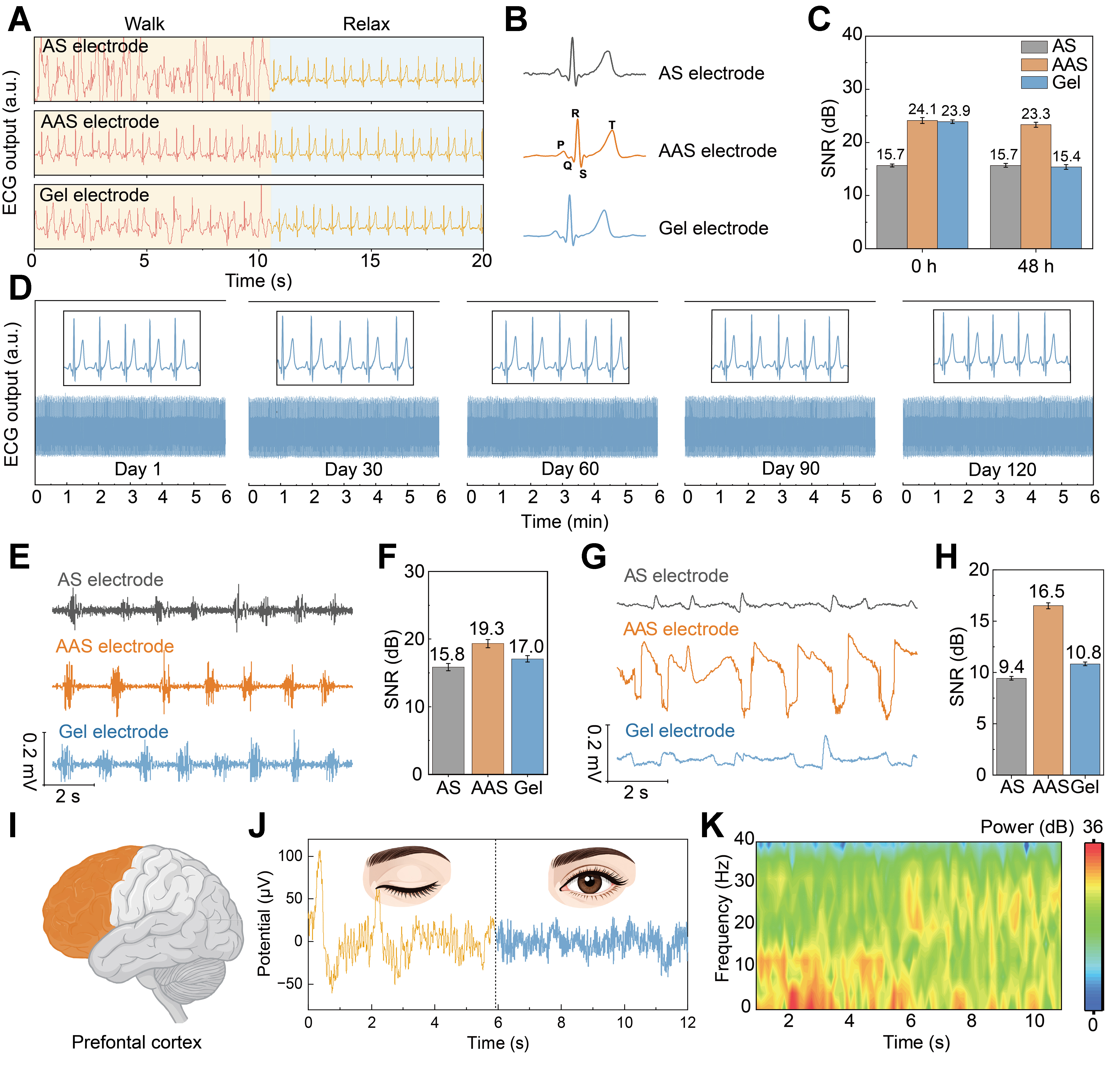
Figure 5. Electrophysiological signal acquisition using the AAS electrode. (A) ECG signals recorded using the AS, AAS, and commercial Ag/AgCl gel electrodes under walking and relaxed conditions; (B) Enlarged ECG waveforms recorded in the relaxed state by different electrodes. Characteristic P, Q, R, S, and T waves are clearly identified from both the AAS electrode and the gel electrode; (C) SNR of ECG signals recorded by the AS, AAS, and gel electrodes at 0 and 48 h. The error bars indicate the SD obtained from repeated tests (n = 5); (D) Long-term storage stability of the AAS electrode was evaluated by ECG recording after storage in air for 1, 30, 60, 90, and 120 days. Insets show enlarged ECG waveforms with well-preserved signal features; (E) EMG signals recorded and (F) SNR values. The error bars indicate the SD obtained from repeated tests (n = 5); (G) EOG signals recorded while looking to the right and (H) SNR values. The error bars indicate the SD obtained from repeated tests (n = 5); (I) The AAS electrode was placed on the forehead to record EEG signals from the prefrontal cortex; (J) EEG signals and (K) corresponding spectrograms recorded using the AAS electrode from a subject with eyes closed (0-6 s) and eyes open with concentration (6-12 s) while awake. AAS: AgNWs/AgCl/SEBS [AgNWs: silver nanowires, SEBS: poly(styrene-ethylene-butylene-styrene)]; ECG: electrocardiography; AS: AgNWs/SEBS; SNR: signal-to-noise ratio; SD: standard deviation; EMG: electromyogram; EOG: electrooculography; EEG: electroencephalogram.





