




fig1
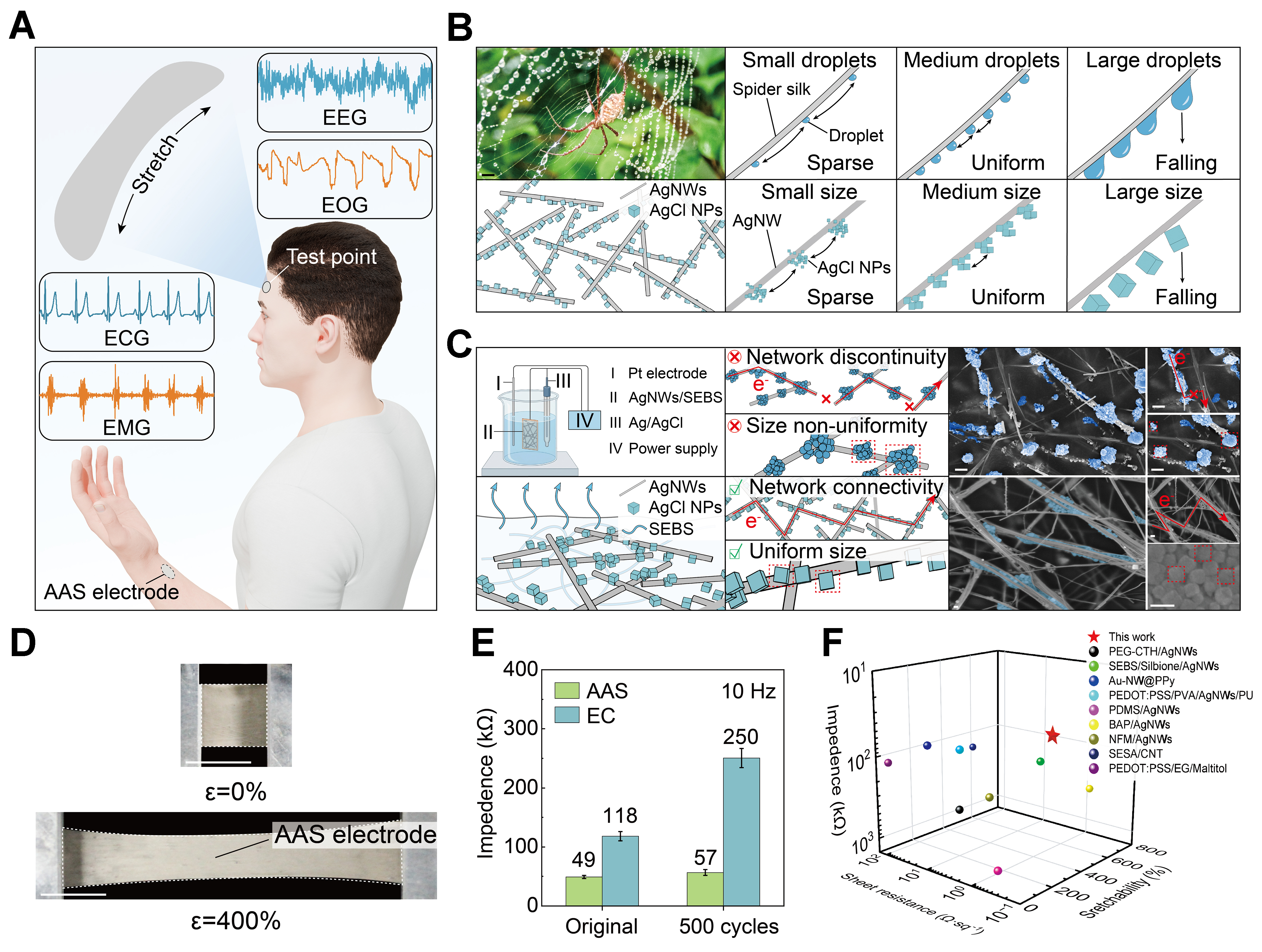
Figure 1. Bioinspired AAS electrode design and performance. (A) Schematic illustration of the AAS electrode for multimodal epidermal electrophysiological monitoring; (B) Bioinspired design concept of bead-on-string architecture inspired by the droplet-on-fiber of spider silk; (C) Structural comparison between electrochemical chlorination (top: EC) and the self-assembly strategy (bottom: AAS), including schematic illustration (left) and corresponding SEM images (right) (scale bars: 500 nm for EC and 100 nm for AAS); (D) Photographs of the AAS electrode under 0% and 400% tensile strain (scale bar: 1 cm); (E) Skin-electrode contact impedance at 10 Hz for AAS and EC electrodes before and after 500 stretching cycles. The error bars indicate the SD obtained from repeated tests (n = 3); (F) Performance comparison of the AAS electrode with recently reported dry electrodes; detailed references are provided in Supplementary Table 1. AAS: AgNWs/AgCl/SEBS [AgNWs: silver nanowires, SEBS: poly(styrene-ethylene-butylene-styrene)]; EC: electrochemically chlorinated; SEM: scanning electron microscope; SD: standard deviation; EEG: electroencephalogram; EOG: electrooculography; ECG: electrocardiography; EMG: electromyogram; AgNWs: silver nanowires; NPs: nanoparticles; SEBS: poly(styrene-ethylene-butylene-styrene); PEG-CTH: polyethylene glycol-tert-Butylcalix arene tetrahydrazide; PEDOT:PSS: poly(3,4-ethylenedioxythiophene)-poly(styrene sulfonic acid); PVA: polyvinyl alcohol; PU: polyurethane; PDMS: polydimethylsiloxane; BAP: bistable adhesive polymer; NFM: nanofibrous membrane; SESA: supramolecular, elastic, self-healing, and adhesive; CNT: carbon-nanotube; EG: ethylene glycol.





