


fig2
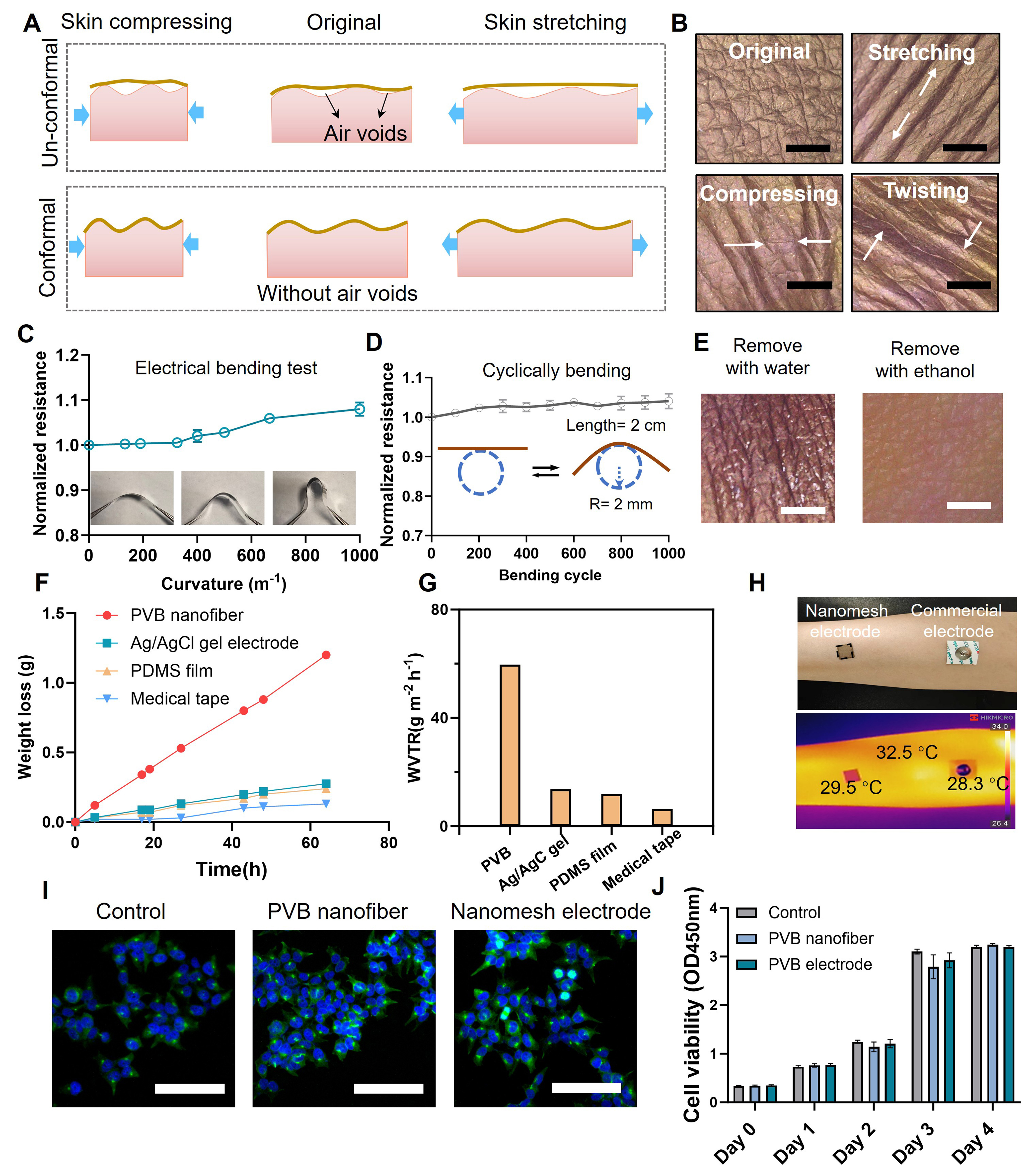
Figure 2. (A) Schematic comparison between a conventional dry electrode and the conformal nanomesh electrode at the electrode-skin interface under skin deformation; (B) Photographs of the nanomesh electrode subjected to undeformed, stretching, compressing and twisting deformation (scale bar: 2 mm); (C) Normalized resistance variation of the nanomesh electrode as a function of bending curvature of 0-1,000 m-1; (D) Resistance variation of the nanomesh electrode during 1,000 bending cycles; (E) Photograph of the nanomesh electrode after removing with water or ethanol. Scale bar: 2 mm; (F) Water permeability curves of the nanomesh electrode under 35 °C conditions compared with Ag/AgCl gel electrode, PDMS film and medical tape, and (G) corresponding WVTR; (H) Optical and infrared thermal images of the nanomesh and Ag/AgCl gel electrode attached onto human forearm; (I) Fluorescence microscopy images of 293T cells cultured with PVB nanofiber network and transferred nanomesh electrode. Scale bar: 100 μm; (J) Cell viability of 293T cells cultured with PVB nanofibers, transferred nanomesh electrode and a blank control. The results in (C, D, and J) are presented as mean ± standard deviation [(C and D): n = 3, (J): n = 6]. PDMS: Polydimethylsiloxane; WVTR: water vapor transmission rate; PVB: poly(vinyl butyral).


