


fig7
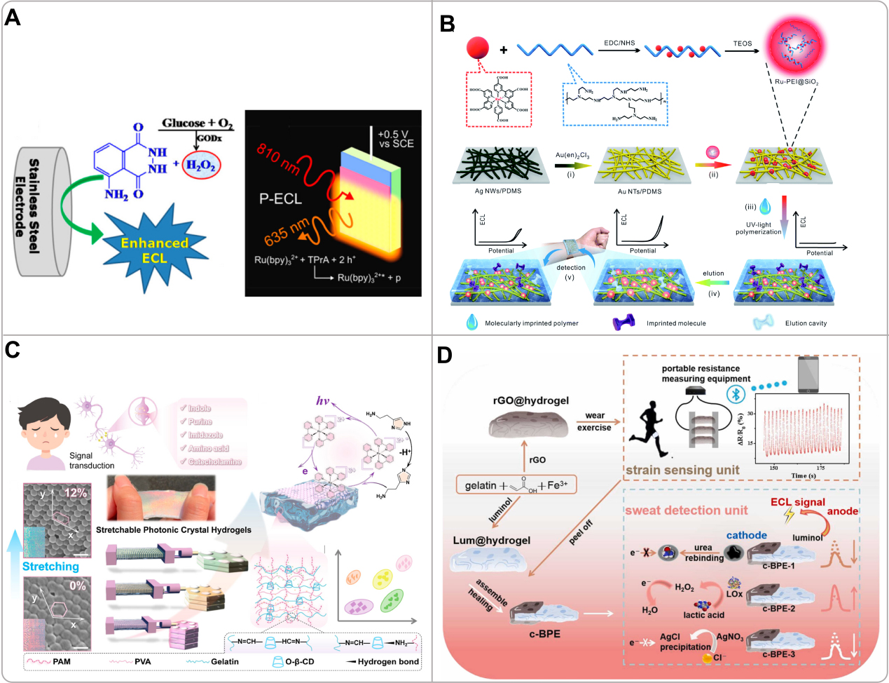
Figure 7. (A) Schematic diagrams of a stainless steel electrode for ECL detection using the luminol/H2O2 system and the mechanism of photoinduced ECL at silicon electrodes protected by nanoscale SiOX and Ni-stabilizing thin films. Reproduced with permission[107]. Copyright 2021, American Chemical Society; (B) Schematic illustration of the Ru-PEI@SiO2 synthesis and the assembly route of the flexible MIP-ECL skin sensor: electrochemical conversion, surface immobilization, MIP layer imprinting, sensor elution, and subsequent detection of epidermal analytes. Reproduced with permission[106]. Copyright 2019, The Royal Society of Chemistry; (C) Schematic diagram of neurotransmitter monitoring using a stretchable photonic-crystal-hydrogel-enhanced ECL platform, together with machine learning-assisted detection and classification. Reproduced with permission[108]. Copyright 2025, American Chemical Society; (D) Schematic depiction of rGO@hydrogel used for monitoring motion behavior and evaluating multiple physiological indicators in sweat, including urea, lactate, and chloride. Reproduced with permission[111]. Copyright 2023, Elsevier. ECL: Electrochemiluminescence; TEOS: tetraethyl orthosilicate; PAM: polyacrylamide; PVA: polyvinyl alcohol; O-β-CD: oxidized β-cyclodextrin; c-BPE: closed bipolar electrode.


