

fig4
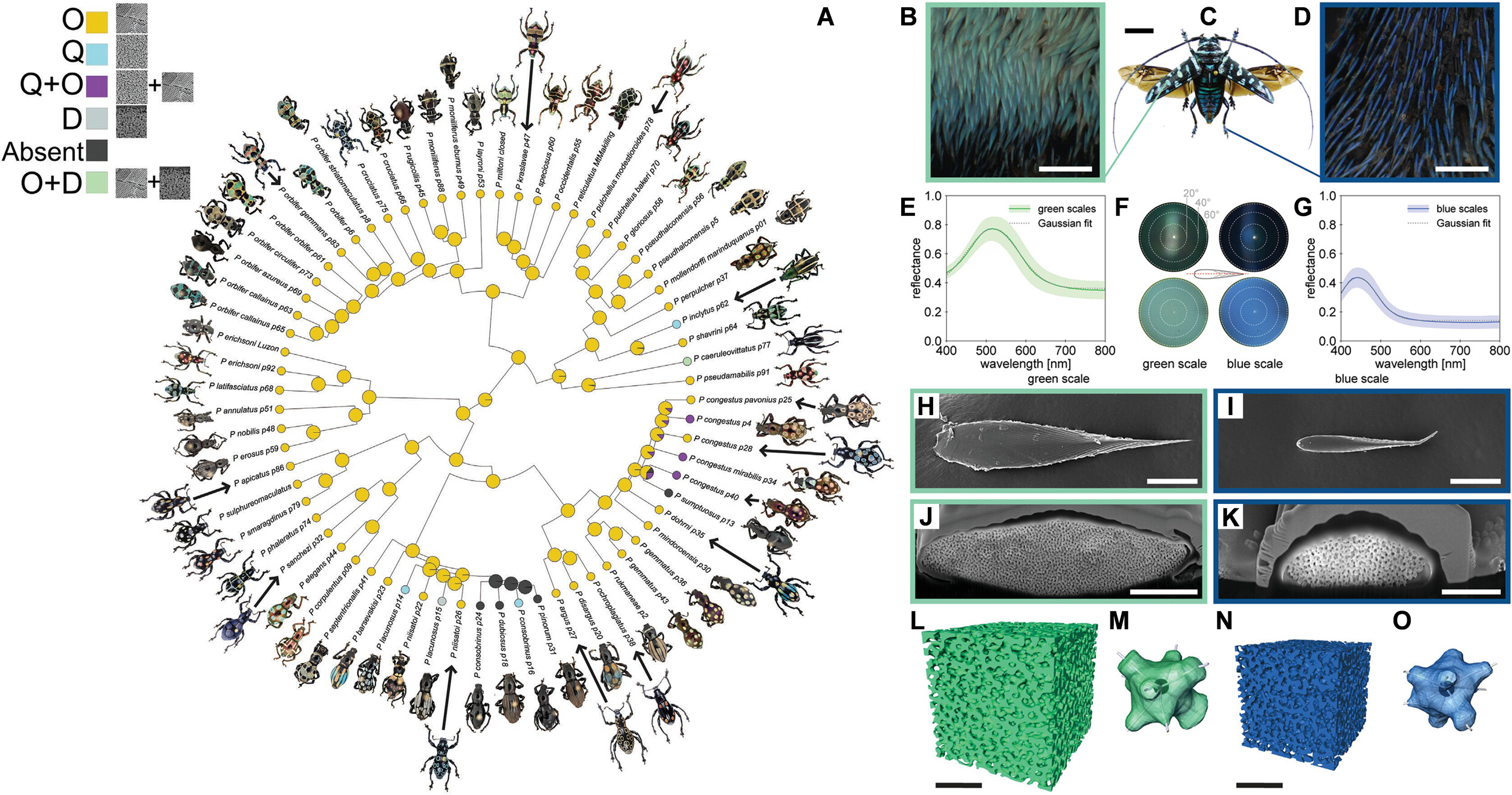
Figure 4. (A) Phylogenetic tree of weevils from the genus Pachyrhynchus, showing the evolutionary distribution of photonic crystal structure types within the scales of different species (O, Q, D denote Ordered, Quasi-ordered and Disordered, respectively). (A) Reproduced under the terms of the CC BY-NC 4.0 License[74]. Copyright 2022, The Authors; (B-O) Optical characterization and ultrastructural analysis of the colored scales of Sternotomis virescens. (B and D) Reflection light micrographs of green (B) and blue (D) scales. (C) Photograph of a male beetle. (E and G) In situ reflectance spectra of the green (E) and blue (G) scales; (F) K-space images of the two scale colors at 100× magnification. (H and I) SEM images showing the seed-like shape of individual green (H) and blue (I) scales. (J and K) Focused ion beam-SEM cross-sectional images showing that both the green (J) and blue (K) scales are amorphous networks surrounded by a thin-film cortex. (L and N) 3D reconstructed networks of the green (L) and blue (N) scales obtained from focused ion beam-SEM tomography. (M and O) A selected sub-unit from the networks of the green (M) and blue (O) scales. Scale bars: (B) and (D) 125 μm, (C) 1 cm, (H) and (I) 20 μm, (J) 5 μm, (K) 2 μm, (L) and (N) 1 μm. (B-O) Reproduced under the terms of the CC BY-NC 4.0 License[78]. Copyright 2023, The Authors. SEM: Scanning electron microscopy.

