

fig3
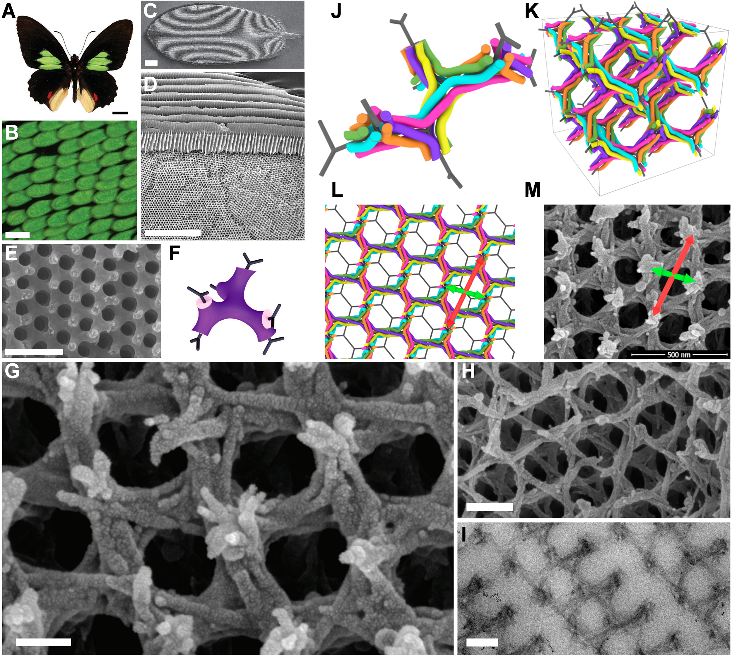
Figure 3. (A-F) SG structure within the green scales of mature Parides sesostris. (A) Photograph of a male Parides sesostris; (B) Light microscopy image of scales containing the SG structure; (C and D) Scanning electron microscopy (SEM) images of the scales; (E) High-magnification SEM image showing the smooth, homogeneous SG structure; (F) Idealized SG model with a chitin volume fraction of 0.35; (G-I) Electron microscopy images of the fibrillar SG structure in the wing scales of developing Parides sesostris pupae. As the pupation period is approximately 18 days, wing scale development was arrested by fixation on day 13 (G, 70% development) and day 15 (H and I, 80% development); (G) Adjacent to the lower lamina of the scale, showing approximately six fibers composing the SG; (H) Adjacent to the upper lamina of the scale, showing 2-3 fibers; (I) Transmission electron microscopy (TEM) image of a cross-section of a scale sampled from the same wing as that shown in (H); (J-M) Geometric model of six helical fibers woven around the SG network; (J) A small section of the model; (K) Approximately 23 unit cells; (L) The full six-fiber weave, with the model confined to a thin slice to simulate the finite observation depth of electron microscopy sections; (M) SEM image of the SG structure at 70% development in Parides sesostris, showing visual similarity to the model in (L). Scale bars: (A) 1 cm, (B) 100 μm, (C) 10 μm, (D) 5 μm, (E) 500 nm, (G) 100 nm, (H) and (I) 200 nm. This figure is reproduced under the terms of the CC BY-NC-ND 4.0 License[67]. Copyright 2025, The Authors. SG: Single gyroid.

