



fig1
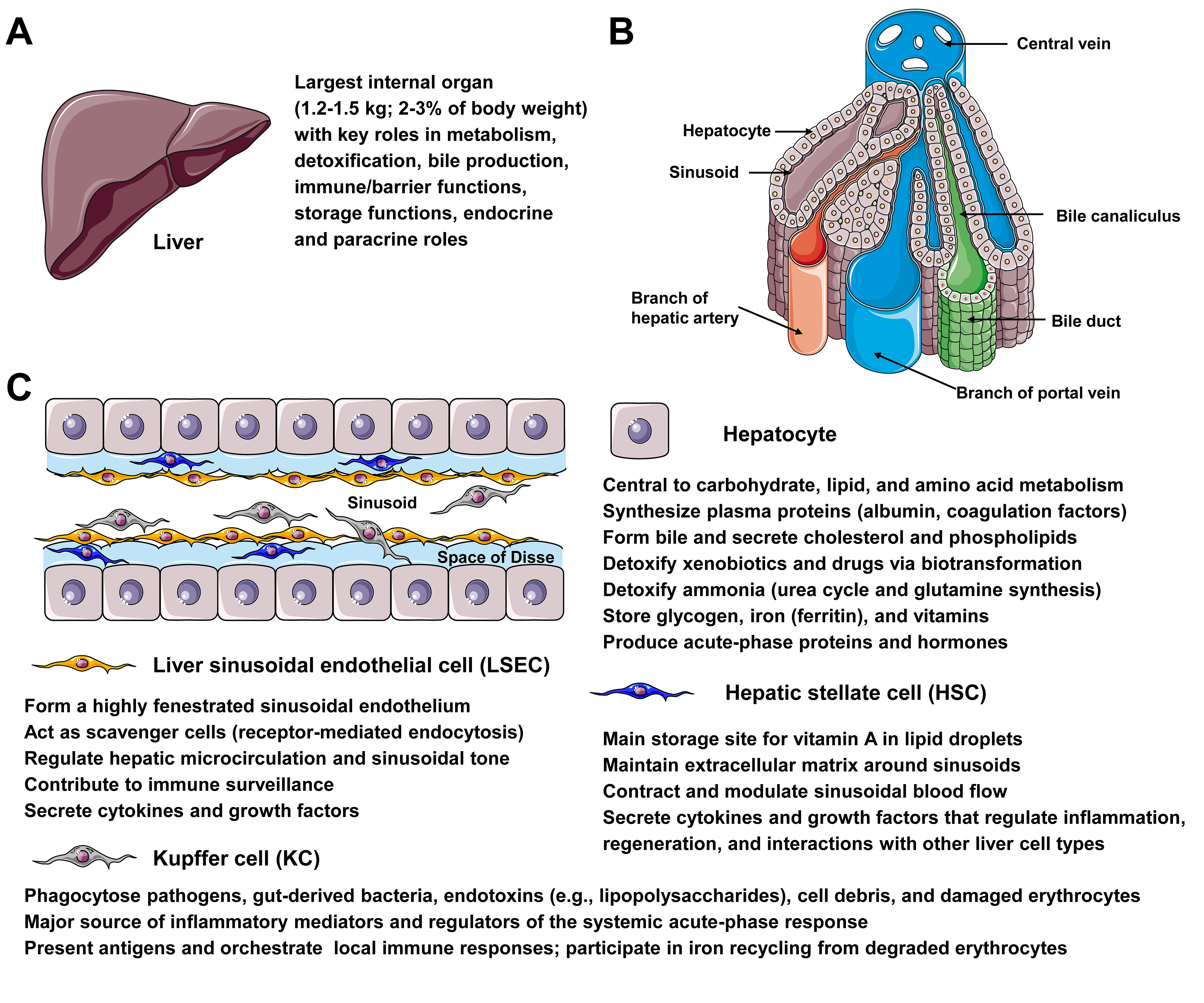
Figure 1. Microanatomy and major liver cell types relevant for gene therapy. (A) The liver, weighing 1.2-1.5 kg and accounting for 2%-3% of body weight, plays vital roles in metabolism, detoxification, bile production, immune/barrier functions, storage, and endocrine/paracrine signaling, making it an attractive target for gene therapy. (B) The diagram shows a hepatic lobule, emphasizing the intralobular (central) vein, sinusoids, bile canaliculi, bile ducts, branches of the portal vein and hepatic artery, and the positioning of hepatocytes. (C) The diagram depicts the architecture of the liver sinusoid, which is lined by fenestrated endothelia and interspersed with Kupffer cells (KCs). Hepatic stellate cells (HSC) are located within the narrow space of Disse, formed by a layer of liver sinusoidal cells (LSECs) and cords of hepatocytes. Hepatocytes play key physiological functions, including central roles in carbohydrate, lipid, and amino acid metabolism; synthesis of plasma proteins (e.g., albumin and coagulation factors); formation of bile and secretion of cholesterol and phospholipids; detoxification of xenobiotics and ammonia; storage of glycogen, iron, and vitamins; and production of acute-phase proteins and hormones. LSECs form a highly fenestrated sinusoidal endothelium, act as scavenger cells via receptor-mediated endocytosis, regulate hepatic microcirculation and sinusoidal tone, and contribute to immune surveillance. HSCs are the main storage site for vitamin A in lipid droplets, maintain the extracellular matrix around sinusoids, contract to modulate sinusoidal blood flow, and secrete cytokines and growth factors that regulate inflammation and regeneration. KCs phagocytose pathogens, gut-derived bacteria, endotoxins, cell debris, and damaged erythrocytes, produce inflammatory mediators that shape the acute-phase response, present antigens, orchestrate local immune responses, and participate in iron recycling.