

fig1
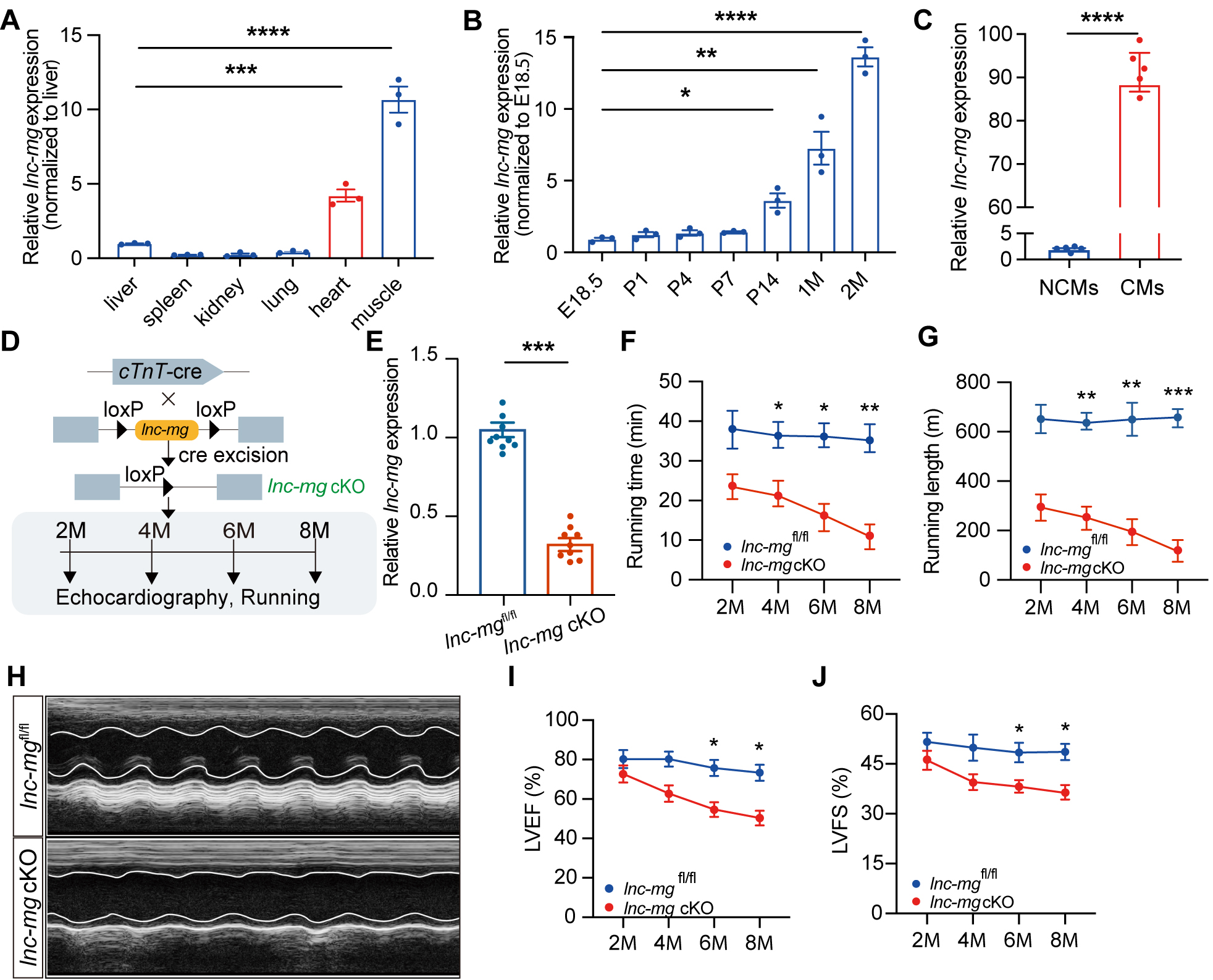
Figure 1. Cardiac expression profile of lnc-mg and phenotypic characterization of cardiomyocyte conditional knockout lnc-mg mice. (A) Relative expression of lnc-mg in different tissues from adult wild-type (WT) mice determined by RT-qPCR. Tissues from three independent mice were analyzed (n = 3 biological replicates). (B) Developmental expression of lnc-mg in mouse hearts at the indicated developmental stages, including embryonic day 18.5 (E18.5), postnatal day 1 (P1), P4, P7, P14, 1 month of age (1M), and 2M. Three independent hearts were analyzed at each developmental stage (n = 3 biological replicates per stage). (C) Relative expression of lnc-mg in isolated cardiomyocytes (CMs) and non-cardiomyocytes (NCMs) from adult mouse hearts. Paired CM and NCM fractions obtained from five independent mice were analyzed (n = 5 biological replicates). (D) Schematic illustrating the generation of lnc-mg cKO mice by crossing lnc-mgfl/fl mice with cTnT-Cre mice and the experimental design for subsequent phenotypic analyses. (E) RT-qPCR validation of lnc-mg deletion in lnc-mg cKO hearts in 2-month-old mice, n = 10 per group. (F and G) Longitudinal assessment of treadmill exercise performance of lnc-mgfl/fl and lnc-mg cKO mice at 2, 4, 6, and 8 months of age. Running time (F) and running distance (G) are shown. Ten independent mice were analyzed (n = 10 biological replicates). (H) Representative M-mode echocardiographic images of lnc-mgfl/fl and lnc-mg cKO mice at 8 months of age. (I and J) Serial echocardiographic assessment of left ventricular ejection fraction (LVEF) (I) and left ventricular fractional shortening (LVFS) (J) in lnc-mgfl/fl and lnc-mg cKO mice from 2 to 8 months of age. Ten independent mice were analyzed




