

fig2
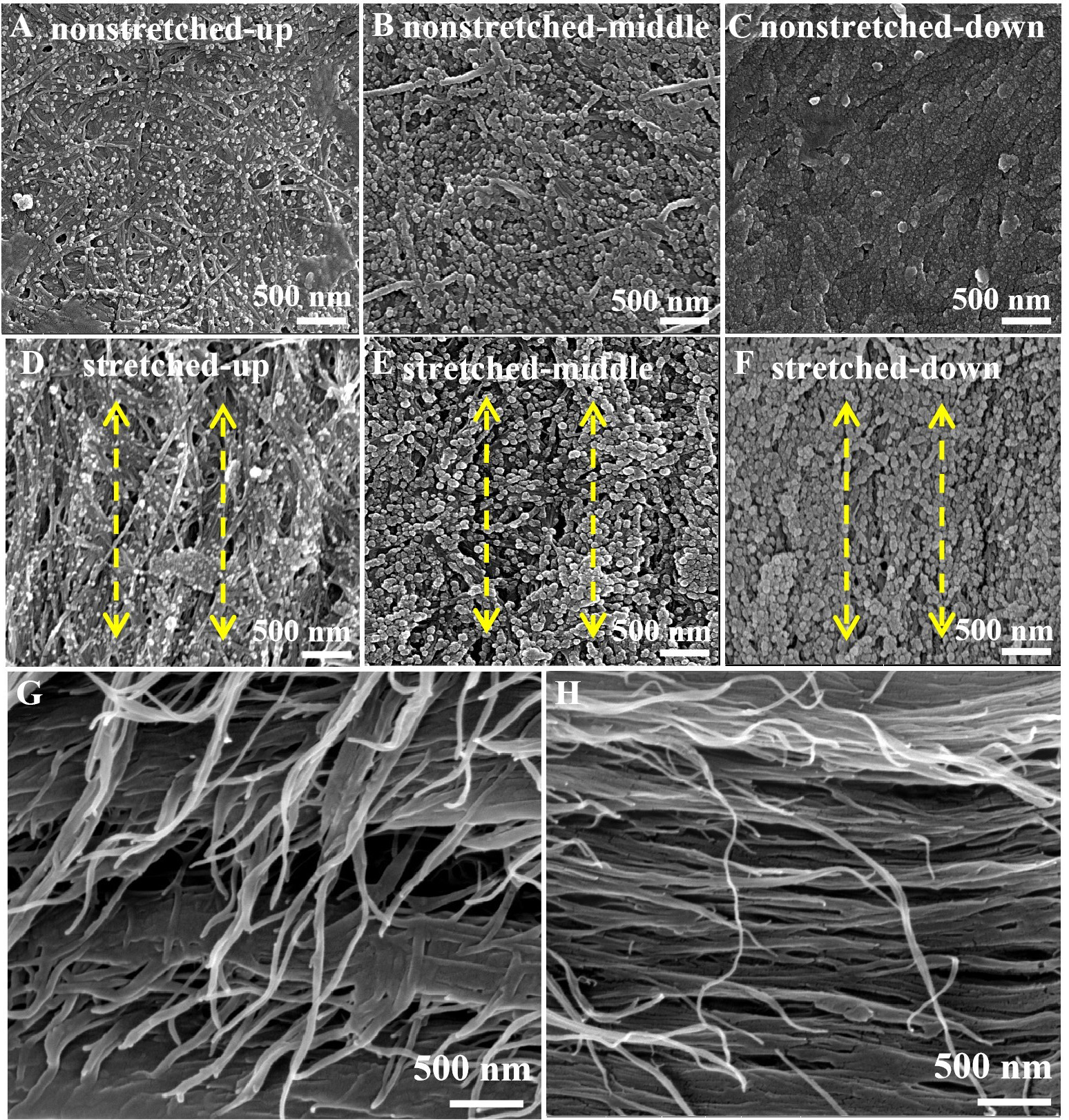
Figure 2. FESEM images of the BC/PPy gradient conductive membranes: (A-C) non-stretched membranes (upper, middle, and lower segments); (D-F) stretched membranes (upper, middle, and lower segments). Cross-sectional images of the stretched lower segment taken perpendicular to the stretching direction (G) and parallel to the stretching direction (H). Scale bar: 500 nm. FESEM: Field-emission scanning electron microscopy; BC: bacterial cellulose; PPy: polypyrrole.

