

fig4
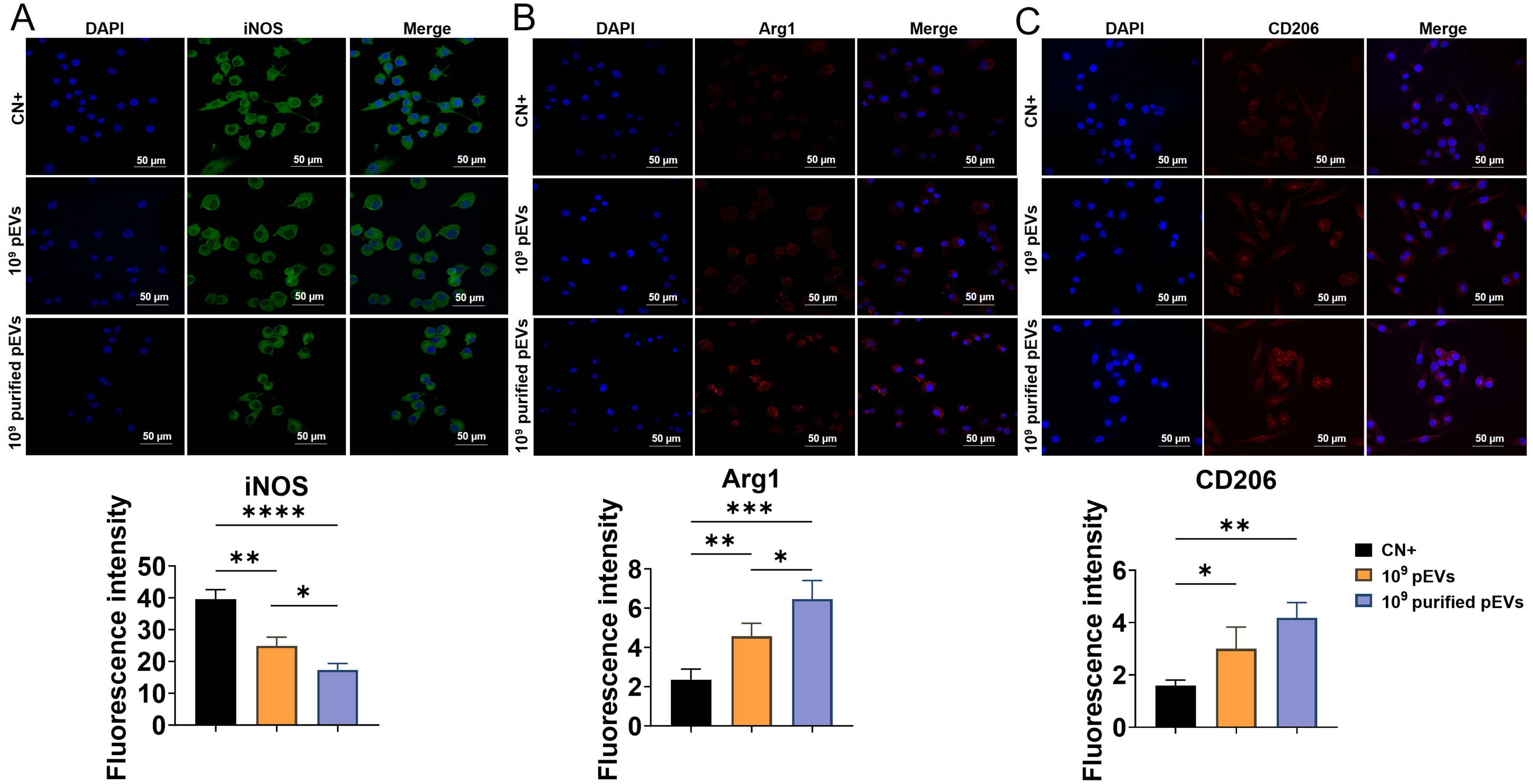
Figure 4. Effects of pEVs and purified pEVs on macrophage phenotype-associated markers. RAW cells were primed with E. coli LPS (1 µg/mL) for 2 h, washed three times with PBS, and subsequently stimulated with 100 ng/mL LPS, either alone or in combination with pEVs or purified pEVs (1 × 10⁹ particles/mL) for 24 h. Representative immunofluorescence images and corresponding quantification of fluorescence intensity for (A) iNOS (green), (B) Arg1 (red), and (C) CD206 (red). Nuclei were stained with DAPI (blue). Data are expressed as the mean ± SD of three independent experiments, each using paired pEV and purified pEV preparations from a different plasma donor (n = 3). Statistical significance is indicated as *P < 0.05; **P < 0.01; ***P < 0.001; ****P < 0.0001. Arg1: Arginase 1; CD206: cluster of differentiation 206; DAPI: 4’,6-diamidino-2-phenylindole; iNOS: inducible nitric oxide synthase; LPS: lipopolysaccharide; pEVs: plasma-derived extracellular vesicles; PBS: phosphate-buffered saline; RAW: RAW264.7 macrophages; SD: standard deviation.