

fig2
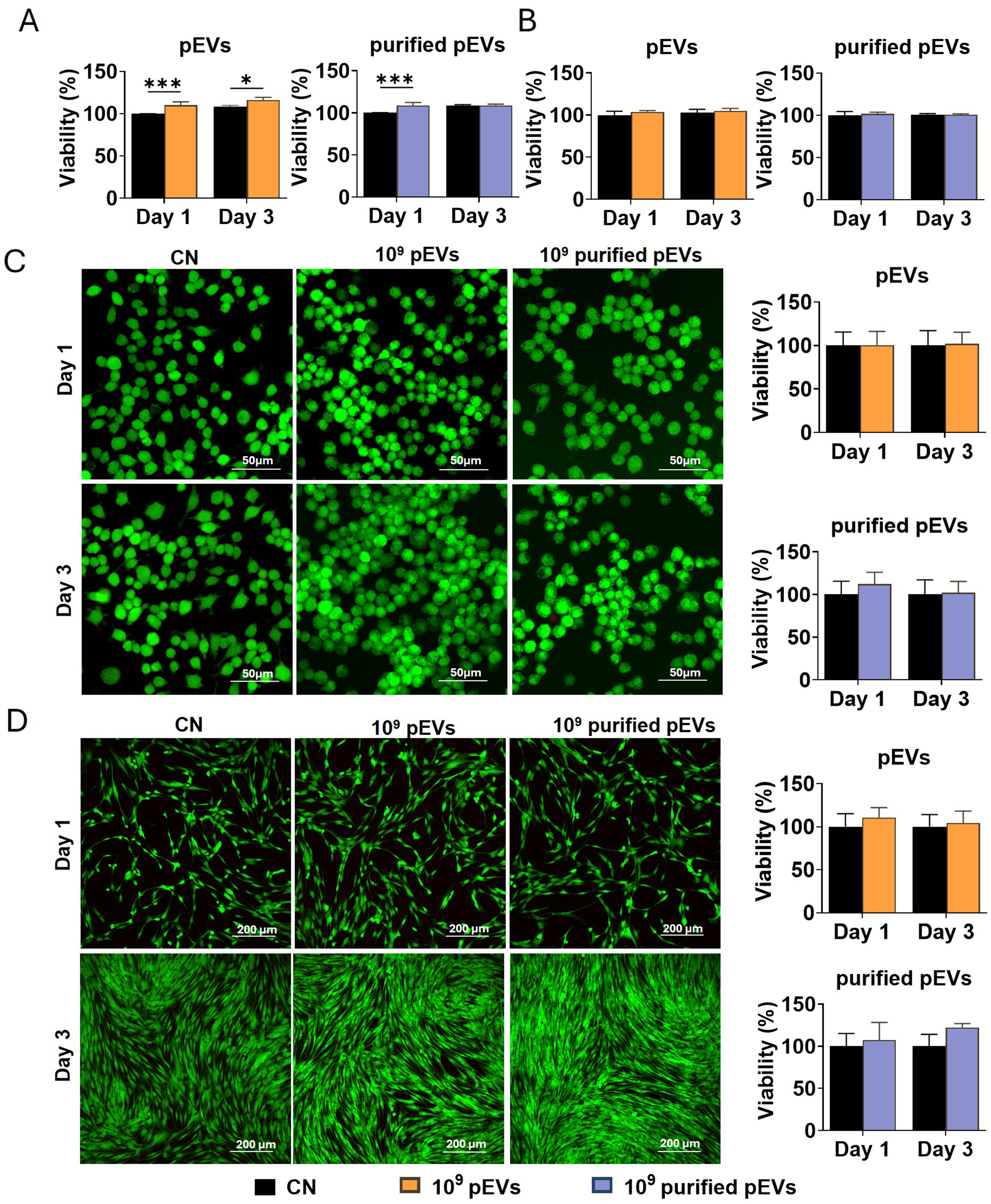
Figure 2. Effects of pEVs and purified pEVs on the viability of RAW cells and hPDLCs. Alamar Blue assays showing the viability of (A) RAW cells and (B) hPDLCs on days 1 and 3 following treatment with pEVs or purified pEVs at 1 × 10⁹ particles/mL. Representative Live/Dead staining images of (C) RAW cells and (D) hPDLCs on days 1 and 3, with FDA labelling live cells in green and PI labelling dead cells in red, accompanied by quantitative analysis of fluorescence intensity. Data are expressed as the mean ± SD of three independent experiments, each using paired pEV and purified pEV preparations from a different plasma donor (n = 3). Statistical significance is indicated as *P < 0.05; **P < 0.01; ***P < 0.001. EVs: Extracellular vesicles; FDA: fluorescein diacetate; hPDLCs: human periodontal ligament cells; pEVs: plasma-derived extracellular vesicles; PI: propidium iodide; RAW: RAW264.7 macrophages; SD: standard deviation.