

Extracellular vesicle surface proteins in tissue recognition, organotropism, biomarkers, and therapeutic engineering
 0
Graphical Abstract
Abstract
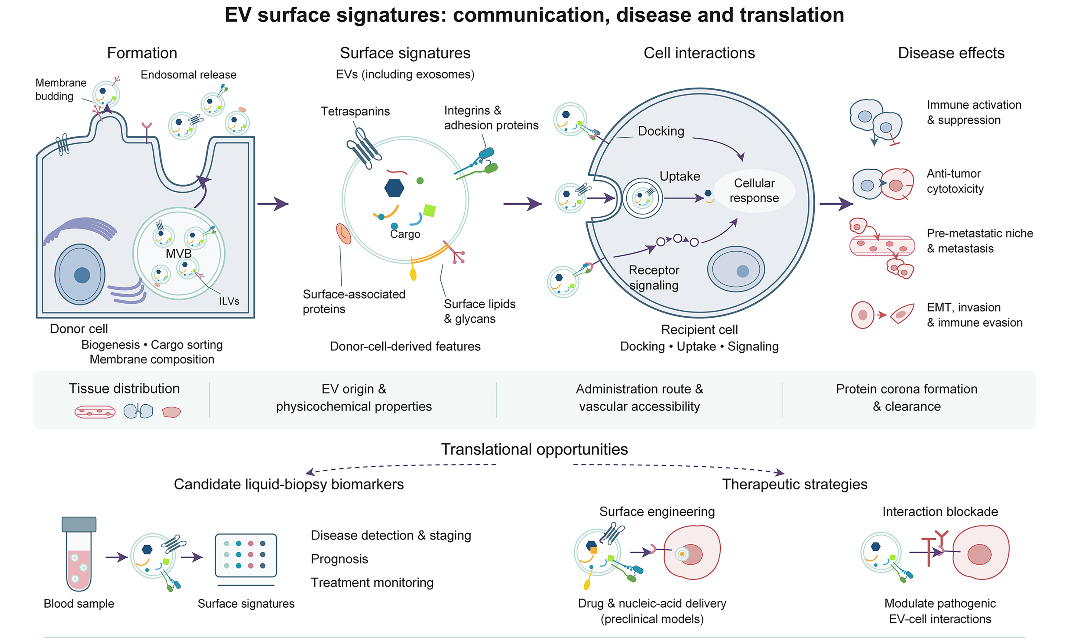
Extracellular vesicles (EVs), including exosomes, carry donor-cell-derived molecular features that influence their interactions with recipient cells. This review links EV biogenesis and the assembly of surface recognition cues to cellular interactions, tissue distribution, and translational applications, with an emphasis on the evidence required to interpret surface localization, biomarker value, and therapeutic outcomes. These cues can contribute to cellular docking, uptake, and receptor signaling and, together with vesicle origin, physicochemical properties, administration route, vascular accessibility, protein corona formation, and clearance, help determine tissue distribution in disease. We discuss how EV-mediated communication participates in immune activation and suppression, anti-tumor cytotoxicity, pre-metastatic niche formation, metastatic progression, epithelial-mesenchymal transition, invasion, and immune evasion. We then summarize the translational potential of EV-associated surface signatures as candidate liquid-biopsy biomarkers for disease detection, staging, prognosis, and treatment monitoring. Surface engineering has been explored in preclinical models, where tissue accumulation, cellular uptake, functional cargo delivery, and therapeutic efficacy are distinct outcomes; modulation of pathogenic EV-cell interactions offers another potential therapeutic avenue. Progress toward clinical application will require standardized isolation and characterization, resolution of EV heterogeneity at the single-vesicle level, reproducible biodistribution and pharmacology, scalable manufacturing, and rigorous evaluation of safety and efficacy. Defining how surface-associated features operate within this multifactorial system may clarify disease mechanisms and guide the development of more precise EV-based diagnostics and therapies.
Keywords


 Download PDF
INTRODUCTION
Extracellular vesicles (EVs) carry proteins, lipids, and nucleic acids between cells and participate in immune regulation, tissue repair, and disease progression[1,2]. Their presence in biofluids also provides a means of examining molecular features of the cells that release them. EVs comprise heterogeneous populations whose composition and functions vary with cellular origin and biogenesis. Throughout this review, EV is used as the general term, whereas “exosome” refers to vesicles with evidence supporting an endosomal origin. The term “small EV” is used when vesicle size has been characterized but the biogenesis pathway has not been established[3].
Surface proteins help determine how EVs interact with their surroundings. Integrins, tetraspanins, adhesion molecules, and receptors can participate in vesicle binding, internalization, or signaling at the recipient-cell membrane[4-7]. Their incorporation into EVs reflects both donor-cell characteristics and the pathways responsible for cargo sorting and membrane budding[8-10]. In tumor models, particular exosomal integrins have been linked to preferential uptake by lung or liver cells and subsequent changes in the local microenvironment[11]. Such findings connect vesicle composition with tissue-associated biological effects.
These interactions have implications for diagnosis and treatment, but their interpretation requires care. Detection of a protein in an EV preparation does not establish that it is exposed on the external vesicle surface or accessible to a recipient-cell receptor[14]. Likewise, increased tissue accumulation or cellular uptake does not, by itself, demonstrate functional cargo delivery[3]. Similar distinctions apply to biomarker studies: a disease-associated change in EV protein abundance provides a starting point for investigation, while diagnostic use requires reproducible measurement and validation in appropriate patient populations. Surface engineering draws on the same recognition mechanisms to improve delivery or modify immune interactions, making the relationship between surface presentation and biological activity relevant to both native and engineered vesicles[15,16].
Previous reviews have examined EV homing and tropism[17,18], as well as the composition, functions, and applications of exosomal membrane proteins[19]. Here, we examine how biogenesis and cellular origin shape EV-associated protein patterns, how surface-exposed molecules contribute to recipient-cell interactions, and how these processes relate to disease-associated functions. We use the surface “global positioning system” (GPS) concept to organize recognition mechanisms within the broader determinants of EV distribution. We then consider the evidence supporting EV-associated proteins as candidate biomarkers and the use of surface engineering for therapeutic applications. Across cancer, immune-related disorders, and other diseases, the emphasis is on connecting proposed mechanisms with the experimental findings needed to support them.
EXOSOME BIOGENESIS
Exosome biogenesis is an endosomal process driven by the host cell that begins when the donor cell’s plasma membrane buds inward to form early endosomes[1,20], which then mature into late endosomes or multivesicular bodies (MVBs) as the endosomal membrane buds inward again to create cargo-rich intraluminal vesicles (ILVs) within the endosomal lumen[21,22]. During the initial stages of early exosome formation, membrane budding can proceed via endocytosis-dependent or endocytosis-independent pathways, ultimately forming these ILVs through three primary molecular pathways, namely, the canonical endosomal sorting complexes required for transport (ESCRT)-dependent pathway, the non-canonical ESCRT-dependent pathway, and the ESCRT-independent pathway. These distinct mechanisms shape the final surface protein composition of the resulting exosomes [Figure 1][2,23]. The exosome biogenesis pathway, summarized in Figure 2, provides a regulatory basis for shaping vesicle composition and function[24]. By influencing the surface protein repertoire, distinct assembly mechanisms can affect subsequent recipient-cell recognition, signaling, and disease-associated activities.
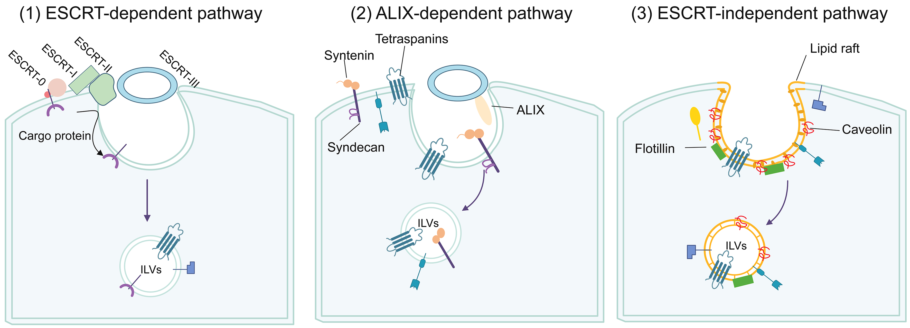
Figure 1. Pathways of intraluminal vesicle formation within multivesicular bodies: (1) canonical ESCRT-dependent; (2) non-canonical ESCRT-dependent (ALIX-dependent); and (3) ESCRT-independent pathways. Purple arrows indicate progression from membrane invagination to ILV formation, and the black curved arrow indicates cargo recruitment. Thin black lines connect labels to the indicated components; colored symbols schematically distinguish the labeled components and cargo. ESCRT-0, -I, -II, and -III denote the corresponding ESCRT complexes. ALIX: Apoptosis-linked gene 2-interacting protein X; ESCRT: endosomal sorting complexes required for transport; ILVs: intraluminal vesicles.
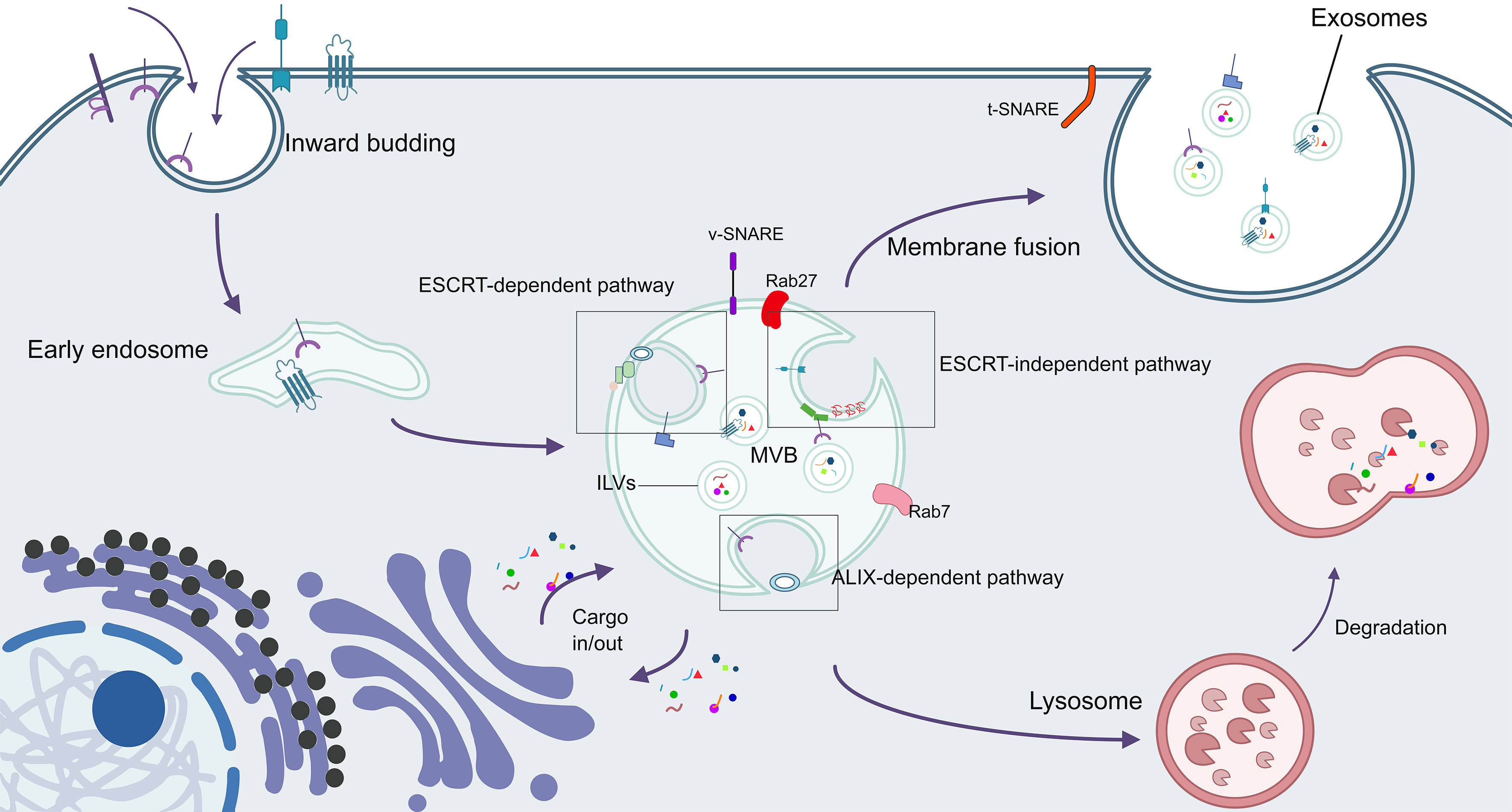
Figure 2. Overview of exosome biogenesis. Purple arrows indicate membrane internalization, endosome maturation, cargo trafficking, and the routes toward exosome release or lysosomal degradation. Thin black lines connect labels to structures, and gray boxes highlight the three ILV-formation pathways; colored symbols represent schematic membrane components and cargo. Rab27 is used as a collective label for Rab27a and Rab27b. ALIX: Apoptosis-linked gene 2-interacting protein X; ESCRT: endosomal sorting complexes required for transport; ILVs: intraluminal vesicles; MVB: multivesicular body; Rab7: Ras-related protein Rab-7; Rab27: Ras-related proteins Rab-27A and Rab-27B; SNARE: soluble N-ethylmaleimide-sensitive factor attachment protein receptor; t-SNARE: target-membrane-associated SNARE; v-SNARE: vesicle-associated SNARE.
EV-ASSOCIATED PROTEIN PATTERNS ARE SHAPED BY DIFFERENT ILV FORMATION PATHWAYS
Interestingly, the pathways that generate ILVs within MVBs can determine which proteins are displayed on exosome surfaces by controlling cargo selection and membrane budding. In other words, the canonical ESCRT-dependent, non-canonical ESCRT-dependent, and ESCRT-independent pathways shape the exosomal surface proteome, and perturbing key components can alter specific surface markers[1,25-29].
Canonical ESCRT-dependent pathway
The canonical pathway utilizes a sequential cascade of complexes, ESCRT-0, -I, -II, -III, and vacuolar protein sorting-associated protein 4 (VPS4), to sort cargo and form ILVs[1]. ESCRT-0 recognizes various cargo proteins via its components, hepatocyte growth factor-regulated tyrosine kinase substrate (HRS) and signal transducing adapter molecule 1 (STAM1)[25]. ESCRT-0 then binds to the ESCRT-I subunit, tumor susceptibility gene 101 (TSG101), to recruit ESCRT-I[30]. As the core component of ESCRT-I, TSG101 attracts ESCRT-II, which recruits charged multivesicular body protein 6 (CHMP6) and charged multivesicular body protein 4B (CHMP4B) to activate ESCRT-III[31]. Then, ESCRT-III drives ILV formation through sequential polymerization, membrane deformation, and cleavage[32]. This cascade is able to shape exosomal surface markers[33]. Studies have shown that depleting HRS reduces secretion of cluster of differentiation 63 (CD63)- and cluster of differentiation 81 (CD81)-positive exosomes in HeLa cells[34] and decreases the exosomal surface expression of programmed death-ligand 1 (PD-L1)[35]. TSG101 actively promotes ILV budding by interacting with HRS to induce ESCRT-III recruitment[36]. In addition, TSG101 can drive reverse-topology budding by recognizing P(T/S)AP short linear motifs via its ubiquitin-conjugating enzyme E2 variant (UEV) domain[4], further influencing which proteins are incorporated into budding ILVs.
Non-canonical ESCRT-dependent pathway
The non-canonical ESCRT-dependent pathway, also known as the apoptosis-linked gene 2-interacting protein X (ALIX)-dependent pathway, bypasses ESCRT-0, -I, and -II. ALIX directly recruits ESCRT-III and VPS4[25]. Proteins in this axis are commonly detected in EV preparations[26]. ALIX serves as a central adaptor for cargo sorting by interacting with tetraspanins[cluster of differentiation 9 (CD9), CD63, CD81] and the syndecan-syntenin complex[8,37], and bridges to CHMP4B via its Bro1 domain[31]. ALIX-tetraspanin binding regulates protein cargo sorting[25] and supports incorporation of tetraspanins into vesicles during ESCRT-III recruitment[38], generating exosomes enriched in the membrane proteins CD63 and syndecan and the associated cytoplasmic adaptor syntenin. Perturbations in this pathway would remodel exosome cargo[39]. Syntenin depletion markedly reduces CD63 recovered in exosome preparations[8]. Reduced or depleted CD63 impairs exosome secretion[40] and reduces the exosomal packaging of latent membrane protein 1 (LMP1)[25,41] and vascular endothelial growth factor (VEGF)[42]. Mechanistically, ALIX drives endosomal membrane budding by binding the three LYPXnL motifs of syntenin[37], engaging ESCRT proteins such as TSG101[8], and interacting with the ESCRT-III subunit sucrose non-fermenting protein 7 (SNF7) to trigger membrane scission[43]. Notably, CD63 participates in biogenesis via ESCRT-dependent mechanisms[40] and can also be incorporated into vesicles through ESCRT-independent, ceramide-mediated budding[9], linking ALIX-dependent and lipid-driven routes.
ESCRT-independent pathway
Cells also form ILVs without ESCRT through mechanisms involving membrane-associated proteins such as caveolin-1 (CAV1) and flotillin[25], as well as glycosylphosphatidylinositol (GPI)-anchored proteins such as prion protein (PrP)[5]. This pathway comprises three steps: ILV formation, avoidance of multivesicular endosome (MVE) degradation, and MVE-plasma membrane fusion[27]. CAV1 and flotillin help sort cargo during ILV production; flotillin is essential to maintain plasma membrane architecture, cargo sorting, and exosome release[44]. Ras-related protein Rab-31 (Rab31) controls this ESCRT-independent pathway and regulates production of exosomal epidermal growth factor receptor (EGFR)[10]. Although ESCRT-dependent machinery can also sort EGFR into ILVs, those receptors are directed to lysosomal degradation rather than exosomal release[45]. Exosomal EGFR is overexpressed in several malignancies, including prostate cancer[46], implying that prostate cancer cells may preferentially utilize ESCRT-independent biogenesis. In the lipid raft-based context, flotillin can function as a membrane-associated cargo-sorting and budding regulator, providing a platform with sphingolipid ceramide and cholesterol. Here, budding is triggered by Rab31[10]. Along with ALIX and TSG101, flotillin serves as an important membrane budding regulator shaping ILV formation within MVBs[47]. Consistent with this lipid-driven pathway, CD63 can also be sorted into ILVs through ceramide-mediated, ESCRT-independent budding[9].
EV-ASSOCIATED PROTEIN PATTERNS CAN BE DETERMINED BY EXOSOME ORIGIN
Besides ILV formation pathways, the types of donor cells also greatly affect the surface proteins expressed on the exosomes[37,48,49]. Some are specific to their cell of origin, whereas others are not. More than 17% of the 271 exosomal proteins are common in the exosomes released from certain cell types such as mast cells and primary bone marrow-derived mast cells (BMMCs)[50]. These EV-associated protein profiles can reflect cellular origin and biogenesis pathways and inform subsequent biological activities[51].
Cell-of-origin-associated EV proteins
Some exosomal surface proteins are associated with their cell of origin, such as major histocompatibility complex (MHC) class I (MHC-I) and class II (MHC-II) molecules[1], cell adhesion molecules (CAMs), syndecans, transferrin receptors (TfRs), integrins, some tetraspanins[37], cluster of differentiation 3 (CD3), the glutamate receptor 2/3 (GluR2/3) subunits and A33 antigen[5]. Examples of other exosomal surface proteins that also reflect features of their donor cells are immune checkpoint proteins such as PD-L1[52] and complement regulators cluster of differentiation 55 (CD55) and cluster of differentiation 59 (CD59)[53]. MHC-II-bearing exosomes have been reported from professional antigen-presenting cells and intestinal epithelial cells[54,55]. TfRs are found in exosomes derived from reticulocytes[5]. These surface proteins can be categorized into adhesion proteins and receptor proteins[6,30,56]. MHC-I, MHC-II, CAMs, some tetraspanins such as CD81, integrins and A33 antigen belong to the adhesion type, mediating cell-to-cell or cell-to-extracellular matrix interactions. The GluR2/3 subunits of glutamate receptors and TfRs belong to the receptor type and bind to specific ligands to initiate cellular signaling pathways.
Cell-of-origin-nonspecific EV-associated proteins
Several EV-associated proteins are shared across donor-cell types, including heat shock proteins [heat shock cognate 70 (HSC70), heat shock protein 90 (HSP90)], tetraspanins (CD9, CD63, CD81), cytoskeletal proteins (actin, tubulin, moesin), ESCRT-related proteins such as TSG101, and trafficking-associated proteins such as flotillin and annexins[1,6,30]. These proteins comprise membrane proteins, membrane-associated components, intraluminal cargo, and factors involved in vesicle biogenesis. Their localization provides a basis for interpreting their roles in vesicle formation, cargo transport, and recipient-cell interactions. External surface localization is established through experiments assessing protein accessibility on intact EVs. Surface-exposed proteins participate in interactions at the vesicle-cell interface, while other EV-associated components contribute to cargo sorting, membrane organization, and intracellular functions. CD9, CD63, and CD81 are widely used EV membrane markers. Other EV-associated proteins, including syntenin-1, epidermal growth factor receptor pathway substrate 15 homology domain-containing protein 4 (EHD4), a disintegrin and metalloproteinase 10 (ADAM10), TSG101, and annexin XI, show differential enrichment across EV populations[57,58].
IMPACTS OF EV-ASSOCIATED PROTEINS ON VESICLE BIOGENESIS
ILV formation pathways affect the exosomal surface protein pattern. In turn, EV-associated membrane proteins and biogenesis factors can affect exosome formation[59]. For example, they orchestrate selective cargo sorting, govern MVB-plasma membrane fusion and secretion, and may contribute to targeting interfaces that influence tissue tropism alongside other determinants and functional outcomes[5,8,10,21,47,60-65].
Involvement of membrane proteins and associated adaptors in selective cargo sorting
Membrane proteins and associated adaptors recruit and sort cargo via multiple, partly intersecting pathways involving syndecans, tetraspanins CD9, CD81, CD63, as well as syntenin, ALIX, and raft-associated proteins such as flotillin, stomatin, and sphingosine 1-phosphate (S1P)[10,60,66]. Among these, the syndecan-syntenin-ALIX axis is the major route. Syndecans sort syntenin into exosomal membranes through binding to syntenin’s two postsynaptic density protein 95/discs large/zona occludens-1 (PDZ) domains[8,67], and syntenin-ALIX interactions then drive syndecan sorting into exosomes[8,67]. ESCRT-dependent sorting also relies on membrane-protein and adaptor interactions, with TSG101 recognizing ubiquitinated cargo through HRS and routing it into ILVs via interactions with the Bro1 domain and proline-rich region (PRR) of ALIX[4,5]. In reticulocytes, ALIX association promotes TfR sorting into exosomes, whereas HSC70 binding reduces this sorting[68].
Raft proteins contribute to cargo recruitment as well. Stomatin mediates the sorting of glucose transporter 1 (GLUT-1) into reticulocyte exosomes[60], while flotillin cooperates with Rab31 to sort EGFR and other receptor tyrosine kinases (RTKs) into ILVs. This flotillin-Rab31 axis bypasses ALIX-dependent pathways and recruits CD9, CD81, CD63, and syntenin-1 into exosomes[10]. Tetraspanins organize detergent-resistant membrane domains that recruit other membrane proteins into ILVs[5]. They sort MHC-II molecules into B-cell-derived exosomes. MHC-II has been observed in lipid-raft-associated domains of B-cell-derived exosomes[60]. Another protein, CD63, mediates the sorting of a number of proteins, including melanoma-related proteins, into ILVs in an ESCRT- and ceramide-independent manner, as well as the sorting of LMP1 into exosomes from Epstein-Barr virus (EBV)-infected cells. CD63 also binds the C-terminal fragment (CTF) of premelanosome protein (PMEL) to facilitate ILV formation and release of PMEL Mα into exosomes[15,40,41]. In parallel, a series of ligands can be recruited into exosomes independently of CD81[15]. These pathways intersect and can compensate for one another. For example, flotillin-Rab31 can bypass ALIX-dependent routes while still recruiting tetraspanins and syntenin, illustrating the overlapping nature of surface protein-driven cargo sorting into exosomes[10,60].
Involvement of membrane and biogenesis factors in MVB-plasma membrane fusion and vesicle secretion
Besides selective cargo loading, EV-associated membrane proteins and biogenesis factors also participate in MVB-plasma membrane fusion. Biogenesis begins with membrane budding within endosomes to form ILVs. As mentioned, this is a process in which ALIX, TSG101, and flotillin act as key regulators[47]. CD63 participates in vesicle formation via both ESCRT-dependent budding[40] and ESCRT-independent, ceramide-mediated mechanisms[9]. Flotillin provides structural platforms within lipid raft domains; triggered by Rab31, flotillin coordinates sphingolipid ceramide and cholesterol to drive budding and ILV formation within MVBs[10]. As MVBs mature, they either fuse with lysosomes for degradation or with the plasma membrane for exosome secretion[61,69,70]. The trafficking and docking steps that bias MVBs toward secretion are controlled by small guanosine triphosphatases (GTPases)[71]. Studies have shown that, in HeLa cells, Ras-related protein Rab-7 (Rab7) inactivation facilitates fusion between MVBs and the plasma membrane and hence promotes exosome release[10], while Ras-related protein Rab-27B (Rab27b) transports MVBs to the cell periphery and Ras-related protein Rab-27A (Rab27a) mediates their docking at the plasma membrane[62].
Fusion at the plasma membrane is initiated when synaptotagmin interacts with syntaxin to activate the soluble N-ethylmaleimide-sensitive factor attachment protein receptor (SNARE) machinery, enabling exosome release through the SNARE complex[21,72,73]. This complex comprises Qa-, Qb-, and Qc-SNARE (glutamine-containing) and R-SNARE (arginine-containing) subfamilies[63]. Membrane proteins and associated factors further modulate these steps: the tetraspanins CD9, CD63, and CD81 contribute to membrane fusion[47], and annexins, especially annexin A2, are raft-associated factors that can recruit ILVs to the MVB limiting membrane by back-fusion, mediate docking of MVBs at the plasma membrane, and promote fusion to facilitate exosome release[5,65]. Consistent with these roles, overexpression of CD63 enhances membrane fusion and changes endosomal morphology in mammalian systems[67]. Following fusion, interaction between TSG101 and ALIX can promote abscission processes that lead to exosome release[8].
THE “GPS” CONCEPT: SURFACE-MEDIATED RECOGNITION, UPTAKE, SIGNALING, AND ORGANOTROPISM
Here, the surface “GPS” concept refers to recognition cues presented by EV-associated surface molecules that contribute to vesicle binding, cellular uptake, and receptor engagement. These interactions support cellular recognition and can contribute to organotropism, the preferential distribution or uptake of vesicles among tissues in vivo. Tumor-derived exosomes (TDEs) bearing integrins α6β4 and α6β1 were reported to show preferential lung uptake, whereas those enriched in αvβ5 showed uptake by liver Kupffer cells and contributed to pre-metastatic niche formation[11]. Biogenesis pathways shape the protein and lipid composition of EVs through cargo sorting and membrane assembly. The syndecan-syntenin-ALIX axis and lipid-dependent budding represent distinct mechanisms contributing to these compositional features[8-10]. Surface-exposed recognition molecules support interactions with recipient cells through receptor binding, cellular uptake, and signaling. Cell-type-specific uptake and receptor-mediated signaling describe interactions at the cellular level; organotropism describes preferential EV distribution or uptake among tissues in vivo. Tissue distribution reflects the combined contributions of surface recognition cues, vesicle origin, physicochemical properties, administration route, vascular accessibility, and clearance[12,74].
Surface-associated EV components mediate uptake by recipient cells
Exosome navigation to target cells can be mediated by the docking of exosomal surface proteins to cell-surface receptors, thereby facilitating exosome uptake[75]. In the human brain microvascular endothelial cell line hCMEC/D3, annexin A2 exposed on glioma-derived EVs interacts with cell-surface heparan sulfate chains to enhance EV uptake[76]. Separately, flotillin-associated membrane domains in recipient cells participate in endocytic pathways involving GPI-anchored proteins[77]. Receptor-mediated routes also operate in other cell types. T-cell-derived exosomes bearing lymphocyte function-associated antigen 1 (LFA-1) and α4β7 integrins engage intercellular adhesion molecule 1 (ICAM-1) and mucosal addressin cell adhesion molecule 1 (MAdCAM-1) on mouse brain endothelial cells to drive uptake[78], and a complex of tetraspanin-8 (TSPAN8) with integrin α4 binds cluster of differentiation 54 (CD54) and facilitates internalization by endothelial cells[4,79]. More broadly in endothelium, tetraspanins can increase exosome uptake and thereby stimulate angiogenesis and vasculogenesis[6]; CD63 can drive tyrosine-based internalization[80]; exosomal integrins can dock the exosomes onto recipient surfaces to promote endocytosis[81]. In immune cell populations, exosomal surface proteins have additional functions. Exosomal LFA-1 interacts with ICAM-1 to mediate the uptake into antigen-presenting cells[82] by facilitating integrin-mediated endocytosis[83]. T-cell immunoglobulin and mucin domain-containing protein 4 (Tim4) on recipient phagocytic cells recognizes phosphatidylserine on EVs[84]. In hepatic stellate cells, integrins αvβ3 and α5β1 on the recipient cells contribute to exosome binding, uptake, and functional cargo transfer[85]. In the pancreas, pancreatic cells similarly internalize exosomes through the TSPAN8-integrin α4 complex on vesicles, which binds CD54 on the recipient surface to promote endocytosis[4]. In the kidney, interactions between exosomal C-X-C motif chemokine receptor 4 (CXCR4) and stromal cell-derived factor-1α direct the uptake of endothelial colony-forming cell-derived exosomes within renal tissues[86].
In the tumor models reported by Hoshino et al., integrin-associated exosome uptake was linked to Src activation and S100 gene expression in recipient cells, with effects on migration and inflammation within the pre-metastatic niche[11]. Across diverse recipient cell types, EV-associated membrane components can also support lipid raft-mediated endocytosis. Heparan sulfate proteoglycans (HSPGs) on recipient cells promote exosome uptake; HSPG-dependent internalization then activates signaling through extracellular signal-regulated kinases 1 and 2 (ERK1/2) to further drive endocytosis[87,88]. These uptake mechanisms involve proteins on both sides of the EV-cell interface: EV-surface annexin A2 can interact with recipient-cell HSPGs[76], whereas recipient-cell flotillin-associated membrane domains and ERK1/2-heat shock protein 27 (HSP27) signaling contribute to endocytosis in specific experimental models[1,77].
Surface-associated EV components activate recipient-cell receptors
Besides facilitating receptor-mediated uptake, the exosomal surface proteins can influence the recipient cells through activation of specific signaling pathways after binding to the cell-surface receptors [Figure 3] without internalization[89,90]. Fibronectin displayed on exosomes binds to integrins on fibroblasts or to HSPGs on myeloma cells[91], hence activating these receptors and triggering specific intracellular signaling pathways[77]. In fibroblasts, fibronectin-integrin engagement activates focal adhesion kinase (FAK) and Src family kinase (SFK) signaling to drive invasion[92]. In myeloma cells, the fibronectin-HSPG binding activates p38 mitogen-activated protein kinase (p38) and phosphorylated extracellular signal-regulated kinase (pERK) pathways and induces Dickkopf-1 (DKK1) and matrix metalloproteinase 9 (MMP-9) expression, promoting myeloma progression[93]. In addition, integrins β1 and β2 on B-cell-derived exosomes promote adhesion to fibroblasts and elicit intracellular Ca2+ signaling. Interferon gamma receptor 1 (IFNGR1) present on exosome surfaces can bind free interferon gamma (IFN-γ) on recipient cells, activating the signal transducer and activator of transcription 1 (STAT1) pathway and facilitating communication between neural stem cells and their microenvironment[23].
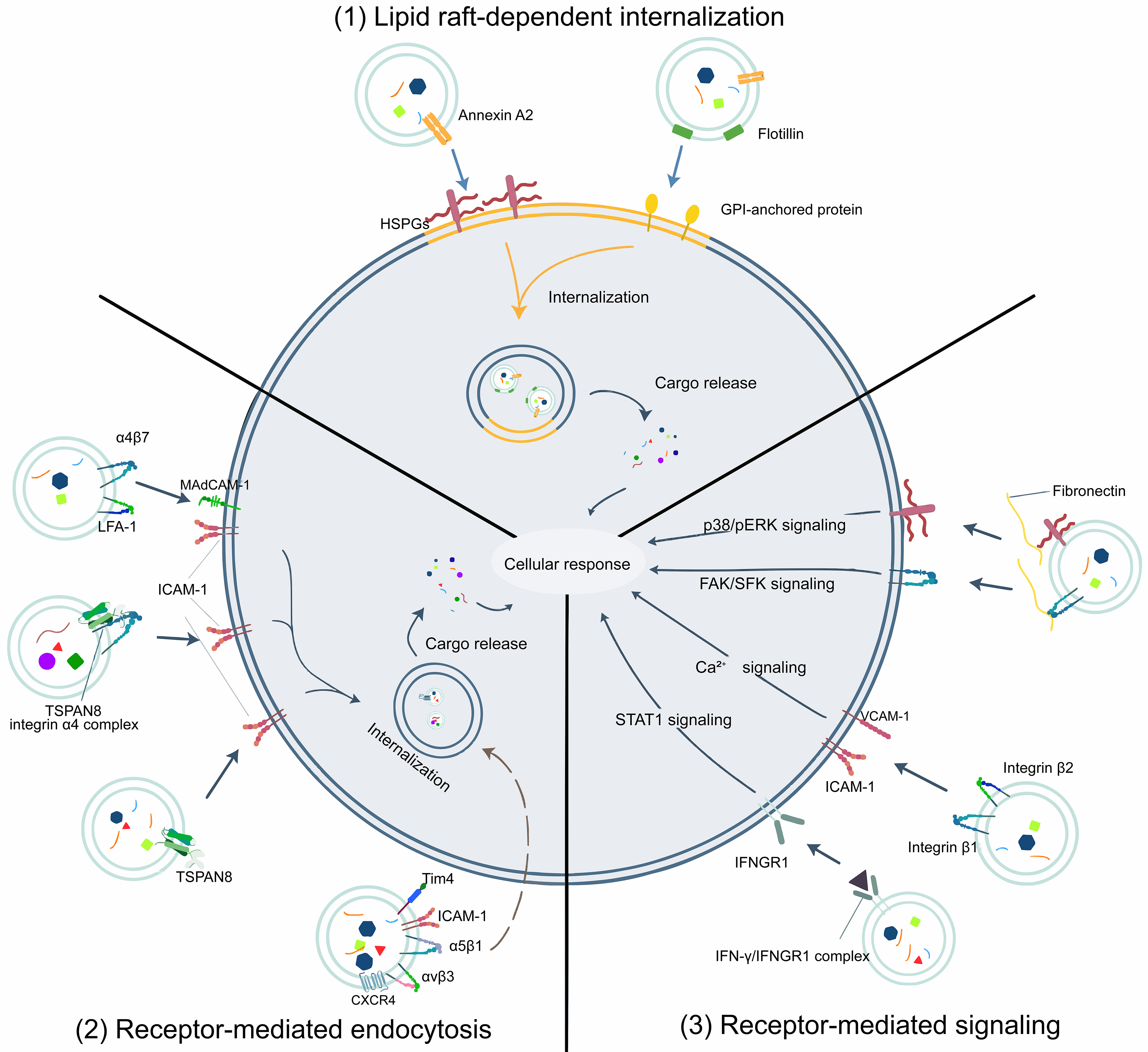
Figure 3. Mechanisms of extracellular vesicle uptake and receptor-mediated signaling in recipient cells: (1) lipid raft-dependent internalization; (2) receptor-mediated endocytosis; and (3) receptor-mediated signaling. Orange arrows indicate lipid raft-dependent internalization; blue-gray arrows indicate the illustrated EV-cell interactions, uptake, cargo release, and signaling. The dashed curved arrow indicates the illustrated uptake route toward the endosomal compartment, while thick black lines separate the three mechanisms. Integrin subunits are designated by Greek letters and numbers. Ca2+: Calcium ion; CXCR4: C-X-C motif chemokine receptor 4; FAK: focal adhesion kinase; GPI: glycosylphosphatidylinositol; HSPGs: heparan sulfate proteoglycans; ICAM-1: intercellular adhesion molecule 1; IFN-γ: interferon gamma; IFNGR1: interferon gamma receptor 1; LFA-1: lymphocyte function-associated antigen 1; MAdCAM-1: mucosal addressin cell adhesion molecule 1; p38: p38 mitogen-activated protein kinase; pERK: phosphorylated extracellular signal-regulated kinase; SFK: Src family kinase; STAT1: signal transducer and activator of transcription 1; Tim4: T-cell immunoglobulin and mucin domain-containing protein 4; TSPAN8: tetraspanin-8; VCAM-1: vascular cell adhesion molecule 1.
Taken together, the engagement of recipient-cell receptors by exosomal surface proteins and the resulting cargo uptake and/or signaling activation can regulate cellular functions and hence influence disease progression[94].
PHYSIOLOGICAL AND PATHOLOGICAL SIGNIFICANCE OF EV SURFACE-MEDIATED COMMUNICATION AND ORGANOTROPIC EFFECTS IN CANCER
Integrins, tetraspanins, adhesion molecules, and receptors on exosomal surfaces form a surface GPS that shapes recipient-cell recognition and, in some models, preferential tissue uptake. In normal physiology, this targeting supports coordinated tissue repair, angiogenesis, immune modulation, and neural-glial communication because the signals can act in receptive niches. However, in pathological conditions, the same machinery can pre-condition distant microenvironments, amplify inflammation or immunosuppression, and facilitate metastatic spread. These dynamics are especially prominent in cancer. Some exosomal surface proteins have tumor-suppressive effects [Table 1], whereas others have tumor-promoting effects [Table 2]. The TDEs can remodel extracellular matrices, instruct stromal and immune cells, and contribute to niche formation and metastatic colonization in experimental models[95].
EV-associated proteins with tumor-suppressive activities
| EV-associated protein(s) | Origin | Reported activity or mechanism | Cancer type | References |
| MHC-II, BAG6 | IFN-γ-stimulated DC-derived EVs | Enhanced NKp30-dependent NK-cell function, correlated with MHC-II and BAG6 expression | NSCLC | [246] |
| IL-15Rα | DC-derived EVs | Immune activation (NK proliferation and IFN-γ secretion) | Multiple | [247] |
| NKG2D ligands | DC-derived EVs | Immune activation (direct NK activation) | Multiple | [247] |
| TNF | DC-derived EVs | Immune activation (stimulates NK-cell IFN-γ) | Multiple | [96] |
| HSP70, BAG4 | Tumor-derived EVs | HSP70-positive EVs enhance NK-cell migration and cytolytic activity | Multiple | [248,249] |
| TNF, FasL, TRAIL | DC-derived EVs | Direct tumor apoptosis (caspase activation) | Multiple | [96] |
| TRAIL | TRAIL-positive EVs | TRAIL-dependent tumor-cell death; DR5-associated apoptosis in SUDHL4 and INT12 | DLBCL (SUDHL4), melanoma (INT12), myeloma (KMS11) | [97] |
EV-associated proteins with tumor-promoting activities
| EV-associated protein(s) | EV source | Reported activity or mechanism | Cancer type | References |
| Integrin α6β4, α6β1 | Lung-tropic breast cancer cell-derived EVs | Preferential lung uptake; lung pre-metastatic niche formation | Breast cancer | [11] |
| Integrin αvβ5 | Liver-tropic pancreatic cancer cell-derived EVs | Preferential uptake by liver Kupffer cells; liver pre-metastatic niche formation | Pancreatic cancer | [11,99] |
| Integrin αvβ6 | Prostate cancer cell-derived EVs | EV-mediated transfer of αvβ6 to recipient prostate cancer cells; enhanced adhesion and migration | Prostate cancer | [101] |
| Integrin αvβ3 | Prostate cancer cell-derived EVs | Tumor progression and metastasis (adhesion/migration) | Prostate cancer | [102] |
| ITGB4, EGFR, MUC1 | Cholangiocarcinoma-derived EVs | EV protein profile associated with cell migration, proliferation and survival pathways | Cholangiocarcinoma | [103] |
| CAV1, MET, S100 family | Metastatic HCC-derived EVs | Tumor progression and metastasis (reprogram hepatocytes) | Hepatocellular carcinoma | [104] |
| PD-L1, N-cadherin | Osteosarcoma-derived EVs | Tumor progression and metastasis | Osteosarcoma | [105] |
| CD44 | Ovarian/breast cancer-derived EVs | Tumor progression and metastasis (invasion, drug resistance) | Ovarian/breast cancer | [106-108] |
| CD97, TSPAN8 | Gastric cancer cell-derived EVs | Tumor progression and metastasis (proliferation/invasion) | Gastric cancer | [109,110] |
| PSGR | Prostate cancer cell-derived EVs | Tumor progression and metastasis; ECM remodeling and EMT (migration, stemness, EMT) | Prostate cancer | [112] |
| ADAM10 | Cancer-associated fibroblast-derived EVs | Tumor progression and metastasis (Notch receptor and RhoA activation) | Multiple | [113] |
| Fibronectin 1, LAMC1 | Gastric cancer-derived EVs | Tumor progression and metastasis (mesothelial adhesion, peritoneal metastasis) | Gastric cancer | [114] |
| B7-H3 | Colorectal cancer-derived EVs | Angiogenesis; tumor progression and metastasis (AKT1/mTOR/VEGFA activation) | Colorectal cancer | [115] |
| PDGFR-β | Melanoma-derived EVs | Therapy resistance (MAPK inhibitor bypass); tumor progression | Melanoma | [116] |
| CD47 | Tumor-derived EVs | Immune evasion ("don’t eat me" signal) | Multiple | [19] |
| PD-L1 | Melanoma, breast cancer and glioblastoma-derived EVs | Immune evasion (T-cell suppression) | Melanoma, breast cancer, glioblastoma | [35,123-125] |
| LMP1, galectin-9 | Nasopharyngeal carcinoma-derived EVs | Transport of viral and immunomodulatory proteins in nasopharyngeal carcinoma EVs | Nasopharyngeal carcinoma | [250,251] |
| TGF-β | Tumor-derived EVs | Immune evasion (downregulate NKG2D on NK/CD8+ T cells) | Multiple | [128] |
| TNF | Melanoma-derived EVs | ROS-mediated redox signaling in recipient cells | Melanoma | [129] |
| HSP72 | Tumor-derived EVs | Immune suppression through IL-6/STAT3 activation in myeloid-derived suppressor cells | Multiple | [252] |
| PD-L1, CD20 | DLBCL-derived EVs | Registered study of EV-associated PD-L1/CD20 and immunotherapy resistance | Diffuse large B-cell lymphoma | [NCT03985696] |
| VEGF, FGF, PDGF, bFGF, TGF-β, TNF-α, syndecan-4, GPC1, IL-8 | Tumor-derived EVs | Angiogenesis | Multiple | [37] |
| VEGF90K | Breast cancer cell-derived EVs | Angiogenesis (VEGFRs activation) | Breast cancer | [131] |
| Dll4 | Dll4-overexpressing U87 glioma cell-derived EVs | Vascular remodeling: Notch activation and capillary sprout retraction | Glioma cell-derived EVs; endothelial sprout model | [132] |
| GPC1, syndecan-4 | Glioblastoma-derived EVs | Angiogenesis (endothelial proliferation/tube formation) | Glioblastoma | [133] |
| TIE2 | Cervical cancer-derived EVs | Angiogenesis (macrophage TIE2-ANG2 pro-angiogenic signaling) | Cervical cancer | [138] |
| E-cadherin | Metastatic ovarian cancer-derived EVs | Angiogenesis | Metastatic ovarian cancer | [134] |
| DPP4 | 5-FU-resistant colon cancer cell-derived EVs | Angiogenesis (periostin upregulation via Twist1 and SMAD) | Colorectal cancer | [139] |
| Carbonic anhydrase 9 | Renal cell carcinoma-derived EV | Angiogenesis | Renal cell carcinoma | [135] |
| Annexin A2 (ANXA2) | Breast cancer-derived EVs | Angiogenesis | Breast cancer | [136] |
| DEL-1 | Mesothelioma cell-derived EVs | DEL-1 detected in EVs; angiogenesis-associated protein | Mesothelioma | [137] |
| Podoplanin | Tumor-derived EVs | Lymphangiogenesis | Multiple | [140] |
| Integrin αvβ5 | Pancreatic cancer cell-derived EVs | Pre-metastatic niche formation (liver tropism via binding Kupffer cells) | Pancreatic cancer | [11] |
| Integrin α6β4, α6β1 | Breast cancer cell-derived EVs | Lung pre-metastatic niche formation through uptake by lung fibroblasts and epithelial cells | Breast cancer | [11] |
| MET | Metastatic melanoma-derived EVs | Pre-metastatic niche formation (via MET pathway in bone marrow progenitor cells) | Metastatic melanoma | [145] |
| CD97 | Gastric cancer-derived EVs | Pre-metastatic niche formation (via lymphatic pathway) | Gastric cancer | [146] |
| MIF | PDAC-derived EVs | Pre-metastatic niche formation (fibronectin induction via Kupffer cell TGF-β) | PDAC | [147] |
| EGFR | Gastric cancer-derived EVs | Liver pre-metastatic niche formation: miR-26a/b suppression and HGF upregulation in hepatic stromal cells | Gastric cancer | [148] |
| TGF-β, IL-6, CAV1 | Tumor-derived EVs | EMT and ECM remodeling; pre-metastatic niche signaling | Multiple cancers | [142,150] |
| Annexin A2 (ANXA2) | Metastatic bladder cancer-derived EVs | EMT | Metastatic bladder cancer | [37] |
| MMP-13 | Nasopharyngeal carcinoma-derived EVs | ECM remodeling and EMT (EMT promotion) | Nasopharyngeal carcinoma | [152] |
| LMP1 | Nasopharyngeal carcinoma-derived EVs | EMT | Nasopharyngeal carcinoma | [154] |
| ITGB2 | Papillary thyroid cancer-derived EVs | EV ITGB2 levels associated with EMT and lymph-node involvement | Papillary thyroid cancer | [155] |
| CD151, TSPAN8 | Rat pancreatic adenocarcinoma-derived EVs | ECM degradation, stromal and hematopoietic-cell reprogramming, and EMT-associated gene expression | Rat pancreatic adenocarcinoma | [156] |
Surface-mediated EV cytotoxicity in tissue contexts
Exosomal surface proteins can directly trigger cancer cell death in a receptor- and tissue-context-dependent manner. For example, tumor necrosis factor (TNF), Fas ligand (FasL), and TNF-related apoptosis-inducing ligand (TRAIL) on exosomes activate caspase-dependent apoptosis in cancer cells[96]. Exosomal TRAIL induces potent apoptosis when engaging death receptor 5 (DR5) on SUDHL4 lymphoma and INT12 melanoma, resulting in 68% and 51% tumor growth inhibition, respectively[97], whereas binding to death receptor 4 (DR4) on KMS11 myeloma predominantly yields necrosis[97]. These observations indicate that TRAIL-positive exosomes can selectively suppress tumor growth depending on the distribution and signaling of death receptors in the target tissues.
Surface protein-mediated metastatic programming and organotropic effects
Exosomal integrins contribute to organotropic metastasis and microenvironmental conditioning[98]. TDEs enriched for α6β4 and α6β1 have been linked to lung metastasis, whereas αvβ5 has been implicated in uptake by Kupffer cells and hepatic niche conditioning[11,99]. Cellular αvβ6 promoted an osteolytic program in prostate-cancer models[100]. Separately, exosomal αvβ6 was transferred between prostate-cancer cells[101]. Beyond guiding tissue targeting, exosomal integrins can be transferred to neighboring tumor cells; lateral acquisition of αvβ6 increases invasiveness and accelerates metastatic outgrowth[101]. Studies showed that exosomal αvβ3 correlates with adhesion and migration in metastatic prostate cancer[102], while integrin β4 (ITGB4) in concert with EGFR and mucin-1 (MUC1) serves as a prognostic module in cholangiocarcinoma by promoting growth and dissemination[103]. Non-integrin EV-associated proteins similarly rewire tumor and stromal behavior. In metastatic hepatocellular carcinoma, exosomal caveolins, hepatocyte growth factor receptor (MET), and S100 family members convert hepatocytes toward an invasive and migratory phenotype by activating phosphoinositide 3-kinase (PI3K)/protein kinase B (AKT) and mitogen-activated protein kinase (MAPK) signaling pathways and elevating their responsiveness to matrix metalloproteinases (MMPs)[104]. Exosomal PD-L1 and N-cadherin enhance migration of osteosarcoma cells that contribute to lung metastasis[105]. Besides, the tetraspanin-linked mechanisms are also prominent. Exosomal cluster of differentiation 44 (CD44) from ovarian cancer can be transferred to peritoneal mesothelial cells to facilitate invasion and metastasis[106]. CD44 engagement also sustains cancer stemness and activates epithelial-mesenchymal transition (EMT) to promote dissemination[107]. It has been reported that CD44 is highly expressed on doxorubicin (Dox)-resistant breast cancer exosomes, linking it to metastasis and drug resistance[108], implying that CD44 can be a therapeutic target to overcome drug resistance. Additional EV-associated mediators include cluster of differentiation 97 (CD97) and TSPAN8, which drive gastric cancer proliferation and invasion[109,110]. At the cellular level, CD63 binds tissue inhibitor of metalloproteinases 1 (TIMP-1) and supports β1 integrin signaling and survival in mammary epithelial cells[111]. Other surface molecules also potentiate metastatic traits and therapeutic resistance. For example, the prostate-specific G protein-coupled receptor (PSGR) augments migration, stemness, and EMT[112]. ADAM10 on cancer-associated fibroblast exosomes activates Notch and Ras homolog family member A (RhoA) to boost motility[113]. Fibronectin 1 and laminin subunit gamma-1 (LAMC1) are associated with gastric cancer exosomes to facilitate peritoneal spread[114]. B7 homolog 3 (B7-H3) on colorectal cancer exosomes activates protein kinase B alpha (AKT1)/mechanistic target of rapamycin (mTOR)/vascular endothelial growth factor A (VEGFA) signaling to promote angiogenesis and metastasis[115]. Melanoma exosomal platelet-derived growth factor receptor beta (PDGFR-β) activates PI3K/AKT to bypass MAPK inhibition and foster resistance to targeted therapy[116]. Collectively, these findings implicate EV-associated proteins in organ targeting, metastatic reprogramming, and niche conditioning, alongside distinct cell-surface signaling mechanisms[117].
Exosomal surface proteins in immune evasion in cancer
Exosomes can suppress anti-tumor immunity through surface-mediated interactions with immune cells[118,119]. Exosomal cluster of differentiation 47 (CD47) engages signal-regulatory protein alpha (SIRPα) on phagocytes to deliver a “don’t eat me” signal that blocks phagocytosis and aids immune escape[19]. Exosomal PD-L1 inhibits T-cell activation and can directly suppress cluster of differentiation 8 (CD8)+ T cells, hence weakening anti-tumor immunity[120]. On the contrary, reducing exosomal PD-L1 enhances anti-tumor responses[121]. Studies have shown that decorating exosomes with anti-programmed cell death protein 1 (PD-1) can activate T cells and inhibit tumor growth[122-125]. Furthermore, the immunosuppressive capacity of exosomal PD-L1 can be therapeutically beneficial in transplantation. Following Wharton’s jelly mesenchymal stem cell (WJMSC) infusion, circulating small-vesicle PD-L1 increased and suppressed T-cell activity
EV surface-mediated angio- and lymphangiogenesis
EV surface-associated signals modulate endothelial and stromal responses involved in angiogenesis and lymphangiogenesis. VEGF signaling is central to tumor angiogenesis[130]. In breast cancer-derived microvesicles, 90-kDa vascular endothelial growth factor (VEGF90K) is associated with surface HSP90 and stimulates endothelial VEGF receptor signaling and angiogenic responses[131]. Exosomal delta-like ligand 4 (Dll4) activated Notch signaling in adjacent endothelial cells and induced capillary-sprout retraction[132]. Glioblastoma exosomes enriched in glypican-1 (GPC1) and syndecan-4 stimulate endothelial proliferation and tube formation, supporting aggressive tumor growth[133]. Exosomal E-cadherin from metastatic ovarian cancer further promotes new vessel formation[134]. Additional surface proteins such as carbonic anhydrase 9 and annexin II augment angiogenic programs[135,136]. Developmental endothelial locus-1 (DEL-1) has also been detected in mesothelioma-derived EV preparations, although its external exposure and EV-associated angiogenic activity were not established in that study[137]. Macrophages also participate in angiogenesis. Cervical cancer exosomes deliver tyrosine kinase with immunoglobulin-like and epidermal growth factor-like domains 2 (TIE2) to macrophages to increase their interaction with angiopoietin-2 (ANG2) and hence amplify pro-angiogenic signaling[138]. Besides, in 5-fluorouracil (5-FU)-resistant colon cancer, exosomal dipeptidyl peptidase 4 (DPP4) upregulates periostin in endothelial cells by inducing twist-related protein 1 (Twist1) nuclear translocation and activating Sma- and Mad-related proteins (SMAD) signaling, thereby driving angiogenesis[139]. Beyond blood vessels, exosomal podoplanin also promotes lymphangiogenesis, expanding lymphatic routes for metastatic dissemination[140].
Pre-metastatic niche establishment by organotropic EVs
Pre-metastatic niche formation is very important in cancer cell metastasis; it can be facilitated by EV-associated proteins through tissue-specific uptake and stromal reprogramming[141]. For example, TGF-β and CAV1 on tumor exosomes initiate niche formation by modulating cellular interactions and inflammatory tone[142,143]. Integrin repertoires can support preferential interactions with recipient cells. In the reported tumor models, exosomal αvβ5 was implicated in uptake by liver Kupffer cells, whereas α6β4 and α6β1 were associated with interactions with lung fibroblasts and epithelial cells and with organ-associated niche formation[11,144]. Metastatic melanoma exosomes that carry MET can activate MET signaling in bone marrow progenitor cells, increasing vascular leakiness and inflammation while enhancing pro-metastatic behavior[145]. Besides, exosomal CD97 facilitates niche preparation and lymphatic dissemination[146]. In pancreatic ductal adenocarcinoma (PDAC), EV-associated macrophage migration inhibitory factor (MIF) activates Kupffer cells to induce TGF-β, which drives fibronectin deposition and creates an immunosuppressive, tumor-permissive microenvironment[147]. The gastric cancer exosomal EGFR suppresses miR-26a/b in hepatic stromal cells, derepressing hepatocyte growth factor and establishing a liver microenvironment conducive to metastatic seeding[148]. Taken together, EV-associated proteins can contribute to metastatic niche formation in multiple cancer models.
EV surface proteins in extracellular matrix remodeling and EMT
EMT is a fundamental biological process that cancer cells hijack to drive invasion and dissemination, thereby facilitating metastasis. EV-associated proteins, including surface-exposed mediators, can remodel the extracellular matrix (ECM) and induce EMT, thereby enhancing motility and invasiveness in target tissues[149]. Interestingly, studies found that exosomal TGF-β and interleukin-6 (IL-6) are potent EMT inducers[150]. Besides, annexin A2 participates in EMT in metastatic bladder cancer[37], and metastatic breast cancer exosomal HSP90 activates matrix metalloproteinase 2 (MMP-2) to remodel ECM and promote invasion[151]. In nasopharyngeal carcinoma, exosomal matrix metalloproteinase 13 (MMP-13) drives EMT[152], consistent with the broader role of MMP activation in releasing matrix-bound growth factors during ECM degradation to facilitate metastasis[153]. Furthermore, the nasopharyngeal carcinoma exosomal LMP1 further triggers EMT and enhances migration and invasion[154]. The elevated exosomal integrin beta-2 (ITGB2) also correlates with EMT extent and lymph node involvement[155]. Other exosomal surface proteins such as the tetraspanins cluster of differentiation 151 (CD151) and TSPAN8 form complexes with integrins and proteases to drive ECM degradation, while reprogramming hematopoietic cells and inducing pro-inflammatory cytokines that help sustain a pro-metastatic niche[156].
PHYSIOLOGICAL AND PATHOLOGICAL SIGNIFICANCE OF EV SURFACE-MEDIATED COMMUNICATION IN IMMUNE-RELATED DISEASES
EV-mediated communication may influence immune niches where vesicles modulate inflammation, antigen presentation, and lymphocyte survival. In physiology, exosome-mediated communication supports interactions between epithelial barriers, dendritic cells (DCs), and lymphoid tissues to coordinate host defense[157]. In pathology, these surface-mediated interactions can sustain chronic inflammation, drive T-cell exhaustion in persistent infections, or enforce immunosuppression that can be either therapeutically desirable in transplantation or detrimental in settings of impaired anti-pathogen responses. Through these mechanisms, exosomal surface proteins influence inflammatory disorders, transplant rejection, viral infections, and autoimmune disease trajectories, as summarized in Table 3.
EV-associated proteins involved in immune modulation
| EV-associated protein(s) | Disease or biological process | Reported activity or study focus | References |
| Fibronectin | Inflammation | Trophoblast-derived EV fibronectin induces macrophage IL-1β production | [159] |
| LFA-1 | Immune activation | LFA-1 is carried by mast-cell EVs that stimulate B- and T-cell proliferation and cytokine production | [160] |
| Bat-3 | HIV-1 infection; chronic inflammation | Registered study of Bat-3/HIV-1 antisense protein-associated EVs and NK-cell activity | [NCT05243381] |
| MHC-I, MHC-II | T-cell activation | MHC-II-associated CD4+ T-cell activation and MHC-I-associated CD8+ T-cell priming | [19,161-163] |
| CD9, CD81, CD82, A33 antigen | Antigen presentation, immune activation | Lower antigen presentation threshold | [54,55] |
| CD80, CD86 | T-cell activation | Activate T cells | [19] |
| ICAM-1 | Immune activation | Stimulates T cells indirectly | [83] |
| PD-L1 | Immune suppression; transplantation-related models | PD-L1-bearing MSC EVs suppress T-cell activity; engineered FGL1/PD-L1 EVs prolong graft survival in mice | [126,127] |
| CD13 | Local neuropeptide metabolism | Microglial EV-associated CD13 cleaves neuropeptides | [165] |
| CD24 | Hyperinflammation in ARDS and COVID-19 | Registered studies evaluating CD24-overexpressing EVs for safety and prevention of clinical deterioration | [NCT05947747, NCT04902183, NCT04747574, NCT04969172] |
| CD63 | Ocular inflammation and immune privilege | CD63-associated EV release from ocular epithelia during inflammation; proposed contribution to ocular immune privilege | [166] |
Surface-mediated EV immune activation in tissue contexts
Exosomal surface proteins can amplify inflammation in a tissue-specific manner by docking to extracellular matrices and leukocytes within inflamed microenvironments[158]. The exosomal fibronectin directly induces interleukin-1 beta (IL-1β) production that enhances inflammation in the target tissues[159]. LFA-1 on exosomes reinforces leukocyte activation through inflammatory signaling pathways, which recruit immune cells and promote effector function at sites where exosomes accumulate[160]. In chronic viral infection, exosome-mediated signaling may perpetuate pathological inflammation. A clinical trial has been conducted to examine whether the exosomal surface protein human leukocyte antigen B-associated transcript 3 (Bat-3) binds the human immunodeficiency virus type 1 (HIV-1) antisense protein (ASP) and interferes with natural killer cell protein 30 (NKp30)-dependent NK cytotoxicity [NCT05243381]. This study may clarify the relationships among viral control, sustained chronic immune activation, T-cell exhaustion, and poor immune reconstitution. Targeted delivery of antigen-presenting machinery further couples epithelial and myeloid compartments to lymphoid niches. Dendritic cell-derived exosomes (Dex) bearing MHC-II activate cluster of differentiation 4 (CD4)+ T cells, while those displaying MHC-I support CD8+ T-cell priming, extending antigen presentation beyond the confines of the cell membrane[19,161,162]. It has been reported that B-cell-derived exosomes also present MHC-I and MHC-II that can augment T-cell responses, thereby significantly broadening the network of cells capable of initiating adaptive immunity[163]. The regulatory effects on immune cells are further enhanced by costimulatory and adhesion molecules on exosomes. For example, DC exosomal cluster of differentiation 80 (CD80) and cluster of differentiation 86 (CD86) promote T-cell activation[19], and ICAM-1 facilitates efficient immune synapse formation and signaling[83]. At mucosal interfaces, local vesicle-mediated communication is evident. Intestinal epithelial cell-derived exosomes enriched in CD9, CD81, cluster of differentiation 82 (CD82), and the A33 antigen communicate with DCs, which can lower antigen presentation thresholds and trigger rapid immune responses[54]. The basolateral exosomes bearing A33 antigen traverse the porous basal membrane to coordinate immune communication across the mucosa[55]. Taken together, these observations illustrate how exosomal surface proteins support antigen presentation and T-cell activation within specific anatomical niches.
Surface-mediated EV immune suppression in tissue contexts
Exosomal surface proteins can mediate immunosuppression to preserve tissue integrity or promote tolerance[164]. PD-L1-bearing small EVs from WJMSCs suppressed T-cell activity in vitro[126]. Microglia-derived exosomes displayed active cluster of differentiation 13 (CD13) capable of degrading neuropeptides, indicating a potential role in local neuropeptide metabolism[165]. Cluster of differentiation 24 (CD24)-overexpressing exosomes have been investigated for anti-inflammatory effects in acute respiratory distress syndrome (ARDS) and severe coronavirus disease 2019 (COVID-19) [NCT05947747, NCT04902183, NCT04747574, NCT04969172], aiming to evaluate their safety and efficacy in preventing clinical deterioration. These examples illustrate how EV-associated surface signals modulate immune responses and local signaling.
EV surface-mediated maintenance of immune privilege
Exosome-mediated communication also preserves immune privilege in barrier tissues. Studies using ARPE-19 cells identified CD63-associated EV release and proposed a possible role for these vesicles in ocular immune privilege[166]. Similar regulatory architectures are likely to operate in other immune-privileged sites such as the brain and placenta, where exosomal surface proteins allow finely tuned crosstalk across anatomical barriers to balance necessary surveillance with the prevention of immunopathology.
PHYSIOLOGICAL AND PATHOLOGICAL SIGNIFICANCE OF EV SURFACE-MEDIATED COMMUNICATION IN OTHER DISEASES
Exosomal surface proteins participate in neural, cardiovascular, and skeletal communication beyond cancer and immunity, with potential roles in disease onset, progression, and repair[167]. By shaping docking, uptake, and signaling within specific tissues, these surface proteins may influence whether exosomes propagate pathology or provide protection in these diseases.
Neural EV transfer and prion propagation
In neurodegeneration, EV-mediated transfer can contribute to spread within neuronal and glial networks. In transmissible spongiform encephalopathies, exosomes that carry normal cellular PrP can foster its conversion to the pathogenic scrapie isoform of PrP (PrPSc), and then transfer this misfolded species to uninfected neuronal and non-neuronal cells, initiating and propagating prion pathology within neural networks[168]. This transfer shows that EV-associated components can increase uptake in neural cells, turning normal protein trafficking into a route for neurodegenerative propagation.
Cardiac and vascular EV signaling in ischemia and cardiovascular diseases
Exosomes interacting with cardiomyocytes and vascular endothelium can activate protective or reparative programs through surface protein-mediated engagement of cardiomyocytes and immune cells. During ischemia-reperfusion, exosomal heat shock protein 70 (HSP70) engages Toll-like receptor 4 (TLR4) on cardiomyocytes to initiate a cytoprotective cascade culminating in HSP27 activation, thereby increasing cell survival by reducing damage under stress[169]. Following cerebral ischemia, exosomes from brain microvascular endothelial cells showed altered surface-protein patterns, including cluster of differentiation 146 (CD146), ephrin type-A receptor 2 (EPHA2), adenosine triphosphate-binding cassette subfamily B member 5 (ABCB5), and ITGB2[170].
Skeletal EV signaling and bone repair
Within the bone microenvironment, EV-associated signals can influence osteoclast and osteoblast responses involved in remodeling and regeneration. CD9 deficiency reduced exosome production and delayed fracture healing in mice; MSC-derived exosomes partially rescued repair[171]. These findings support a role for MSC-derived exosomes in fracture repair, while the contribution of specific surface proteins to skeletal targeting and their therapeutic relevance to osteoporosis require further evaluation.
EV-ASSOCIATED SURFACE PROTEINS AS CANDIDATE DISEASE BIOMARKERS
Surface proteins that contribute to exosome recognition and tissue distribution also form disease-associated profiles that can be sampled noninvasively from biofluids[172]. These profiles may contain information about donor-cell states and disease processes, supporting their investigation for diagnosis, prognosis, and response monitoring across cancer and nonmalignant disorders. Elevated or altered expression levels of defined exosomal surface proteins can serve as promising candidate circulating biomarkers[173].
Diagnostic readouts of disease-associated pathology
Multiple EV-associated proteins exhibit disease-associated changes that are being investigated for diagnostic use[174]. For example, in non-small cell lung cancer (NSCLC), a multiplex EV protein panel distinguished patients with advanced disease from patients with suspected but unconfirmed lung cancer in a cross-validated study[175]. Proteomic analysis of pleural-effusion EVs provided additional candidate proteins[176]. TSPAN8 expression was associated with gastric cancer progression in tissue and experimental EV studies[110]. In breast cancer, cell-line EV proteomics identified candidate proteins including GLUT-1, GPC1, and ADAM10[177], while CD24 was detected in patient-derived EV preparations[178]. Tetraspanin profiles are also under investigation. Increased EV-associated CD63 was reported in melanoma patient samples[179], while prostate and breast cancer cell-line studies showed variable CD63 expression across EV preparations[180]. CD63 is a broadly expressed EV marker, so these findings do not establish a cancer-specific diagnostic signal. In lung cancer, patients display increased exosomal cluster of differentiation 171 (CD171), CD151, and tetraspanin expression[181]. Among GPI-anchored proteins, exosomal GPC1 was reported to detect early-stage PDAC and discriminate malignant pancreatic cancer from chronic pancreatitis[182,183]. In melanoma, caveolae-associated CAV1 is elevated in exosomes from patients[179]. EV-associated receptors and cytokines provide further candidate signals. EGFR and TGF-β were detected in serum EV preparations from brain tumor patients[184], and elevated EV-associated EGFR was reported in five of nine lung cancer patients in a pilot study[185]. Other proteins such as human epidermal growth factor receptor 2 (HER2) and C-C motif chemokine receptor 6 (CCR6) are increased on exosomes from gastric cancer patients[98,186]. Cell adhesion and tight junction proteins are similarly informative. EV-associated CD24 and claudin-4 have been investigated as candidate ovarian cancer markers in patient cohorts and pilot assays[187,188], while exosomal CD44 and tumor-associated calcium signal transducer 2 (TACSTD2) increase in bladder cancer, alongside cluster of differentiation 36 (CD36), basigin, and cluster of differentiation 73 (CD73) on tumor-derived vesicles[98,189]. Tumor-associated carbohydrate antigens captured on vesicles, including carcinoembryonic antigen (CEA), cancer antigen 15-3 (CA15-3), and trophoblast glycoprotein (5T4), increase with disease progression in breast and bladder cancers[189,190]. Interestingly, immune checkpoint profiling reveals that exosomal PD-L1 is elevated in head and neck squamous cell carcinoma, PDAC, and gastric cancer[191-193]. Heat shock proteins (HSPs) are also informative. HSP27, heat shock protein 60 (HSP60), and HSP90 were detected by Western blotting in plasma EV preparations from patients with thyroid cancer[194]. A separate study protocol proposed evaluating circulating EV-associated HSP70 in patients with solid tumors, including ovarian cancer[195]. Higher serum EV-associated gamma-glutamyltransferase activity was reported in prostate cancer than in benign prostatic hyperplasia (BPH), supporting investigation of its diagnostic potential[196]. Urinary EV-associated leucine-rich alpha-2-glycoprotein 1 (LRG1) was identified as a candidate NSCLC marker[197], and EV-associated fetuin-A was investigated as a candidate acute kidney injury marker in animal models and a small patient series[198].
Several EV-associated proteins show disease-associated changes in abundance or detectability[199]. CD81 in the serum exosome-enriched fraction was elevated in patients with chronic hepatitis C and elevated alanine aminotransferase (ALT)[200]. Plasma EV α-synuclein differed between patients with Parkinson’s disease and comparison groups in two cohorts[201]; epidermal growth factor receptor variant III (EGFRvIII) mRNA was detected by nested RT-PCR in serum EV preparations from patients with glioblastoma[202], and exosomal PrPSc was detected in transmissible spongiform encephalopathies[168]. These disease-associated signatures provide a basis for developing EV-associated proteins and RNA as candidate circulating biomarkers[203].
Studies of EV-associated protein markers have reported diagnostic performance in defined patient cohorts. The original exosomal GPC1 study reported 100% specificity for PDAC in its study cohort[183]. A separate serum study reported specificities of 79.49% for carbohydrate antigen 19-9 (CA19-9) and 82.14% for serum GPC1[204]. These estimates describe performance within the respective study populations and analytical settings. An EV-associated DEL-1 study reported 94.7% sensitivity and 86.36% specificity for breast cancer detection in its test cohort, with corresponding estimates of 92.31% and 86.62% in a separate validation cohort[205]. Serum EV-fraction CEA has also been investigated for its clinical associations in colorectal cancer[206].
Reported findings for GPC1 include associations with tumor burden and postoperative changes, together with variation in diagnostic discrimination across study cohorts[207,208]. Cohort composition, comparator populations, sample handling, EV isolation, antibodies, and detection platforms provide the methodological context for interpreting these findings. Clinical development of EV-associated protein biomarkers involves standardized analytical workflows and evaluation in independent cohorts with clinically relevant comparator groups. Biofluids contain EVs from multiple cell types and vesicle subpopulations, while shared surface proteins can limit assignment of tissue origin and disease specificity[3,209,210]. Candidate discovery, diagnostic validation for a defined clinical use, and routine clinical implementation therefore represent distinct stages; the studies reviewed here include exploratory findings and cohort validation but do not establish routine clinical use.
Dynamic markers of disease progression and invasion
EV-associated proteins are also being investigated as indicators of disease progression[211]. In pancreatic cancer, exosomal GPC1 correlates with disease burden. Its levels have been associated with tumor size and metastatic load[183,207]. Elevated exosomal PD-L1 is found to be associated with aggressiveness of head and neck squamous cell carcinoma and the extent of lymph node invasion[191]. Cellular and tumor-tissue ITGB4 expression has been associated with triple-negative breast cancer aggressiveness, providing biological context rather than direct evidence for an EV biomarker[212]. In patients with chronic hepatitis C, higher CD81 levels in the serum exosome-enriched fraction were associated with severe liver fibrosis[200]. CD24 was detected in EVs from ovarian cancer ascites, but its detection was not correlated with tumor-tissue CD24 expression in that study[213]. However, not all markers increase with malignancy. Exosomal survivin-2B, which counteracts pro-tumor survivin and survivin-ΔEx3 and promotes cancer cell death, exhibits a negative correlation with tumor grade, supporting further investigation as a candidate marker of disease severity[214].
Prognosis and treatment monitoring through EV-associated protein signatures
Exosomal surface proteins have been investigated as possible correlates of outcomes and treatment response. In gastric cancer, co-expression of MHC-I with exosomal PD-L1 promotes immune exhaustion through T-cell suppression, linking higher exosomal PD-L1 levels with poorer immune status and worse survival[193]. In pancreatic cancer, EV-associated GPC1 levels and their postoperative changes were associated with survival in a longitudinal patient cohort[183]. Detection of CD24 in ovarian cancer ascites EVs did not establish an EV-based survival association[213]. Survivin-2B remains a candidate prognostic marker requiring longitudinal validation[214].
EV-associated protein profiles can change in response to treatment[215]. Studies show that patients with colorectal cancer exhibit marked reductions in exosomal GPC1 after surgery, with decreases exceeding the baseline differences between patients and healthy controls[216]. Postsurgical thyroid cancer patients exhibit notable declines in exosomal HSP27, HSP60, and HSP90 compared with preoperative levels[194]. These changes suggest that EV-associated protein measurements may inform longitudinal treatment monitoring; their relationship to therapeutic efficacy requires validation.
THERAPEUTIC APPLICATIONS OF SURFACE-ENGINEERED EVs
Native exosomal surface proteins mediate docking and uptake across tissues. However, compared with synthetic carriers, the intrinsic targeting ability of exosomes is less selective. Moreover, exosomes can be cleared rapidly in vivo, limiting their therapeutic efficacy. Engineering the exosomal surface to display defined ligands, receptors, and peptides can improve target-cell engagement and delivery in specific preclinical models[217,218]. Tissue accumulation, cellular uptake, functional cargo delivery, and therapeutic efficacy are distinct outcomes: accumulation or uptake alone does not establish cargo activity or treatment benefit[3].
Surface-engineered EV delivery of chemotherapeutics
Surface engineering has been explored to improve EV targeting and drug delivery in tumor and ischemia models[219]. In one study, fusion of the exosomal scaffold lysosome-associated membrane protein 2 isoform B (LAMP2B) with the αv integrin-binding internalizing arginine-glycine-aspartic acid peptide (iRGD) on Dex-directed exosomes to αv integrin-positive breast cancer cells for therapeutic delivery of doxorubicin[220]. In a transient middle-cerebral-artery-occlusion model, exosomes chemically conjugated with the cyclic arginine-glycine-aspartic acid-D-tyrosine-lysine peptide c(RGDyK) targeted ischemic brain lesions, and curcumin-loaded vesicles reduced inflammation and apoptosis[221]. These results support enhanced targeting of ischemic brain lesions and local anti-inflammatory and anti-apoptotic effects.
Engineering EV recognition for siRNA therapeutics
The engineered exosomes can also deliver siRNA for therapy[222,223]. Exosomes expressing a fusion of LAMP2B with designed ankyrin repeat protein (DARPin) G3 can bind to HER2/Neu on breast cancer cells and deliver tumor protein D52 (TPD52)-targeting siRNA that achieved 70% knockdown in SKBR3 cells[224]. LAMP2B fused to iRGD can bring siRNA-loaded exosomes to lung tumors to knock down Kirsten rat sarcoma viral oncogene homolog (KRAS) and suppress tumor growth[225]. Fusing LAMP2B with interleukin-3 (IL-3) targets the IL-3 receptor on chronic myeloid leukemia cells, enabling exosomes to deliver breakpoint cluster region-Abelson tyrosine kinase (BCR-ABL) siRNA to suppress chronic myeloid leukemia progression[226]. For central nervous system delivery, dendritic cell-derived exosomes displaying LAMP2B fused to a rabies virus glycoprotein (RVG) peptide delivered GAPDH siRNA to neurons, microglia, and oligodendrocytes. In a separate experiment, siRNA targeting beta-site amyloid precursor protein-cleaving enzyme 1 (BACE1) reduced its mRNA and protein in the brains of wild-type mice, demonstrating functional RNAi delivery to a target relevant to Alzheimer’s disease[227]. These studies show that engineered surface ligands can support recipient-cell engagement and functional siRNA delivery, with therapeutic benefit demonstrated only in models where disease outcomes were directly evaluated.
miRNA delivery through ligand-directed EV targeting
Engineered exosomes can reprogram tumor and hepatic niches by delivering therapeutic microRNAs to recipient cells[228]. For example, a platelet-derived growth factor receptor (PDGFR)-GE11 fusion enhances binding to EGFR-positive breast cancer, allowing exosomal transfer of let-7a miRNA and consequent tumor growth inhibition[229]. Exosomes displaying a CD63-apolipoprotein A-I fusion bound scavenger receptor class B type 1 on HepG2 cells and delivered miR-26a, reducing proliferation and migration in vitro[230]. These designs exploit receptor-ligand biology to improve recipient-cell engagement and cargo delivery in specific preclinical models.
Engineered EVs for regenerative tissue targeting
Surface engineering can steer exosomes to injured musculoskeletal and cardiac tissues to enhance repair[231]. In the knee joint, LAMP2B fused with MSC-binding peptide E7 on exosomes can deliver small-molecule cargo such as kartogenin (KGN) to promote chondrogenic differentiation and cartilage regeneration[232]. Besides, ischemic myocardium-targeting peptide (IMTP)-expressing exosomes can promote vasculogenesis and reduce fibrosis and inflammation[233]. Furthermore, cardiosphere-derived exosomes displaying a cardiomyocyte-targeting peptide (WLSEAGPVVTVRALRGTGSW) fused to LAMP2B showed increased myocardial retention 2 h after intramyocardial injection in non-infarcted mice and anti-apoptotic effects in cultured cardiomyocytes[234]. These studies show that engineered surface motifs can improve tissue-associated retention or recipient-cell engagement in selected preclinical models[235,236].
Surface protein engineering for immunomodulation and vaccination
Rewriting immune interactions at disease sites can also be achieved by modifying exosomes with immunologically active surface proteins. Given that the well-known CD47-SIRPα engagement delivers a “don’t eat me” signal to macrophages that protects tumor cells[237], SIRPα overexpressed on exosomes can act as a decoy to disrupt this axis and reduce tumor resistance to phagocytosis, thereby enhancing innate clearance through a surface-mediated interaction rather than requiring intracellular cargo delivery[238]. For vaccination, a DNA construct encoding a CD63-ovalbumin (OVA) fusion generated OVA-bearing EVs in transfected cells; purified EVs induced antigen-specific T-cell responses. Separately, vaccination with the pCD63-OVA DNA construct elicited OVA-specific CD8+ T-cell responses and inhibited tumor growth in mice[239]. Fusing exosomal scaffolds such as LAMP2B, PDGFR, and CD63 to peptides, cytokine domains, and high-affinity binders can improve recipient-cell engagement and delivery in preclinical models. Surface stability, reproducible biodistribution, and clearance remain challenges for translation[16,240].
Recent studies extend these foundational engineering approaches. Cardiomyocyte-binding-peptide-engineered EVs improved cardiac targeting, retention, and functional outcomes after intravenous delivery in a mouse myocardial-infarction model[241]. A self-cleaving mini-intein loading system combined with fusogenic vesicular stomatitis virus glycoprotein G (VSV-G) improved functional intracellular delivery of proteins and genome-editing cargoes; brain recombination followed intracerebroventricular administration[242].
Administration route and dose shape circulation, tissue exposure, and clearance[12,16]. Free label or transfer of labeled membrane components can influence measured biodistribution[3,12]. During scale-up, purification, and storage, maintaining surface ligand display, biological potency, and batch consistency remains a manufacturing challenge[16,243]. Minimal Information for Studies of Extracellular Vesicles 2023 (MISEV2023) provides guidance for vesicle characterization and functional controls, while clinical development also depends on validated manufacturing processes and indication-specific evaluation[3,16,244].
FUTURE PROSPECTS AND CHALLENGES
Resolving EV heterogeneity for surface-guided recognition and delivery
The surface “GPS” codes contribute to recipient-cell recognition and targeting as part of a multifactorial system, with potential applications in disease detection and drug delivery. Biodistribution also reflects vesicle origin, physicochemical properties, administration route, vascular accessibility, and clearance by mononuclear phagocytes; labeling, isolation, dose, and observation time influence its measurement[12]. An atlas linking EV biogenesis pathways, experimentally characterized surface features, and cell or tissue interactions could help identify context-dependent recognition patterns and guide their evaluation for diagnostic and therapeutic use.
Translating a surface-recognition framework into diagnostic and therapeutic applications requires evidence for each proposed clinical use. On the diagnostic front, we still have to resolve the issue of exosomal heterogeneity[245]. Single-vesicle and spatially resolved proteomics, paired with machine learning, may help investigate relationships between EV surface profiles, tissue distribution, and donor-cell states, provided that inferred signatures are validated in relevant samples. Longitudinal sampling and dynamic tracking are other strategies to link the exosomal surface complexity with treatment responses, disease progression, and prognosis. Although modular surface engineering offers a path to programmable recognition and delivery, additional strategies such as aptamers could refine cell- and tissue-specific recognition for drug delivery. Besides, microenvironment-responsive motifs could refine delivery to hypoxic, inflamed, or fibrotic niches. Addressing pharmacology is also essential. Strategies to extend circulation, reduce opsonization and rapid clearance, and enhance endosomal escape could improve functional delivery.
Disrupting pathological EV tissue targeting and recipient-cell interactions
Another therapeutic strategy is to disrupt tissue targeting or recipient-cell interactions of pathogenic exosomes. Strategies such as blocking exosomal adhesion modules, neutralizing pathogenic vesicle populations, inhibiting specific biogenesis routes, or transiently depleting circulating exosomes may prevent niche conditioning, angiogenesis, and immune escape in cancer and chronic inflammation.
CONCLUSION
Exosome biogenesis shapes cargo sorting and the surface protein repertoire, which contributes to recipient-cell recognition and receptor engagement and, together with other vesicle and host factors, influences tissue distribution in physiology and disease. Surface profiles are being investigated as noninvasive biomarkers for detection, prognosis, and treatment monitoring, with clinical value dependent on assay validation and disease specificity. Surface engineering has improved targeting or biological effects in selected preclinical models, but tissue accumulation, cellular uptake, functional cargo delivery, and therapeutic efficacy require distinct evidence. By connecting biogenesis and recognition mechanisms with these diagnostic and therapeutic evidence requirements, this review provides a framework for interpreting EV surface proteins and prioritizing further validation.
DECLARATIONS
Authors’ contributions
Performed the literature search: Fong HL, Leung CY, Qin T, Jiang W, Yang H, Ye Y, Peng M, Wang J
Prepared the original draft and figures and tables: Fong HL, Leung CY, Qin T
Conceived and designed the review: Xu K, Kwan HY
Critically revised and edited the manuscript: Qin T, Jia B, Xu K, Kwan HY
All authors read and approved the final version of the manuscript.
Availability of data and materials
Not applicable.
AI and AI-assisted tools statement
Not applicable.
Financial support and sponsorship
This work was partially supported by the Musketeers Education and Culture Charitable Foundation, Vincent and Lily Woo Foundation (No. 42.4621.179636), Health and Medical Research Fund (No. 08193596; No. 13242866), Initial Grant for Faculty Niche Research Areas (No. RC-FNRA-IG/23-24/SCM/01), Seed Funding for Collaborative Research Grants (No. RC-SFCRG/23-24/SCM/02), Natural Science Foundation of Guangdong Province (No. 2023A1515011811), and Chinese Medicine Task Force Donation (TFD2026-P06) to Kwan HY; National Natural Science Foundation for Youth of China (No. 82503193), the Science and Technology Development Fund, Macau SAR (No. 0107/2025/ITP2), and the Faculty Research Grant of the Macau University of Science and Technology (No. FRG-25-078-FC) to Xu K.
Conflicts of interest
All authors declared that there are no conflicts of interest.
Ethical approval and consent to participate
Not applicable.
Consent for publication
Not applicable.
Copyright
© The Author(s) 2026.
REFERENCES
1. Gurung S, Perocheau D, Touramanidou L, Baruteau J. The exosome journey: from biogenesis to uptake and intracellular signalling. Cell Commun Signal. 2021;19:47.
2. Kalluri R, LeBleu VS. The biology, function, and biomedical applications of exosomes. Science. 2020;367:eaau6977.
3. Welsh JA, Goberdhan DCI, O’Driscoll L, et al. ; MISEV Consortium. Minimal information for studies of extracellular vesicles (MISEV2023): from basic to advanced approaches. J Extracell Vesicles. 2024;13:e12404.
4. Li XX, Yang LX, Wang C, Li H, Shi DS, Wang J. The roles of exosomal proteins: classification, function, and applications. Int J Mol Sci. 2023;24:3061.
5. van Niel G, Porto-Carreiro I, Simoes S, Raposo G. Exosomes: a common pathway for a specialized function. J Biochem. 2006;140:13-21.
6. Pant S, Hilton H, Burczynski ME. The multifaceted exosome: biogenesis, role in normal and aberrant cellular function, and frontiers for pharmacological and biomarker opportunities. Biochem Pharmacol. 2012;83:1484-94.
7. Ripoll L, Zickler AM, Vader P, El Andaloussi S, Verweij FJ, van Niel G. Biology and therapeutic potential of extracellular vesicle targeting and uptake. Nat Rev Mol Cell Biol. 2026;27:358-76.
8. Baietti MF, Zhang Z, Mortier E, et al. Syndecan-syntenin-ALIX regulates the biogenesis of exosomes. Nat Cell Biol. 2012;14:677-85.
9. Trajkovic K, Hsu C, Chiantia S, et al. Ceramide triggers budding of exosome vesicles into multivesicular endosomes. Science. 2008;319:1244-7.
10. Wei D, Zhan W, Gao Y, et al. RAB31 marks and controls an ESCRT-independent exosome pathway. Cell Res. 2021;31:157-77.
11. Hoshino A, Costa-Silva B, Shen TL, et al. Tumour exosome integrins determine organotropic metastasis. Nature. 2015;527:329-35.
12. Kang M, Jordan V, Blenkiron C, Chamley LW. Biodistribution of extracellular vesicles following administration into animals: a systematic review. J Extracell Vesicles. 2021;10:e12085.
13. Heidarzadeh M, Zarebkohan A, Rahbarghazi R, Sokullu E. Protein corona and exosomes: new challenges and prospects. Cell Commun Signal. 2023;21:64.
14. Försönits AI, Tóth EÁ, Jezsoviczky S, et al. Improved accessibility of extracellular vesicle surface molecules upon partial removal of the protein corona by high ionic strength. J Extracell Vesicles. 2025;14:e70124.
15. Liang Y, Duan L, Lu J, Xia J. Engineering exosomes for targeted drug delivery. Theranostics. 2021;11:3183-95.
16. Xu G, Jin J, Fu Z, et al. Extracellular vesicle-based drug overview: research landscape, quality control and nonclinical evaluation strategies. Signal Transduct Target Ther. 2025;10:255.
17. Edelmann MJ, Kima PE. Current understanding of extracellular vesicle homing/tropism. Zoonoses. 2022;2:14.
18. Choi W, Park DJ, Eliceiri BP. Defining tropism and activity of natural and engineered extracellular vesicles. Front Immunol. 2024;15:1363185.
19. Xu F, Luo S, Lu P, Cai C, Li W, Li C. Composition, functions, and applications of exosomal membrane proteins. Front Immunol. 2024;15:1408415.
20. Dixson AC, Dawson TR, Di Vizio D, Weaver AM. Context-specific regulation of extracellular vesicle biogenesis and cargo selection. Nat Rev Mol Cell Biol. 2023;24:454-76.
21. Liu J, Ren L, Li S, et al. The biology, function, and applications of exosomes in cancer. Acta Pharm Sin B. 2021;11:2783-97.
22. Arya SB, Collie SP, Parent CA. The ins-and-outs of exosome biogenesis, secretion, and internalization. Trends Cell Biol. 2024;34:90-108.
23. He C, Zheng S, Luo Y, Wang B. Exosome theranostics: biology and translational medicine. Theranostics. 2018;8:237-55.
24. Ferreira JV, da Rosa Soares A, Ramalho J, et al. LAMP2A regulates the loading of proteins into exosomes. Sci Adv. 2022;8:eabm1140.
25. Han QF, Li WJ, Hu KS, et al. Exosome biogenesis: machinery, regulation, and therapeutic implications in cancer. Mol Cancer. 2022;21:207.
26. Noonin C, Thongboonkerd V. Exosome-inflammasome crosstalk and their roles in inflammatory responses. Theranostics. 2021;11:4436-51.
27. van Niel G, D’Angelo G, Raposo G. Shedding light on the cell biology of extracellular vesicles. Nat Rev Mol Cell Biol. 2018;19:213-28.
28. Lee YJ, Shin KJ, Chae YC. Regulation of cargo selection in exosome biogenesis and its biomedical applications in cancer. Exp Mol Med. 2024;56:877-89.
29. Marie PP, Fan SJ, Mason J, et al. Accessory ESCRT-III proteins are conserved and selective regulators of Rab11a-exosome formation. J Extracell Vesicles. 2023;12:e12311.
31. Vietri M, Radulovic M, Stenmark H. The many functions of ESCRTs. Nat Rev Mol Cell Biol. 2020;21:25-42.
32. Pfitzner AK, Mercier V, Jiang X, et al. An ESCRT-III polymerization sequence drives membrane deformation and fission. Cell. 2020;182:1140-55.e18.
33. Lee YJ, Shin KJ, Jang HJ, et al. GPR143 controls ESCRT-dependent exosome biogenesis and promotes cancer metastasis. Dev Cell. 2023;58:320-34.e8.
34. Mathieu M, Martin-Jaular L, Lavieu G, Théry C. Specificities of secretion and uptake of exosomes and other extracellular vesicles for cell-to-cell communication. Nat Cell Biol. 2019;21:9-17.
35. Chen G, Huang AC, Zhang W, et al. Exosomal PD-L1 contributes to immunosuppression and is associated with anti-PD-1 response. Nature. 2018;560:382-6.
36. Colombo M, Moita C, van Niel G, et al. Analysis of ESCRT functions in exosome biogenesis, composition and secretion highlights the heterogeneity of extracellular vesicles. J Cell Sci. 2013;126:5553-65.
37. Mashouri L, Yousefi H, Aref AR, Ahadi AM, Molaei F, Alahari SK. Exosomes: composition, biogenesis, and mechanisms in cancer metastasis and drug resistance. Mol Cancer. 2019;18:75.
38. Larios J, Mercier V, Roux A, Gruenberg J. ALIX- and ESCRT-III-dependent sorting of tetraspanins to exosomes. J Cell Biol. 2020;219:e201904113.
39. Castro-Cruz M, Hyka L, Daaboul G, et al. PDZ scaffolds regulate extracellular vesicle production, composition, and uptake. Proc Natl Acad Sci U S A. 2023;120:e2310914120.
40. van Niel G, Charrin S, Simoes S, et al. The tetraspanin CD63 regulates ESCRT-independent and -dependent endosomal sorting during melanogenesis. Dev Cell. 2011;21:708-21.
41. Hurwitz SN, Nkosi D, Conlon MM, et al. CD63 regulates epstein-barr virus LMP1 exosomal packaging, enhancement of vesicle production, and noncanonical NF-κB signaling. J Virol. 2017;91:e02251-16.
42. Ma S, Mangala LS, Hu W, et al. CD63-mediated cloaking of VEGF in small extracellular vesicles contributes to anti-VEGF therapy resistance. Cell Rep. 2021;36:109549.
43. Li X, Li C, Zhang L, et al. The significance of exosomes in the development and treatment of hepatocellular carcinoma. Mol Cancer. 2020;19:1.
44. Phuyal S, Hessvik NP, Skotland T, Sandvig K, Llorente A. Regulation of exosome release by glycosphingolipids and flotillins. FEBS J. 2014;281:2214-27.
45. Katzmann DJ, Babst M, Emr SD. Ubiquitin-dependent sorting into the multivesicular body pathway requires the function of a conserved endosomal protein sorting complex, ESCRT-I. Cell. 2001;106:145-55.
46. Kharmate G, Hosseini-Beheshti E, Caradec J, Chin MY, Tomlinson Guns ES. Epidermal growth factor receptor in prostate cancer derived exosomes. PLoS One. 2016;11:e0154967.
47. Lai JJ, Chau ZL, Chen SY, et al. Exosome processing and characterization approaches for research and technology development. Adv Sci. 2022;9:e2103222.
48. Théry C, Zitvogel L, Amigorena S. Exosomes: composition, biogenesis and function. Nat Rev Immunol. 2002;2:569-79.
49. Garcia-Martin R, Brandao BB, Thomou T, Altindis E, Kahn CR. Tissue differences in the exosomal/small extracellular vesicle proteome and their potential as indicators of altered tissue metabolism. Cell Rep. 2022;38:110277.
50. Valadi H, Ekström K, Bossios A, Sjöstrand M, Lee JJ, Lötvall JO. Exosome-mediated transfer of mRNAs and microRNAs is a novel mechanism of genetic exchange between cells. Nat Cell Biol. 2007;9:654-9.
51. Song L, Tang S, Han X, et al. KIBRA controls exosome secretion via inhibiting the proteasomal degradation of Rab27a. Nat Commun. 2019;10:1639.
52. Gurunathan S, Kang MH, Kim JH. A comprehensive review on factors influences biogenesis, functions, therapeutic and clinical implications of exosomes. Int J Nanomedicine. 2021;16:1281-312.
53. Li J, Wang X, Chen Y, et al. Exosome-mediated lectin pathway and resistin-MIF-AA metabolism axis drive immune dysfunction in immune thrombocytopenia. Adv Sci. 2025;12:e2412378.
54. Mallegol J, Van Niel G, Lebreton C, et al. T84-intestinal epithelial exosomes bear MHC class II/peptide complexes potentiating antigen presentation by dendritic cells. Gastroenterology. 2007;132:1866-76.
55. Van Niel G, Mallegol J, Bevilacqua C, et al. Intestinal epithelial exosomes carry MHC class II/peptides able to inform the immune system in mice. Gut. 2003;52:1690-7.
56. Lin W, Fang J, Wei S, et al. Extracellular vesicle-cell adhesion molecules in tumours: biofunctions and clinical applications. Cell Commun Signal. 2023;21:246.
57. Kowal J, Arras G, Colombo M, et al. Proteomic comparison defines novel markers to characterize heterogeneous populations of extracellular vesicle subtypes. Proc Natl Acad Sci U S A. 2016;113:E968-77.
58. Lischnig A, Bergqvist M, Ochiya T, Lässer C. Quantitative proteomics identifies proteins enriched in large and small extracellular vesicles. Mol Cell Proteomics. 2022;21:100273.
59. Palmulli R, Couty M, Piontek MC, et al. CD63 sorts cholesterol into endosomes for storage and distribution via exosomes. Nat Cell Biol. 2024;26:1093-109.
60. de Gassart A, Geminard C, Fevrier B, Raposo G, Vidal M. Lipid raft-associated protein sorting in exosomes. Blood. 2003;102:4336-44.
61. Hessvik NP, Llorente A. Current knowledge on exosome biogenesis and release. Cell Mol Life Sci. 2018;75:193-208.
62. Ostrowski M, Carmo NB, Krumeich S, et al. Rab27a and Rab27b control different steps of the exosome secretion pathway. Nat Cell Biol. 2010;12:19-30.
63. Jahn R, Cafiso DC, Tamm LK. Mechanisms of SNARE proteins in membrane fusion. Nat Rev Mol Cell Biol. 2024;25:101-18.
64. Gross JC, Chaudhary V, Bartscherer K, Boutros M. Active Wnt proteins are secreted on exosomes. Nat Cell Biol. 2012;14:1036-45.
65. Valapala M, Vishwanatha JK. Lipid raft endocytosis and exosomal transport facilitate extracellular trafficking of annexin A2. J Biol Chem. 2011;286:30911-25.
66. Rädler J, Gupta D, Zickler A, Andaloussi SE. Exploiting the biogenesis of extracellular vesicles for bioengineering and therapeutic cargo loading. Mol Ther. 2023;31:1231-50.
68. Géminard C, De Gassart A, Blanc L, Vidal M. Degradation of AP2 during reticulocyte maturation enhances binding of hsc70 and Alix to a common site on TFR for sorting into exosomes. Traffic. 2004;5:181-93.
69. Yeat NY, Liu LH, Chang YH, Lai CP, Chen RH. Bro1 proteins determine tumor immune evasion and metastasis by controlling secretion or degradation of multivesicular bodies. Dev Cell. 2025;60:2114-30.e9.
70. Liu DA, Tao K, Wu B, et al. A phosphoinositide switch mediates exocyst recruitment to multivesicular endosomes for exosome secretion. Nat Commun. 2023;14:6883.
71. Verweij FJ, Bebelman MP, George AE, et al. ER membrane contact sites support endosomal small GTPase conversion for exosome secretion. J Cell Biol. 2022;221.
72. Liu C, Liu D, Wang S, Gan L, Yang X, Ma C. Identification of the SNARE complex that mediates the fusion of multivesicular bodies with the plasma membrane in exosome secretion. J Extracell Vesicles. 2023;12:e12356.
73. Matsui T, Sakamaki Y, Hiragi S, Fukuda M. VAMP5 and distinct sets of cognate Q-SNAREs mediate exosome release. Cell Struct Funct. 2023;48:187-98.
74. Magoling BJA, Wu AY, Chen YJ, et al. Membrane protein modification modulates big and small extracellular vesicle biodistribution and tumorigenic potential in breast cancers in vivo. Adv Mater. 2023;35:e2208966.
75. Elsharkasy OM, de Voogt WS, Tognoli ML, et al. Integrin beta 1 and fibronectin mediate extracellular vesicle uptake and functional RNA delivery. J Biol Chem. 2025;301:108305.
76. Song YX, Li X, Nie SD, et al. Extracellular vesicles released by glioma cells are decorated by Annexin A2 allowing for cellular uptake via heparan sulfate. Cancer Gene Ther. 2023;30:1156-66.
77. Mulcahy LA, Pink RC, Carter DR. Routes and mechanisms of extracellular vesicle uptake. J Extracell Vesicles. 2014;3:24641.
78. Soe ZY, Prajuabjinda O, Myint PK, et al. Talin-2 regulates integrin functions in exosomes. Biochem Biophys Res Commun. 2019;512:429-34.
79. Malla RR, Pandrangi S, Kumari S, Gavara MM, Badana AK. Exosomal tetraspanins as regulators of cancer progression and metastasis and novel diagnostic markers. Asia Pac J Clin Oncol. 2018;14:383-91.
80. Charrin S, Jouannet S, Boucheix C, Rubinstein E. Tetraspanins at a glance. J Cell Sci. 2014;127:3641-8.
81. Soe ZY, Park EJ, Shimaoka M. Integrin regulation in immunological and cancerous cells and exosomes. Int J Mol Sci. 2021;22:2193.
82. Andreu Z, Yáñez-Mó M. Tetraspanins in extracellular vesicle formation and function. Front Immunol. 2014;5:442.
83. Segura E, Nicco C, Lombard B, et al. ICAM-1 on exosomes from mature dendritic cells is critical for efficient naive T-cell priming. Blood. 2005;106:216-23.
84. Miyanishi M, Tada K, Koike M, Uchiyama Y, Kitamura T, Nagata S. Identification of Tim4 as a phosphatidylserine receptor. Nature. 2007;450:435-9.
85. Chen L, Brigstock DR. Integrins and heparan sulfate proteoglycans on hepatic stellate cells (HSC) are novel receptors for HSC-derived exosomes. FEBS Lett. 2016;590:4263-74.
86. Viñas JL, Spence M, Gutsol A, et al. Receptor-ligand interaction mediates targeting of endothelial colony forming cell-derived exosomes to the kidney after ischemic injury. Sci Rep. 2018;8:16320.
87. Tian T, Zhu YL, Zhou YY, et al. Exosome uptake through clathrin-mediated endocytosis and macropinocytosis and mediating miR-21 delivery. J Biol Chem. 2014;289:22258-67.
88. Christianson HC, Svensson KJ, van Kuppevelt TH, Li JP, Belting M. Cancer cell exosomes depend on cell-surface heparan sulfate proteoglycans for their internalization and functional activity. Proc Natl Acad Sci U S A. 2013;110:17380-5.
89. Clayton A, Turkes A, Dewitt S, Steadman R, Mason MD, Hallett MB. Adhesion and signaling by B cell-derived exosomes: the role of integrins. FASEB J. 2004;18:977-9.
91. Ginini L, Billan S, Fridman E, Gil Z. Insight into extracellular vesicle-cell communication: from cell recognition to intracellular fate. Cells. 2022;11:1375.
92. Chanda D, Otoupalova E, Hough KP, et al. Fibronectin on the surface of extracellular vesicles mediates fibroblast invasion. Am J Respir Cell Mol Biol. 2019;60:279-88.
93. Purushothaman A, Bandari SK, Liu J, Mobley JA, Brown EE, Sanderson RD. Fibronectin on the surface of myeloma cell-derived exosomes mediates exosome-cell interactions. J Biol Chem. 2016;291:1652-63.
94. Zhang J, Liu H, Wu Q, et al. Exosomal ANXA2 facilitates ovarian cancer peritoneal metastasis by activating peritoneal mesothelial cells through binding with TLR2. Cell Commun Signal. 2024;22:616.
95. Hánělová K, Raudenská M, Masařík M, Balvan J. Protein cargo in extracellular vesicles as the key mediator in the progression of cancer. Cell Commun Signal. 2024;22:25.
96. Munich S, Sobo-Vujanovic A, Buchser WJ, Beer-Stolz D, Vujanovic NL. Dendritic cell exosomes directly kill tumor cells and activate natural killer cells via TNF superfamily ligands. Oncoimmunology. 2012;1:1074-83.
97. Rivoltini L, Chiodoni C, Squarcina P, et al. TNF-related apoptosis-inducing ligand (TRAIL)-armed exosomes deliver proapoptotic signals to tumor site. Clin Cancer Res. 2016;22:3499-512.
98. Li W, Li C, Zhou T, et al. Role of exosomal proteins in cancer diagnosis. Mol Cancer. 2017;16:145.
99. Casari I, Howard JA, Robless EE, Falasca M. Exosomal integrins and their influence on pancreatic cancer progression and metastasis. Cancer Lett. 2021;507:124-34.
100. Dutta A, Li J, Lu H, et al. Integrin αvβ6 promotes an osteolytic program in cancer cells by upregulating MMP2. Cancer Res. 2014;74:1598-608.
101. Fedele C, Singh A, Zerlanko BJ, Iozzo RV, Languino LR. The αvβ6 integrin is transferred intercellularly via exosomes. J Biol Chem. 2015;290:4545-51.
102. Singh A, Fedele C, Lu H, Nevalainen MT, Keen JH, Languino LR. Exosome-mediated transfer of αvβ3 integrin from tumorigenic to nontumorigenic cells promotes a migratory phenotype. Mol Cancer Res. 2016;14:1136-46.
103. Arbelaiz A, Azkargorta M, Krawczyk M, et al. Serum extracellular vesicles contain protein biomarkers for primary sclerosing cholangitis and cholangiocarcinoma. Hepatology. 2017;66:1125-43.
104. He M, Qin H, Poon TC, et al. Hepatocellular carcinoma-derived exosomes promote motility of immortalized hepatocyte through transfer of oncogenic proteins and RNAs. Carcinogenesis. 2015;36:1008-18.
105. Wang J, Zhang H, Sun X, et al. Exosomal PD-L1 and N-cadherin predict pulmonary metastasis progression for osteosarcoma patients. J Nanobiotechnology. 2020;18:151.
106. Nakamura K, Sawada K, Kinose Y, et al. Exosomes promote ovarian cancer cell invasion through transfer of CD44 to peritoneal mesothelial cells. Mol Cancer Res. 2017;15:78-92.
107. Skandalis SS, Karalis TT, Chatzopoulos A, Karamanos NK. Hyaluronan-CD44 axis orchestrates cancer stem cell functions. Cell Signal. 2019;63:109377.
108. Wang X, Cheng K, Zhang G, et al. Enrichment of CD44 in exosomes from breast cancer cells treated with doxorubicin promotes chemoresistance. Front Oncol. 2020;10:960.
109. Li C, Liu DR, Li GG, et al. CD97 promotes gastric cancer cell proliferation and invasion through exosome-mediated MAPK signaling pathway. World J Gastroenterol. 2015;21:6215-28.
110. Anami K, Oue N, Noguchi T, et al. TSPAN8, identified by Escherichia coli ampicillin secretion trap, is associated with cell growth and invasion in gastric cancer. Gastric Cancer. 2016;19:370-80.
111. Jung KK, Liu XW, Chirco R, Fridman R, Kim HR. Identification of CD63 as a tissue inhibitor of metalloproteinase-1 interacting cell surface protein. EMBO J. 2006;25:3934-42.
112. Li Y, Li Q, Li D, et al. Exosome carrying PSGR promotes stemness and epithelial-mesenchymal transition of low aggressive prostate cancer cells. Life Sci. 2021;264:118638.
113. Shimoda M, Principe S, Jackson HW, et al. Loss of the Timp gene family is sufficient for the acquisition of the CAF-like cell state. Nat Cell Biol. 2014;16:889-901.
114. Zheng Z, Zhai Y, Yan X, et al. Functions and clinical applications of exosomes in gastric cancer. Int J Biol Sci. 2025;21:2330-45.
115. Wu R, Zhang Y, Xu X, et al. Exosomal B7-H3 facilitates colorectal cancer angiogenesis and metastasis through AKT1/mTOR/VEGFA pathway. Cell Signal. 2023;109:110737.
116. Vella LJ, Behren A, Coleman B, Greening DW, Hill AF, Cebon J. Intercellular resistance to BRAF inhibition can be mediated by extracellular vesicle-associated PDGFRβ. Neoplasia. 2017;19:932-40.
117. Li K, Xue W, Lu Z, et al. Tumor-derived exosomal ADAM17 promotes pre-metastatic niche formation by enhancing vascular permeability in colorectal cancer. J Exp Clin Cancer Res. 2024;43:59.
118. Zhang W, Zhong W, Wang B, et al. ICAM-1-mediated adhesion is a prerequisite for exosome-induced T cell suppression. Dev Cell. 2022;57:329-43.e7.
119. Turiello R, Capone M, Morretta E, et al. Exosomal CD73 from serum of patients with melanoma suppresses lymphocyte functions and is associated with therapy resistance to anti-PD-1 agents. J Immunother Cancer. 2022;10:e004043.
120. Ma F, Liu X, Zhang Y, et al. Tumor extracellular vesicle-derived PD-L1 promotes T cell senescence through lipid metabolism reprogramming. Sci Transl Med. 2025;17:eadm7269.
121. Shen DD, Pang JR, Bi YP, et al. LSD1 deletion decreases exosomal PD-L1 and restores T-cell response in gastric cancer. Mol Cancer. 2022;21:75.
122. Lu Y, Ye L, Jian X, et al. Integrated microfluidic system for isolating exosome and analyzing protein marker PD-L1. Biosens Bioelectron. 2022;204:113879.
123. Chen J, Song Y, Miao F, et al. PDL1-positive exosomes suppress antitumor immunity by inducing tumor-specific CD8+ T cell exhaustion during metastasis. Cancer Sci. 2021;112:3437-54.
124. Yang Y, Li CW, Chan LC, et al. Exosomal PD-L1 harbors active defense function to suppress T cell killing of breast cancer cells and promote tumor growth. Cell Res. 2018;28:862-4.
125. Ricklefs FL, Alayo Q, Krenzlin H, et al. Immune evasion mediated by PD-L1 on glioblastoma-derived extracellular vesicles. Sci Adv. 2018;4:eaar2766.
126. Li M, Soder R, Abhyankar S, et al. WJMSC-derived small extracellular vesicle enhance T cell suppression through PD-L1. J Extracell Vesicles. 2021;10:e12067.
127. Tsai HI, Wu Y, Liu X, et al. Engineered small extracellular vesicles as a FGL1/PD-L1 dual-targeting delivery system for alleviating immune rejection. Adv Sci. 2022;9:e2102634.
128. Clayton A, Mitchell JP, Court J, Linnane S, Mason MD, Tabi Z. Human tumor-derived exosomes down-modulate NKG2D expression. J Immunol. 2008;180:7249-58.
129. Söderberg A, Barral AM, Söderström M, Sander B, Rosén A. Redox-signaling transmitted in trans to neighboring cells by melanoma-derived TNF-containing exosomes. Free Radic Biol Med. 2007;43:90-9.
130. Hicklin DJ, Ellis LM. Role of the vascular endothelial growth factor pathway in tumor growth and angiogenesis. J Clin Oncol. 2005;23:1011-27.
131. Feng Q, Zhang C, Lum D, et al. A class of extracellular vesicles from breast cancer cells activates VEGF receptors and tumour angiogenesis. Nat Commun. 2017;8:14450.
132. Sharghi-Namini S, Tan E, Ong LL, Ge R, Asada HH. Dll4-containing exosomes induce capillary sprout retraction in a 3D microenvironment. Sci Rep. 2014;4:4031.
133. Monteforte A, Lam B, Sherman MB, et al. *Glioblastoma exosomes for therapeutic angiogenesis in peripheral ischemia. Tissue Eng Part A. 2017;23:1251-61.
134. Tang MKS, Yue PYK, Ip PP, et al. Soluble E-cadherin promotes tumor angiogenesis and localizes to exosome surface. Nat Commun. 2018;9:2270.
135. Horie K, Kawakami K, Fujita Y, et al. Exosomes expressing carbonic anhydrase 9 promote angiogenesis. Biochem Biophys Res Commun. 2017;492:356-61.
136. Maji S, Chaudhary P, Akopova I, et al. Exosomal annexin II promotes angiogenesis and breast cancer metastasis. Mol Cancer Res. 2017;15:93-105.
137. Hegmans JP, Bard MP, Hemmes A, et al. Proteomic analysis of exosomes secreted by human mesothelioma cells. Am J Pathol. 2004;164:1807-15.
138. Du S, Qian J, Tan S, et al. Tumor cell-derived exosomes deliver TIE2 protein to macrophages to promote angiogenesis in cervical cancer. Cancer Lett. 2022;529:168-79.
139. Zheng X, Liu J, Li X, et al. Angiogenesis is promoted by exosomal DPP4 derived from 5-fluorouracil-resistant colon cancer cells. Cancer Lett. 2021;497:190-201.
140. Carrasco-Ramírez P, Greening DW, Andrés G, et al. Podoplanin is a component of extracellular vesicles that reprograms cell-derived exosomal proteins and modulates lymphatic vessel formation. Oncotarget. 2016;7:16070-89.
141. Li XQ, Zhang R, Lu H, Yue XM, Huang YF. Extracellular vesicle-packaged CDH11 and ITGA5 induce the premetastatic niche for bone colonization of breast cancer Cells. Cancer Res. 2022;82:1560-74.
142. Syn N, Wang L, Sethi G, Thiery JP, Goh BC. Exosome-mediated metastasis: from epithelial-mesenchymal transition to escape from immunosurveillance. Trends Pharmacol Sci. 2016;37:606-17.
143. Wang Y, Li Y, Zhong J, et al. Tumor-derived Cav-1 promotes pre-metastatic niche formation and lung metastasis in breast cancer. Theranostics. 2023;13:1684-97.
144. Paolillo M, Schinelli S. Integrins and exosomes, a dangerous liaison in cancer progression. Cancers. 2017;9:95.
145. Peinado H, Alečković M, Lavotshkin S, et al. Melanoma exosomes educate bone marrow progenitor cells toward a pro-metastatic phenotype through MET. Nat Med. 2012;18:883-91.
146. Liu D, Li C, Trojanowicz B, et al. CD97 promotion of gastric carcinoma lymphatic metastasis is exosome dependent. Gastric Cancer. 2016;19:754-66.
147. Costa-Silva B, Aiello NM, Ocean AJ, et al. Pancreatic cancer exosomes initiate pre-metastatic niche formation in the liver. Nat Cell Biol. 2015;17:816-26.
148. Zhang H, Deng T, Liu R, et al. Exosome-delivered EGFR regulates liver microenvironment to promote gastric cancer liver metastasis. Nat Commun. 2017;8:15016.
149. Zhu Y, Tao Z, Chen Y, et al. Exosomal MMP-1 transfers metastasis potential in triple-negative breast cancer through PAR1-mediated EMT. Breast Cancer Res Treat. 2022;193:65-81.
150. Whiteside TL. The role of tumor-derived exosomes in epithelial mesenchymal transition (EMT). Transl Cancer Res. 2017;6:S90-2.
151. Hendrix A, Maynard D, Pauwels P, et al. Effect of the secretory small GTPase Rab27B on breast cancer growth, invasion, and metastasis. J Natl Cancer Inst. 2010;102:866-80.
152. You Y, Shan Y, Chen J, et al. Matrix metalloproteinase 13-containing exosomes promote nasopharyngeal carcinoma metastasis. Cancer Sci. 2015;106:1669-77.
153. Kim HS, Kim HJ, Lee MR, Han I. EMMPRIN expression is associated with metastatic progression in osteosarcoma. BMC Cancer. 2021;21:1059.
154. Yoshizaki T, Kondo S, Wakisaka N, et al. Pathogenic role of Epstein-Barr virus latent membrane protein-1 in the development of nasopharyngeal carcinoma. Cancer Lett. 2013;337:1-7.
155. Luo D, Zhan S, Xia W, Huang L, Ge W, Wang T. Proteomics study of serum exosomes from papillary thyroid cancer patients. Endocr Relat Cancer. 2018;25:879-91.
156. Yue S, Mu W, Erb U, Zöller M. The tetraspanins CD151 and Tspan8 are essential exosome components for the crosstalk between cancer initiating cells and their surrounding. Oncotarget. 2015;6:2366-84.
157. Kalluri R. The biology and function of extracellular vesicles in immune response and immunity. Immunity. 2024;57:1752-68.
158. Buzas EI. The roles of extracellular vesicles in the immune system. Nat Rev Immunol. 2023;23:236-50.
159. Atay S, Gercel-Taylor C, Taylor DD. Human trophoblast-derived exosomal fibronectin induces pro-inflammatory IL-1β production by macrophages. Am J Reprod Immunol. 2011;66:259-69.
160. Skokos D, Le Panse S, Villa I, et al. Mast cell-dependent B and T lymphocyte activation is mediated by the secretion of immunologically active exosomes. J Immunol. 2001;166:868-76.
161. Théry C, Duban L, Segura E, Véron P, Lantz O, Amigorena S. Indirect activation of naïve CD4+ T cells by dendritic cell-derived exosomes. Nat Immunol. 2002;3:1156-62.
162. André F, Chaput N, Schartz NE, et al. Exosomes as potent cell-free peptide-based vaccine. I. Dendritic cell-derived exosomes transfer functional MHC class I/peptide complexes to dendritic cells. J Immunol. 2004;172:2126-36.
163. Raposo G, Nijman HW, Stoorvogel W, et al. B lymphocytes secrete antigen-presenting vesicles. J Exp Med. 1996;183:1161-72.
164. Wang W, Qiao S, Kong X, Zhang G, Cai Z. The role of exosomes in immunopathology and potential therapeutic implications. Cell Mol Immunol. 2025;22:975-95.
165. Potolicchio I, Carven GJ, Xu X, et al. Proteomic analysis of microglia-derived exosomes: metabolic role of the aminopeptidase CD13 in neuropeptide catabolism. J Immunol. 2005;175:2237-43.
166. McKechnie NM, Copland D, Braun G. Hr44 secreted with exosomes: loss from ciliary epithelium in response to inflammation. Invest Ophthalmol Vis Sci. 2003;44:2650-6.
167. Alvarez-Erviti L, Seow Y, Schapira AH, et al. Lysosomal dysfunction increases exosome-mediated alpha-synuclein release and transmission. Neurobiol Dis. 2011;42:360-7.
168. Vella LJ, Sharples RA, Lawson VA, Masters CL, Cappai R, Hill AF. Packaging of prions into exosomes is associated with a novel pathway of PrP processing. J Pathol. 2007;211:582-90.
169. Vicencio JM, Yellon DM, Sivaraman V, et al. Plasma exosomes protect the myocardium from ischemia-reperfusion injury. J Am Coll Cardiol. 2015;65:1525-36.
170. Yang D, Li Z, Gao G, et al. Combined analysis of surface protein profile and microRNA expression profile of exosomes derived from brain microvascular endothelial cells in early cerebral ischemia. ACS Omega. 2021;6:22410-21.
171. Furuta T, Miyaki S, Ishitobi H, et al. Mesenchymal stem cell-derived exosomes promote fracture healing in a mouse model. Stem Cells Transl Med. 2016;5:1620-30.
172. Wang X, Huang J, Chen W, Li G, Li Z, Lei J. The updated role of exosomal proteins in the diagnosis, prognosis, and treatment of cancer. Exp Mol Med. 2022;54:1390-400.
173. Xu X, Zhang Y, Liu J, et al. Concurrent detection of protein and miRNA at the single extracellular vesicle level using a digital dual CRISPR-Cas assay. ACS Nano. 2025;19:1271-85.
174. Zhai C, Long J, He J, et al. Precise identification and profiling of surface proteins of ultra rare tumor specific extracellular vesicle with dynamic quantitative plasmonic imaging. ACS Nano. 2023;17:16656-67.
175. Jakobsen KR, Paulsen BS, Bæk R, Varming K, Sorensen BS, Jørgensen MM. Exosomal proteins as potential diagnostic markers in advanced non-small cell lung carcinoma. J Extracell Vesicles. 2015;4:26659.
176. Park JO, Choi DY, Choi DS, et al. Identification and characterization of proteins isolated from microvesicles derived from human lung cancer pleural effusions. Proteomics. 2013;13:2125-34.
177. Risha Y, Minic Z, Ghobadloo SM, Berezovski MV. The proteomic analysis of breast cell line exosomes reveals disease patterns and potential biomarkers. Sci Rep. 2020;10:13572.
178. Rupp AK, Rupp C, Keller S, et al. Loss of EpCAM expression in breast cancer derived serum exosomes: role of proteolytic cleavage. Gynecol Oncol. 2011;122:437-46.
179. Logozzi M, De Milito A, Lugini L, et al. High levels of exosomes expressing CD63 and caveolin-1 in plasma of melanoma patients. PLoS One. 2009;4:e5219.
180. Yoshioka Y, Konishi Y, Kosaka N, Katsuda T, Kato T, Ochiya T. Comparative marker analysis of extracellular vesicles in different human cancer types. J Extracell Vesicles. 2013;2:20424.
181. Sandfeld-Paulsen B, Jakobsen KR, Bæk R, et al. Exosomal proteins as diagnostic biomarkers in lung cancer. J Thorac Oncol. 2016;11:1701-10.
182. Yin T, Xu L, Gil B, et al. Graphene sensor arrays for rapid and accurate detection of pancreatic cancer exosomes in patients’ blood plasma samples. ACS Nano. 2023;17:14619-31.
183. Melo SA, Luecke LB, Kahlert C, et al. Glypican-1 identifies cancer exosomes and detects early pancreatic cancer. Nature. 2015;523:177-82.
184. Graner MW, Alzate O, Dechkovskaia AM, et al. Proteomic and immunologic analyses of brain tumor exosomes. FASEB J. 2009;23:1541-57.
185. Yamashita T, Kamada H, Kanasaki S, et al. Epidermal growth factor receptor localized to exosome membranes as a possible biomarker for lung cancer diagnosis. Pharmazie. 2013;68:969-73.
186. Baran J, Baj-Krzyworzeka M, Weglarczyk K, et al. Circulating tumour-derived microvesicles in plasma of gastric cancer patients. Cancer Immunol Immunother. 2010;59:841-50.
187. Li J, Sherman-Baust CA, Tsai-Turton M, Bristow RE, Roden RB, Morin PJ. Claudin-containing exosomes in the peripheral circulation of women with ovarian cancer. BMC Cancer. 2009;9:244.
188. Zhao Z, Yang Y, Zeng Y, He M. A microfluidic ExoSearch chip for multiplexed exosome detection towards blood-based ovarian cancer diagnosis. Lab Chip. 2016;16:489-96.
189. Welton JL, Khanna S, Giles PJ, et al. Proteomics analysis of bladder cancer exosomes. Mol Cell Proteomics. 2010;9:1324-38.
190. Toth B, Nieuwland R, Liebhardt S, et al. Circulating microparticles in breast cancer patients: a comparative analysis with established biomarkers. Anticancer Res. 2008;28:1107-12.
191. Theodoraki MN, Yerneni SS, Hoffmann TK, Gooding WE, Whiteside TL. Clinical significance of PD-L1+ exosomes in plasma of head and neck cancer patients. Clin Cancer Res. 2018;24:896-905.
192. Lux A, Kahlert C, Grützmann R, Pilarsky C. c-Met and PD-L1 on circulating exosomes as diagnostic and prognostic markers for pancreatic cancer. Int J Mol Sci. 2019;20:3305.
193. Fan Y, Che X, Qu J, et al. Exosomal PD-L1 retains immunosuppressive activity and is associated with gastric cancer prognosis. Ann Surg Oncol. 2019;26:3745-55.
194. Caruso Bavisotto C, Cipolla C, Graceffa G, et al. Immunomorphological pattern of molecular chaperones in normal and pathological thyroid tissues and circulating exosomes: potential use in clinics. Int J Mol Sci. 2019;20:4496.
195. Chanteloup G, Cordonnier M, Isambert N, et al. Membrane-bound exosomal HSP70 as a biomarker for detection and monitoring of malignant solid tumours: a pilot study. Pilot Feasibility Stud. 2020;6:35.
196. Kawakami K, Fujita Y, Matsuda Y, et al. Gamma-glutamyltransferase activity in exosomes as a potential marker for prostate cancer. BMC Cancer. 2017;17:316.
197. Li Y, Zhang Y, Qiu F, Qiu Z. Proteomic identification of exosomal LRG1: a potential urinary biomarker for detecting NSCLC. Electrophoresis. 2011;32:1976-83.
198. Zhou H, Pisitkun T, Aponte A, et al. Exosomal Fetuin-A identified by proteomics: a novel urinary biomarker for detecting acute kidney injury. Kidney Int. 2006;70:1847-57.
199. Yan S, Jiang C, Janzen A, et al. Neuronally derived extracellular vesicle α-synuclein as a serum biomarker for individuals at risk of developing parkinson disease. JAMA Neurol. 2024;81:59-68.
200. Welker MW, Reichert D, Susser S, et al. Soluble serum CD81 is elevated in patients with chronic hepatitis C and correlates with alanine aminotransferase serum activity. PLoS One. 2012;7:e30796.
201. Stuendl A, Kraus T, Chatterjee M, et al. α-Synuclein in plasma-derived extracellular vesicles is a potential biomarker of Parkinson’s disease. Mov Disord. 2021;36:2508-18.
202. Skog J, Würdinger T, van Rijn S, et al. Glioblastoma microvesicles transport RNA and proteins that promote tumour growth and provide diagnostic biomarkers. Nat Cell Biol. 2008;10:1470-6.
203. Cai Y, Chen T, Cai Y, et al. Surface protein profiling and subtyping of extracellular vesicles in body fluids reveals non-CSF biomarkers of Alzheimer’s disease. J Extracell Vesicles. 2024;13:e12432.
204. Zhou CY, Dong YP, Sun X, et al. High levels of serum glypican-1 indicate poor prognosis in pancreatic ductal adenocarcinoma. Cancer Med. 2018;7:5525-33.
205. Moon PG, Lee JE, Cho YE, et al. Identification of developmental endothelial Locus-1 on circulating extracellular vesicles as a novel biomarker for early breast cancer detection. Clin Cancer Res. 2016;22:1757-66.
206. Yokoyama S, Takeuchi A, Yamaguchi S, et al. Clinical implications of carcinoembryonic antigen distribution in serum exosomal fraction-Measurement by ELISA. PLoS One. 2017;12:e0183337.
207. Frampton AE, Prado MM, López-Jiménez E, et al. Glypican-1 is enriched in circulating-exosomes in pancreatic cancer and correlates with tumor burden. Oncotarget. 2018;9:19006-13.
208. Lucien F, Lac V, Billadeau DD, Borgida A, Gallinger S, Leong HS. Glypican-1 and glycoprotein 2 bearing extracellular vesicles do not discern pancreatic cancer from benign pancreatic diseases. Oncotarget. 2019;10:1045-55.
209. Karimi N, Dalirfardouei R, Dias T, Lötvall J, Lässer C. Tetraspanins distinguish separate extracellular vesicle subpopulations in human serum and plasma - Contributions of platelet extracellular vesicles in plasma samples. J Extracell Vesicles. 2022;11:e12213.
210. Carney RP, Mizenko RR, Bozkurt BT, et al. Harnessing extracellular vesicle heterogeneity for diagnostic and therapeutic applications. Nat Nanotechnol. 2025;20:14-25.
211. Li H, Chiang CL, Kwak KJ, et al. Extracellular vesicular analysis of glypican 1 mRNA and protein for pancreatic cancer diagnosis and prognosis. Adv Sci. 2024;11:e2306373.
212. Bierie B, Pierce SE, Kroeger C, et al. Integrin-β4 identifies cancer stem cell-enriched populations of partially mesenchymal carcinoma cells. Proc Natl Acad Sci U S A. 2017;114:E2337-46.
213. Runz S, Keller S, Rupp C, et al. Malignant ascites-derived exosomes of ovarian carcinoma patients contain CD24 and EpCAM. Gynecol Oncol. 2007;107:563-71.
214. Khan S, Bennit HF, Turay D, et al. Early diagnostic value of survivin and its alternative splice variants in breast cancer. BMC Cancer. 2014;14:176.
215. de Miguel-Perez D, Russo A, Arrieta O, et al. Extracellular vesicle PD-L1 dynamics predict durable response to immune-checkpoint inhibitors and survival in patients with non-small cell lung cancer. J Exp Clin Cancer Res. 2022;41:186.
216. Li J, Chen Y, Guo X, et al. GPC1 exosome and its regulatory miRNAs are specific markers for the detection and target therapy of colorectal cancer. J Cell Mol Med. 2017;21:838-47.
217. Zhang M, Hu S, Liu L, et al. Engineered exosomes from different sources for cancer-targeted therapy. Signal Transduct Target Ther. 2023;8:124.
218. Bergqvist M, Park KS, Karimi N, Yu L, Lässer C, Lötvall J. Extracellular vesicle surface engineering with integrins (ITGAL & ITGB2) to specifically target ICAM-1-expressing endothelial cells. J Nanobiotechnology. 2025;23:64.
219. Mohammadi AH, Ghazvinian Z, Bagheri F, Harada M, Baghaei K. Modification of extracellular vesicle surfaces: an approach for targeted drug delivery. BioDrugs. 2023;37:353-74.
220. Tian Y, Li S, Song J, et al. A doxorubicin delivery platform using engineered natural membrane vesicle exosomes for targeted tumor therapy. Biomaterials. 2014;35:2383-90.
221. Tian T, Zhang HX, He CP, et al. Surface functionalized exosomes as targeted drug delivery vehicles for cerebral ischemia therapy. Biomaterials. 2018;150:137-49.
222. Zhuo Y, Luo Z, Zhu Z, et al. Direct cytosolic delivery of siRNA via cell membrane fusion using cholesterol-enriched exosomes. Nat Nanotechnol. 2024;19:1858-68.
223. Xie M, Wu Y, Zhang Y, et al. Membrane fusion-mediated loading of therapeutic siRNA into exosome for tissue-specific application. Adv Mater. 2024;36:e2403935.
224. Limoni SK, Moghadam MF, Moazzeni SM, Gomari H, Salimi F. Engineered exosomes for targeted transfer of siRNA to HER2 positive breast cancer cells. Appl Biochem Biotechnol. 2019;187:352-64.
225. Zhou Y, Yuan Y, Liu M, Hu X, Quan Y, Chen X. Tumor-specific delivery of KRAS siRNA with iRGD-exosomes efficiently inhibits tumor growth. ExRNA. 2019;1:28.
226. Bellavia D, Raimondo S, Calabrese G, et al. Interleukin 3- receptor targeted exosomes inhibit in vitro and in vivo Chronic Myelogenous Leukemia cell growth. Theranostics. 2017;7:1333-45.
227. Alvarez-Erviti L, Seow Y, Yin H, Betts C, Lakhal S, Wood MJ. Delivery of siRNA to the mouse brain by systemic injection of targeted exosomes. Nat Biotechnol. 2011;29:341-5.
228. Xu K, Wu Q, Lingyun Z, et al. Extracellular vesicles as a promising platform of precision medicine in liver cancer. Pharmacol Res. 2025;217:107800.
229. Ohno S, Takanashi M, Sudo K, et al. Systemically injected exosomes targeted to EGFR deliver antitumor microRNA to breast cancer cells. Mol Ther. 2013;21:185-91.
230. Liang G, Kan S, Zhu Y, Feng S, Feng W, Gao S. Engineered exosome-mediated delivery of functionally active miR-26a and its enhanced suppression effect in HepG2 cells. Int J Nanomedicine. 2018;13:585-99.
231. Zhang H, Yan W, Wang J, et al. Surface functionalization of exosomes for chondrocyte-targeted siRNA delivery and cartilage regeneration. J Control Release. 2024;369:493-505.
232. Xu X, Liang Y, Li X, et al. Exosome-mediated delivery of kartogenin for chondrogenesis of synovial fluid-derived mesenchymal stem cells and cartilage regeneration. Biomaterials. 2021;269:120539.
233. Wang X, Chen Y, Zhao Z, et al. Engineered exosomes with ischemic myocardium-targeting peptide for targeted therapy in myocardial infarction. J Am Heart Assoc. 2018;7:e008737.
234. Mentkowski KI, Lang JK. Exosomes engineered to express a cardiomyocyte binding peptide demonstrate improved cardiac retention in vivo. Sci Rep. 2019;9:10041.
235. Gu J, You J, Liang H, Zhan J, Gu X, Zhu Y. Engineered bone marrow mesenchymal stem cell-derived exosomes loaded with miR302 through the cardiomyocyte specific peptide can reduce myocardial ischemia and reperfusion (I/R) injury. J Transl Med. 2024;22:168.
236. Meng WT, Zhu J, Wang YC, et al. Targeting delivery of miR-146a via IMTP modified milk exosomes exerted cardioprotective effects by inhibiting NF-κB signaling pathway after myocardial ischemia-reperfusion injury. J Nanobiotechnology. 2024;22:382.
237. Cho E, Nam GH, Hong Y, et al. Comparison of exosomes and ferritin protein nanocages for the delivery of membrane protein therapeutics. J Control Release. 2018;279:326-35.
238. Koh E, Lee EJ, Nam GH, et al. Exosome-SIRPα, a CD47 blockade increases cancer cell phagocytosis. Biomaterials. 2017;121:121-9.
239. Kanuma T, Yamamoto T, Kobiyama K, et al. CD63-mediated antigen delivery into extracellular vesicles via DNA vaccination results in robust CD8+ T cell responses. J Immunol. 2017;198:4707-15.
240. Zhang Y, Bi J, Huang J, Tang Y, Du S, Li P. Exosome: a review of its classification, isolation techniques, storage, diagnostic and targeted therapy applications. Int J Nanomedicine. 2020;15:6917-34.
241. Mentkowski KI, Tarvirdizadeh T, Manzanero CA, Eagler LA, Lang JK. Surface engineering enhances the therapeutic potential of systemically delivered extracellular vesicles following acute myocardial infarction. FASEB J. 2024;38:e70070.
242. Liang X, Gupta D, Xie J, et al. Engineering of extracellular vesicles for efficient intracellular delivery of multimodal therapeutics including genome editors. Nat Commun. 2025;16:4028.
243. Ahn SH, Ryu SW, Choi H, You S, Park J, Choi C. Manufacturing therapeutic exosomes: from bench to industry. Mol Cells. 2022;45:284-90.
244. Wang CK, Tsai TH, Lee CH. Regulation of exosomes as biologic medicines: Regulatory challenges faced in exosome development and manufacturing processes. Clin Transl Sci. 2024;17:e13904.
245. Saftics A, Abuelreich S, Romano E, et al. Single extracellular vesicle nanoscopy. J Extracell Vesicles. 2023;12:e12346.
246. Besse B, Charrier M, Lapierre V, et al. Dendritic cell-derived exosomes as maintenance immunotherapy after first line chemotherapy in NSCLC. Oncoimmunology. 2016;5:e1071008.
247. Viaud S, Terme M, Flament C, et al. Dendritic cell-derived exosomes promote natural killer cell activation and proliferation: a role for NKG2D ligands and IL-15Ralpha. PLoS One. 2009;4:e4942.
248. Gastpar R, Gehrmann M, Bausero MA, et al. Heat shock protein 70 surface-positive tumor exosomes stimulate migratory and cytolytic activity of natural killer cells. Cancer Res. 2005;65:5238-47.
249. Lv LH, Wan YL, Lin Y, et al. Anticancer drugs cause release of exosomes with heat shock proteins from human hepatocellular carcinoma cells that elicit effective natural killer cell antitumor responses in vitro. J Biol Chem. 2012;287:15874-85.
250. Keryer-Bibens C, Pioche-Durieu C, Villemant C, et al. Exosomes released by EBV-infected nasopharyngeal carcinoma cells convey the viral latent membrane protein 1 and the immunomodulatory protein galectin 9. BMC Cancer. 2006;6:283.
251. Flanagan J, Middeldorp J, Sculley T. Localization of the Epstein-Barr virus protein LMP 1 to exosomes. J Gen Virol. 2003;84:1871-9.
Cite This Article
 Open Access
How to Cite
Fong HL, Leung CY, Qin T, Jiang W, Yang H, Ye Y, Peng M, Wang J, Jia B, Xu K, Kwan HY. Extracellular vesicle surface proteins in tissue recognition, organotropism, biomarkers, and therapeutic engineering. Extracell Vesicles Circ Nucleic Acids. 2026;7:2017-48. https://dx.doi.org/10.20517/evcna.2026.197
Download Citation
If you have the appropriate software installed, you can download article citation data to the citation manager of your choice. Simply select your manager software from the list below and click on download.
Export Citation File
Type of Import
Tips on Downloading Citation
Citation Manager File Format
Type of Import
Direct Import: When the Direct Import option is selected (the default state), a dialogue box will give you the option to Save or Open the downloaded citation data. Choosing Open will either launch your citation manager or give you a choice of applications with which to use the metadata. The Save option saves the file locally for later use.
Indirect Import: When the Indirect Import option is selected, the metadata is displayed and may be copied and pasted as needed.
About This Article
Copyright

Data & Comments
Data
0










Comments
Comments must be written in English. Spam, offensive content, impersonation, and private information will not be permitted. If any comment is reported and identified as inappropriate content by OAE staff, the comment will be removed without notice. If you have any queries or need any help, please contact us at [email protected].