
fig4
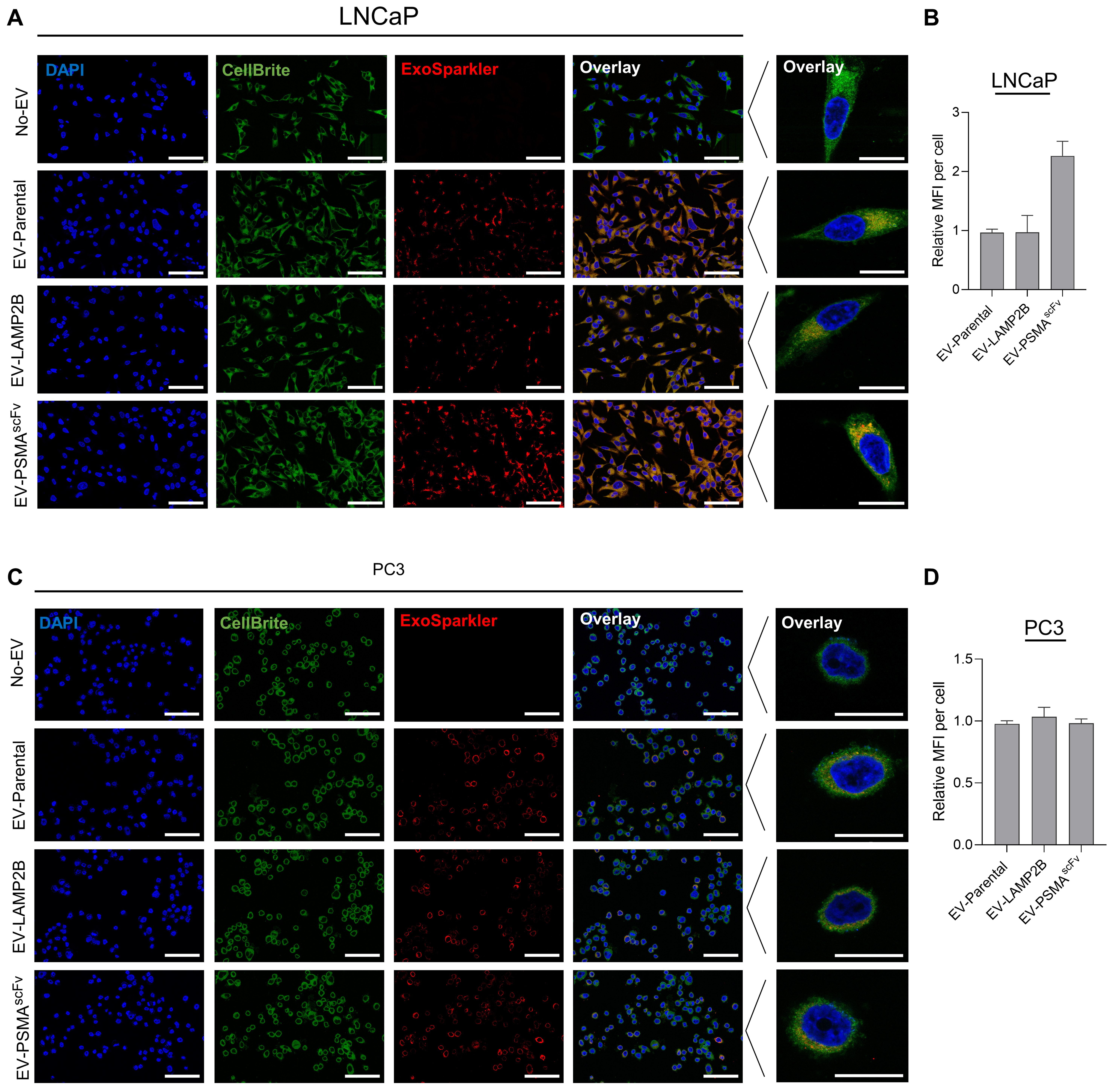
Figure 4. Intracellular uptake of EV-PSMAscFv in prostate cancer cells. (A and C) Confocal microscopy analysis of EV uptake in PSMA-positive LNCaP (A) and PSMA-negative PC3 (C) cells following incubation with ExoSparkler-labeled Parental, LAMP2B, or PSMAscFv EVs. Nuclei were stained with DAPI (blue), cell membranes with CellBriteTM Green (green), and EVs are shown in red. Representative merged images and high-magnification single-cell images are shown. Scale bars: 100 μm (merged images) and 20 μm (high-magnification images); (B and D) Quantitative analysis of EV-associated fluorescence intensity per cell in LNCaP (B) and PC3 (D) cells following exposure to labeled EVs. Fluorescence intensity was quantified across three fields of view from a single experimental sample using one EV preparation. Data are presented as mean ± SD of the three fields of view. Representative confocal microscopy images are shown. EVs: Extracellular vesicles; PSMA: prostate-specific membrane antigen; scFv: single-chain variable fragment; LAMP2B: lysosome-associated membrane protein 2B; DAPI: 4’,6-diamidino-2-phenylindole; SD: standard deviation; MFI: mean fluorescence intensity; EV-PSMAscFv: extracellular vesicles displaying PSMA-targeting single-chain variable fragments.