

fig1
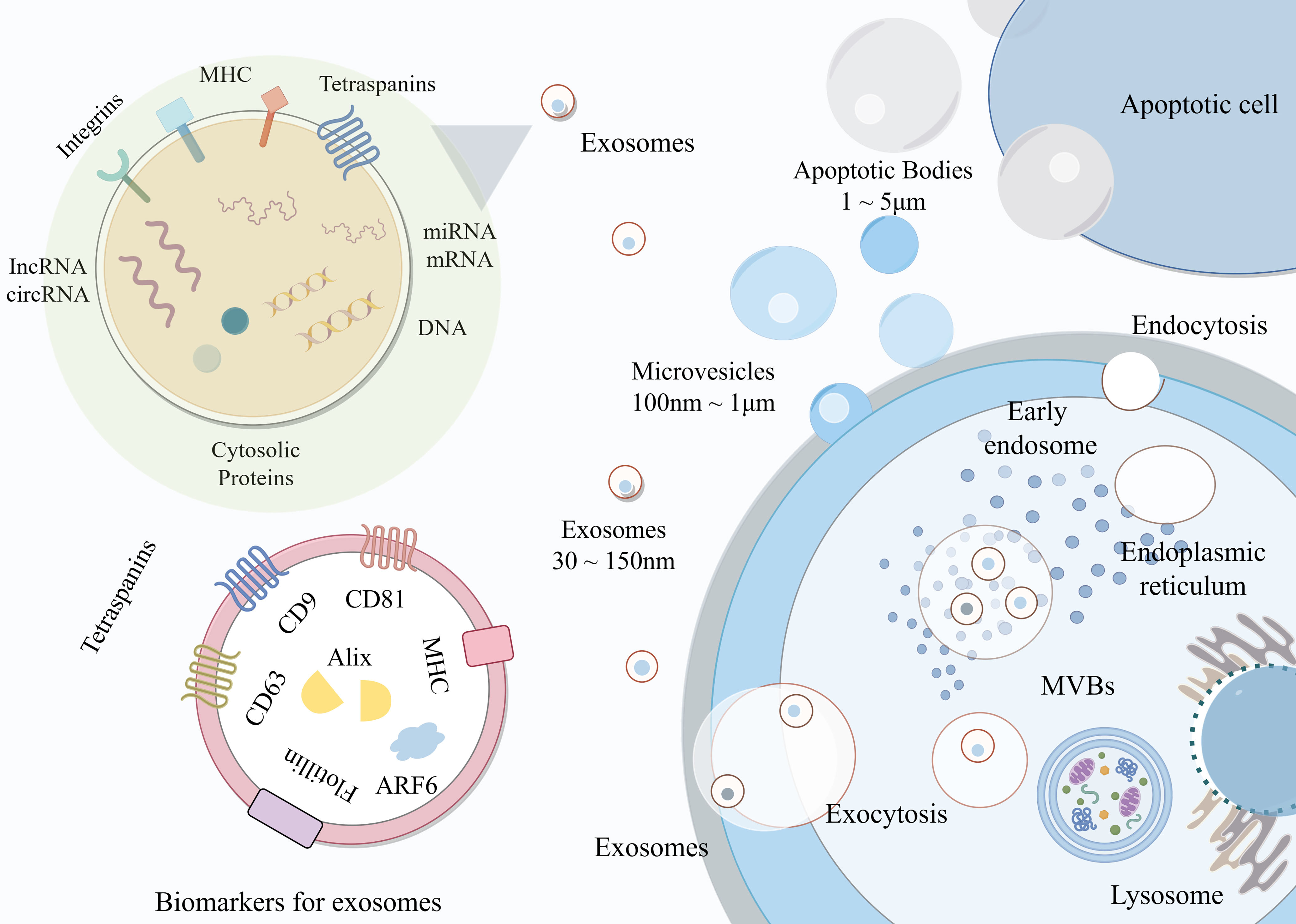
Figure 1. Schematic illustration of EV biogenesis, composition, and secretion. This schematic illustrates the heterogeneity and biogenesis of EVs, which are classified by size into apoptotic bodies, microvesicles, and exosomes. The left panels detail the structural composition of exosomes, highlighting various cargoes. The right panel maps the cellular biogenesis pathway, where endocytosis leads to the formation of early endosomes and subsequent MVBs. MVBs either fuse with the plasma membrane to release exosomes via exocytosis or are directed toward lysosomes for degradation. This figure was drawn with Figdraw and is an original work of the authors. Figdraw ID: WSWOUbbd28. EV: Extracellular vesicle; circRNA: circular RNA; lncRNA: long non-coding RNA; MHC: major histocompatibility complex; miRNA: microRNA; mRNA: messenger RNA; MVB: multivesicular body.