


fig4
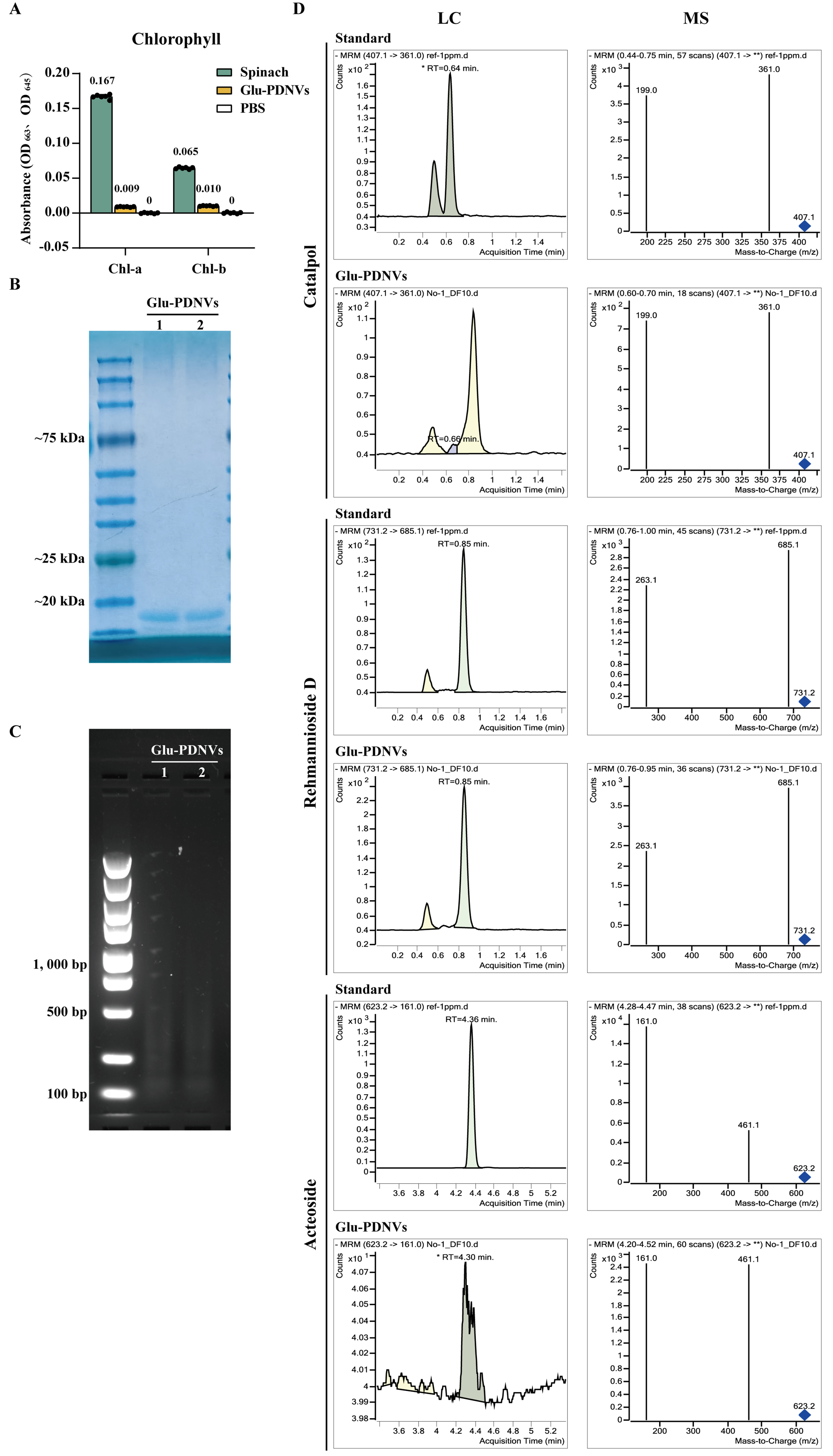
Figure 4. Compositional Analysis of Glu-PDNVs. (A) Chlorophyll content analysis of Glu-PDNV preparations. Absorbance was measured at 663 and 645 nm, the characteristic wavelengths for chlorophyll a and b, respectively. n = 3. Data are presented as mean ± SD; (B) Protein composition of Glu-PDNVs was evaluated by Coomassie Blue staining; (C) Nucleic acid composition was analyzed by agarose gel electrophoresis; (D) Representative LC-MS chromatograms for catalpol, rehmannioside D, and acteoside in Glu-PDNVs. PDNVs: Plant-derived nanovesicles; Glu-PDNVs: Rehmannia glutinosa-derived nanovesicles; LC-MS: liquid chromatography-mass spectrometry; MRM: multiple reaction monitoring; RT: retention time; m/z: mass-to-charge ratio; OD: optical density; Chl-a: chlorophyll a; Chl-b: chlorophyll b; PBS: phosphate-buffered saline; SD: standard deviation; kDa: kilodalton; bp: base pair.


