
fig5
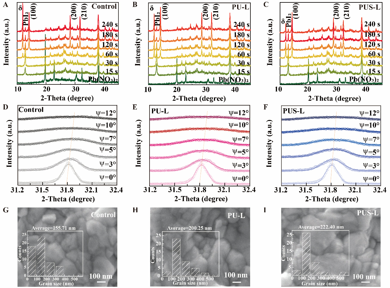
Figure 5. Time-dependent XRD patterns of (A) control, (B) PU-L, (C) PUS-L precursor films incubated in OAS solution for different durations. Tilt-angle XRD patterns of the perovskite (210) reflection for (D) control, (E) PU-L, and (F) PUS-L films at different ψ angles. Surface SEM and grain size statistics of (G) control, (H) PU-L and (I) PUS-L. XRD: X-ray diffraction; OAS: organic ammonium salts; SEM: scanning electron microscopy; PU-L: sample prepared using a Pb(NO3)2 precursor containing 0.3 mg/mL urea; PUS-L: the corresponding sample after additional heat treatment at 150 °C for 20 min.