

Key ion channels in mechanical stress-induced cardiac remodeling: from mechanotransduction to therapeutic targets
 0
0  ,
, Graphical Abstract
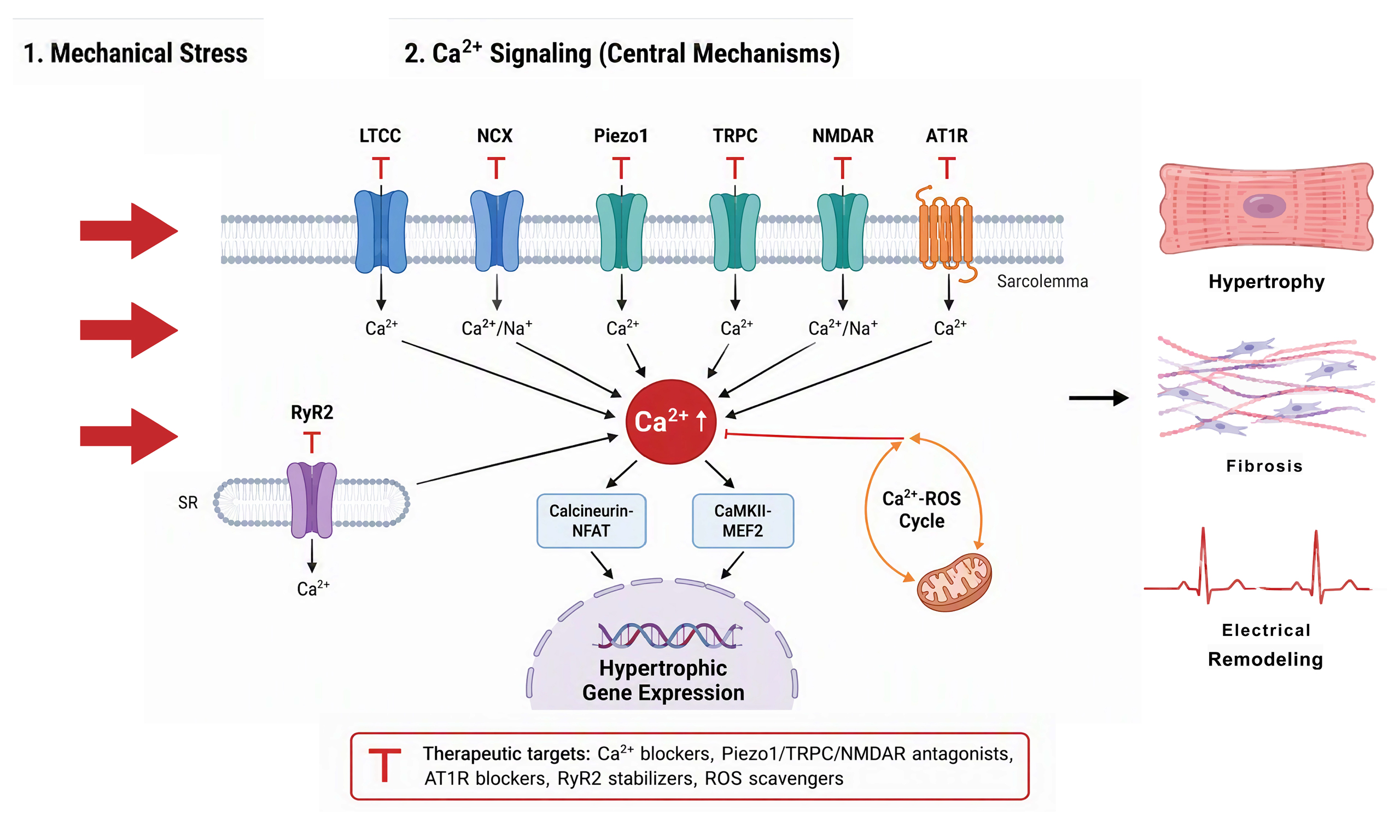
Abstract
Mechanical stress—including pressure overload and volume overload—is a core pathophysiological stimulus driving cardiac remodeling, and Ca2+ signaling is the central common pathway mediating this process. Traditional research has primarily focused on L-type Ca2+ channels (LTCC) and the renin-angiotensin-aldosterone system, whereas the synergistic contributions of mechanosensitive channels (transient receptor potential (TRP), Piezo), canonical excitation-contraction coupling channels [voltage-gated Na+ channels, LTCC, the sarcoplasmic reticulum Ca2+ release channel type 2 ryanodine receptor (RyR2), and the Na+/Ca2+ exchanger (NCX)], non-canonical Ca2+ channels (NMDAR), and the mechanosensitive angiotensin II type 1 receptor are increasingly being recognized. This review systematically integrates the mechanisms by which these distinct ion channel families transduce mechanical stimuli into intracellular Ca2+ signals and cooperatively regulate cardiac remodeling, with particular emphasis on their cell-type-specific distribution and function in cardiomyocytes, endothelial cells, vascular smooth muscle cells, cardiac fibroblasts, and perivascular sensory neurons and macrophages. We highlight the Ca2+-Na+-reactive oxygen species (ROS) signaling triad, which forms a self-reinforcing vicious cycle that drives cardiomyocyte hypertrophy, fibrosis, and electrical remodeling, and we elucidate how the relevant channels are differentially regulated under pressure overload versus volume overload. Key findings include the direct mechanosensor function of Piezo1, the multimodal sensing capacity of TRP channels, the non-canonical cardiac expression of NMDAR and its proarrhythmic effects, the paradoxical hypertrophic response triggered by reduced LTCC activity, and the pivotal role of RyR2 as a disease-modifying node. From a therapeutic perspective, we review classical calcium channel blockers and emerging strategies—including mechanosensitive channel modulators, NMDAR antagonists, RyR2 stabilizers, NCX modulation, and ROS-targeted interventions—stratified by disease context and level of evidence. This integrated mechano-calcium signaling framework provides a new theoretical basis and potential therapeutic targets for cardiac remodeling-related diseases.
Keywords


 Download PDF
INTRODUCTION
Mechanical stress (including pressure overload and volume overload) is a core pathophysiological stimulus driving the initiation and progression of cardiac remodeling[1-3], and Ca2+ signaling is the central common pathway mediating the transduction of mechanical stress into cardiac remodeling, playing an indispensable regulatory role in processes such as cardiomyocyte hypertrophy, fibrosis, and electrical remodeling. For a long time, traditional research in this field has primarily focused on excitation-contraction coupling (ECC) mediated by L-type Ca2+ channels (LTCC)[4,5] and the indirect regulation of calcium signaling by the renin-angiotensin-aldosterone system (RAAS), establishing the classical regulatory theory of cardiac remodeling centered on LTCC and RAAS[6]. However, the synergistic effects of different types of ion channels in cardiac remodeling have been severely underestimated[7-9]. Mechanosensitive channels (transient receptor potential (TRP), Piezo), voltage/excitation-related channels (LTCC, Na+ channels), and non-canonical Ca2+ channels (NMDAR) do not act independently; rather, they form complex signaling networks under mechanical stress to jointly regulate Ca2+ homeostasis and the progression of cardiac remodeling. The traditional single-pathway research perspective cannot fully elucidate the underlying pathological mechanisms. On this basis, this review aims to systematically integrate the mechanistic roles of different types of ion channels under mechanical stress, clarify the synergistic regulatory network of these channels in cardiac remodeling, and provide a new theoretical basis and potential therapeutic targets for the treatment of cardiac remodeling-related diseases (such as hypertensive heart disease and heart failure).
MECHANICAL STRESS AND ION CHANNELS: FROM MECHANICAL FORCE TO ELECTROCHEMICAL SIGNALS
Mechanical stress and mechanical stress-mediated cardiac remodeling
Mechanical stress is one of the most fundamental physiological and pathological stimuli for cardiomyocytes and a key driver of cardiac remodeling initiation and progression. According to its mode of action on the ventricular wall, mechanical stress can be divided into pressure overload and volume overload, which differ significantly in their pathophysiological characteristics and ion channel activation patterns. The two activate ion channels through distinct modes of mechanosensing, converting mechanical signals into electrochemical signals and ultimately regulating the process of cardiac remodeling[10,11].
Pressure overload refers to sustained mechanical stimulation caused by elevated ventricular afterload, which is commonly seen in conditions such as hypertension, aortic valve stenosis, and pulmonary hypertension. Its core mechanical feature is increased ventricular wall stress; cardiomyocytes are mainly subjected to the combined effects of radial compression and circumferential stretch, inducing concentric cardiac hypertrophy, myocardial fibrosis, and electrical remodeling[10,11]. Under pressure overload, mechanosensitive ion channels (such as Piezo1 and TRPC1/6), acting as the primary mechanosensors, can directly sense changes in membrane tension and become rapidly activated, mediating Ca2+ influx[1,2]. Meanwhile, voltage-gated ion channels (such as LTCC and Na+ channels) amplify calcium signals through ECC and cooperate with mechanosensitive channels to drive the activation of hypertrophic signaling pathways[12,13]. Traditional research has focused mainly on LTCC and the RAAS, whereas the critical role of mechanosensitive channels—including stretch-activated ion channels and the mechanosensitive angiotensin II type 1 receptor (AGTR1)—in the sensing of pressure overload has only gradually been recognized in recent years[1,14].
Volume overload refers to sustained stretch stimulation caused by elevated ventricular preload, which is commonly seen in conditions such as aortic regurgitation, mitral regurgitation, and arteriovenous fistula. Its core mechanical feature is excessive ventricular wall stretch; cardiomyocytes are mainly subjected to axial stretch, inducing eccentric cardiac hypertrophy, ventricular dilation, and wall thinning[10,11]. Under volume overload, mechanosensitive channels such as TRPV4 are considered to play important roles; among them, TRPV4 can directly sense stretch stress and mediate non-canonical Ca2+ influx[15]. Some scholars have also proposed that NMDAR may participate in volume overload-related Ca2+ signaling regulation, but there is currently no experimental evidence that volume overload directly activates NMDAR; traditional LTCC play a relatively minor role in this context[9,16].
Fundamental modes of mechanotransduction
Mechanotransduction is the core process by which cells sense and respond to extracellular mechanical stimuli. Its fundamental mode relies on key nodes such as changes in cell membrane tension, cytoskeletal rearrangement, and activation of mechanosensitive channels, efficiently converting physical signals into intracellular biochemical signals and participating in the regulation of diverse biological functions, including cell proliferation, differentiation, migration, and the maintenance of tissue homeostasis[16,17].
The cell membrane is the first interface for sensing mechanical stimuli, and changes in lipid bilayer tension constitute the initial step of mechanotransduction. External forces such as blood flow-induced shear stress and extracellular matrix traction can directly alter cell membrane tension and curvature. Such physical changes can either directly regulate membrane protein conformation through the lipid tension model or trigger downstream signaling cascades by altering membrane fluidity, thereby laying the foundation for subsequent signal transduction[18]. Studies have shown that cell membrane tension can serve as a physical bridge connecting extracellular mechanical stimuli to intracellular signaling pathways, occupying a central position in mechanical signal sensing[19].
The cytoskeleton is mainly composed of actin filaments, microtubules, and intermediate filaments, and is connected to the cell membrane through structures such as focal adhesions and integrins. The cell membrane rapidly transmits mechanical signals to the cytoskeleton via membrane-localized focal adhesions and integrins, triggering rapid rearrangement of cytoskeletal components. Tension signals are transmitted through the actin-myosin system, activating the Rho/ROCK pathway and promoting stress fiber formation. Meanwhile, focal adhesion complexes (such as FAK and talin) bind integrins outward to engage the extracellular matrix and connect to the cytoskeleton inward. Through their own deformation, they transmit mechanical stretch signals from the extracellular space to the cytoskeleton and further inward to the nucleus, achieving signal transmission across compartments[20]. Previous studies have systematically elucidated the three-tiered regulatory mechanism of the cytoskeleton in mechanotransduction, encompassing focal adhesion assembly, stress fiber formation, and nucleoskeletal signal transduction, and have confirmed that the cytoskeleton can participate in the activation of mechanosensitive channels through the tether model, thereby solidifying the structural basis of mechanical signal transmission[21]. For example, Viola et al.[22] reported that LTCC are coupled to mitochondria via the cytoskeleton (F-actin and β-tubulin)[22].
Mechanosensitive G protein-coupled receptor: the AGTR1
In addition to ion channels, G protein-coupled receptors (GPCRs) can also serve as mechanosensors, particularly the AGTR1 within the RAAS[14,23]. Using cardiomyocytes from angiotensinogen-knockout mice, Zou et al.[14] demonstrated that even in the complete absence of angiotensin II, mechanical stretch can still activate AGTR1 and initiate downstream ERK and hypertrophic signaling, establishing AGTR1 as a bona fide mechanosensor[14]. Subsequent biophysical analyses revealed that membrane stretch can stabilize an active conformation of AGTR1, causing it to preferentially couple to β-arrestin-biased signaling, indicating that mechanical force is an allosteric modulator of AGTR1 signaling bias[23].
These findings have two important implications. First, they provide a molecular basis for the observation that "mechanical overload can locally activate RAAS-dependent hypertrophic programs without requiring systemic angiotensin II generation." Second, they offer a mechanistic explanation for the efficacy of angiotensin receptor blockers (ARBs) with inverse agonist properties under pressure overload conditions: by stabilizing the inactive conformation of AGTR1, these drugs can simultaneously block both ligand-dependent and mechanically evoked receptor activation[14,23]. Mechanosensitive AGTR1 signaling converges with multiple pathways activated by mechanosensitive ion channels (including Ca2+ mobilization, reactive oxygen species (ROS) generation, and calcineurin/nuclear factor of activated T cells (NFAT) activation), highlighting the close interplay between receptor- and channel-mediated mechanotransduction in cardiac remodeling[24,25].
Ion channels and cardiac remodeling: the Ca2+-Na+-ROS signaling triad
The transmembrane influx of Ca2+ and Na+ mediated by the activation of mechanosensitive ion channels, together with the generation of ROS, constitutes the core signal-integration network downstream of mechanotransduction. Through the interplay between ion homeostasis regulation and redox signaling, these signals convert mechanical signals into diverse cellular effects, playing key roles in mechanosensing and disease regulation in the cardiovascular system[24,25].
Mechanical stimulation triggers the opening of mechanosensitive channels (such as Piezo1 and TRPV4), first mediating Ca2+ influx, which is the most central second-messenger event downstream of mechanotransduction. Elevated intracellular Ca2+ concentration can directly activate calmodulin (CaM), calmodulin-dependent protein kinase (CaMK), and the calcineurin/NFAT pathway, triggering the expression of hypertrophy-related genes; meanwhile, Piezo1-mediated Ca2+ influx in vascular endothelial cells can further activate eNOS signaling to regulate vascular tone, whereas mechanical stretch of cardiomyocytes participates in the pathological process of cardiac hypertrophy through Ca2+ signaling mediated by TRPC channels. In addition, Ca2+ influx can also activate NADPH oxidases (NOX), providing a critical trigger signal for subsequent ROS generation.
Na+ signaling cooperates with Ca2+ signaling to indirectly regulate the activity of voltage-gated Ca2+ channels (VGCCs) by altering the cell membrane potential, forming a Na+-Ca2+ signal amplification loop: Na+ influx mediated by mechanosensitive sodium channels (such as members of the ENaC family and the nonselective cation currents through Piezo channels) causes cell membrane depolarization, promoting VGCC opening to further increase Ca2+ influx; meanwhile, elevated intracellular Na+ concentration can activate the NCX in its reverse mode to transport Ca2+ into the cell, amplifying the calcium signal. This synergistic regulation is highly significant for cardiomyocyte mechanical responses and arrhythmogenesis[26,27].
ROS serve as an integration hub connecting ionic signals with redox signals in mechanotransduction and exert bidirectional regulatory effects: on the one hand, Ca2+ influx promotes ROS generation by activating NOX enzymes; on the other hand, ROS can enhance the sensitivity of mechanosensitive channels (such as Piezo1 and TRPV4) to mechanical stimuli through redox modification of their cysteine residues, forming a positive feedback loop. This reciprocal regulation plays a key role in vascular endothelial dysfunction and myocardial ischemia-reperfusion injury, in which ROS can amplify mechanical signals and thereby aggravate the pathological process[24,25].
Ca2+, Na+, and ROS signals do not exist independently but achieve functional integration through multiple feedback loops: Na+ influx amplifies Ca2+ signals through membrane potential regulation, Ca2+ further induces ROS generation, and ROS, in turn, regulates ion channel activity through redox modification. Together, these three components constitute the signaling network downstream of mechanotransduction, determining the direction of cellular responses to mechanical stimuli and participating in the development and progression of cardiovascular diseases.
At the molecular level, the feedback of ROS on ion channel activity is mediated by well-defined oxidative modifications. ROS oxidize critical methionine residues (Met281/282) of calmodulin-dependent protein kinase II (CaMKII), locking the kinase in a constitutively active state that persists even after the initial Ca2+ signal has subsided[28]. Oxidative modification of type 2 ryanodine receptor (RyR2) increases sarcoplasmic reticulum (SR) Ca2+ leak, amplifies cytosolic and mitochondrial Ca2+ oscillations, and further stimulates ROS generation in chronic heart failure[29]. Reactive nitrogen species can S-nitrosylate TRP channels and enhance their activity, providing direct redox gating of mechanosensitive Ca2+ influx[30]. In cardiomyocytes, TRPC3 forms a signaling complex with NADPH oxidase 2 (Nox2); stretch-induced TRPC3-mediated Ca2+ influx stabilizes Nox2 and increases ROS generation, while ROS, in turn, sustains TRPC3 activity—an amplification loop that drives adverse remodeling[31]. Finally, elevated cytosolic Na+ in failing cardiomyocytes reduces mitochondrial Ca2+ uptake, impairs NADH regeneration and antioxidant capacity, and thereby increases mitochondrial ROS formation, directly linking Na+ channel remodeling to oxidative stress[27].
These channel-ROS feedback mechanisms contribute differentially to the three major features of adverse remodeling. First, hypertrophic growth is primarily driven by sustained activation of Ca2+-dependent calcineurin/NFAT and CaMKII/histone deacetylase (HDAC) signaling downstream of mechanosensitive Ca2+ influx[6,32]. Second, myocardial fibrosis arises from mechanical stress activation of mechanosensitive channels in cardiac fibroblasts (such as TRPM7, TRPV4, and Piezo1), which couple Ca2+ influx to profibrotic transcription, myofibroblast differentiation, and extracellular matrix deposition[33-35]. Third, electrical remodeling stems from redox- and Ca2+-dependent modifications of ion channels—including increased late sodium current, oxidized RyR2-mediated SR Ca2+ leak, and NMDAR-mediated Nav1.5 downregulation—which together constitute the substrate for arrhythmogenesis[26,29,36]. This mechanistic stratification provides a framework for understanding how targeting different channels can modify specific aspects of the remodeling phenotype [Figure 1].
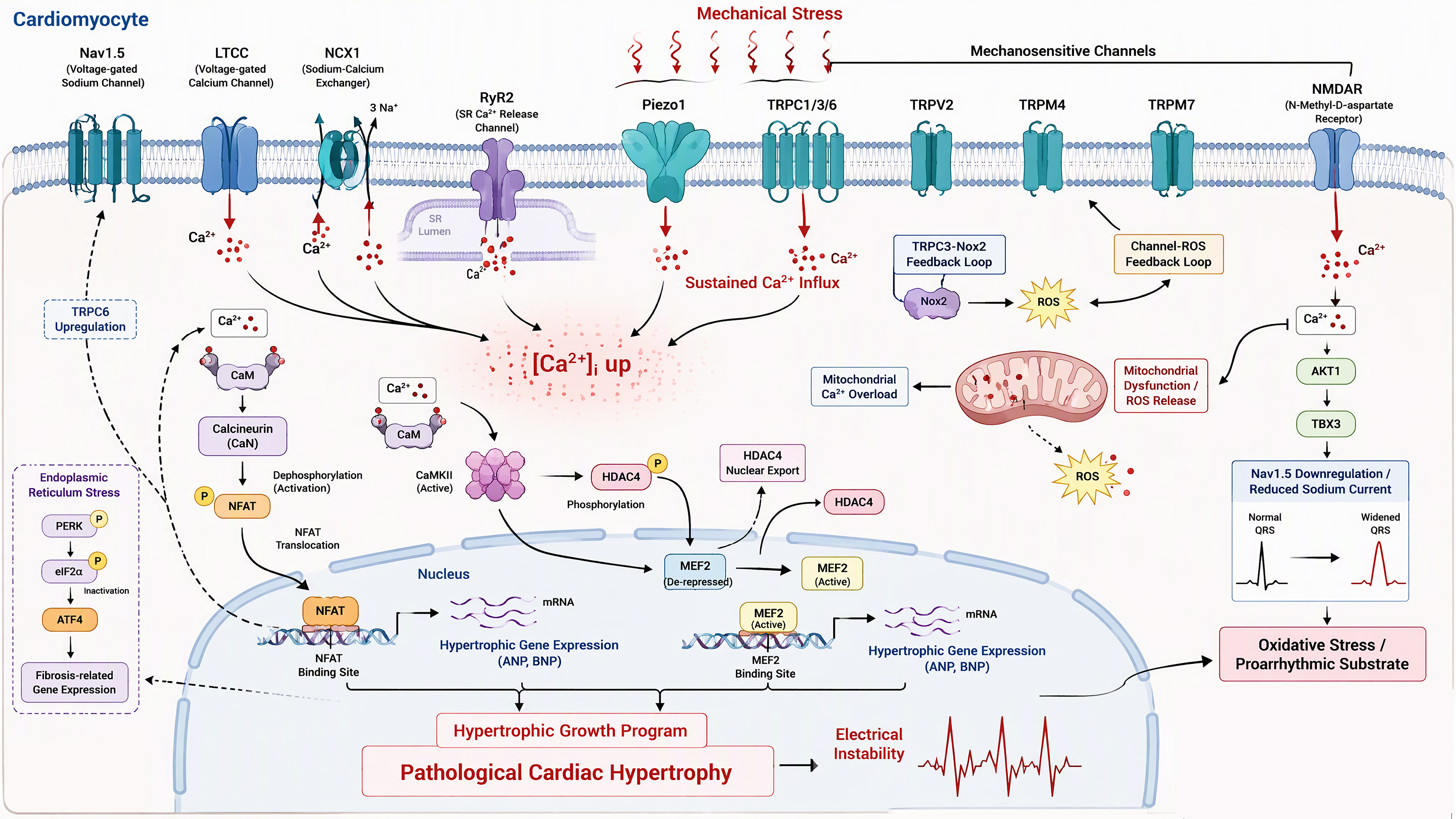
Figure 1. Cell-type-specific ion channel and receptor mechanisms of mechanical stress-induced cardiac remodeling. Mechanical stress activates mechanosensors (Piezo1, TRP channels, NMDAR, AGTR1) and dysregulates cardiomyocyte excitation-contraction coupling channels (LTCC, Nav1.5, RyR2, NCX1), driving Ca2+ signaling and ROS feedback that culminate in hypertrophy, fibrosis, and electrical remodeling. The figure was created with Matplotlib (Python) and Adobe Illustrator.
CONVENTIONAL SIGNALING PATHWAYS
The ion channels discussed in this section—such as voltage-gated Na+ channels, LTCC, RyR2, and the NCX—are mainly expressed in cardiomyocytes and participate in ECC. ECC is the central process that converts the action potential into mechanical contraction, and Ca2+-induced Ca2+ release (CICR) is the fundamental mode of ECC. The transient elevation of cytosolic Ca2+ is the direct driving force of contraction, but extracellular Ca2+ influx alone is insufficient to trigger effective contraction. Signal cascade amplification must rely on Na+ channels, LTCC, and RyR2[4,5].
Canonical pathways
Early studies showed that cardiac hypertrophy is regulated by a series of canonical and complex intracellular signaling pathways that mediate physiological or pathological cardiac growth, depending on the nature of the stimulus. Among these, GPCRs are the core initiating elements that sense neurohumoral factors (such as angiotensin II, endothelin-1, and catecholamines) and induce pathological cardiac hypertrophy, activating multiple downstream pathways. Members of the mitogen-activated protein kinase (MAPK) family exert distinct roles: the MEK1-ERK1/2 pathway can induce compensatory hypertrophy, whereas p38 and JNK generally negatively regulate the hypertrophic response, and their activation is more closely associated with cardiomyopathy and cardiac dysfunction. The calcineurin-NFAT pathway is the central pathway of pathological hypertrophy: Ca2+-activated calcineurin dephosphorylates NFAT, promoting its nuclear translocation and initiating hypertrophic gene expression. The IGF-I-PI3K-AKT/PKB-mTOR pathway plays a dual role in regulating physiological hypertrophy, growth, and development, in which AKT/PKB is essential for cardiac growth, but its excessive activation is deleterious. In addition, the HDAC pathway is equally critical: class I HDACs (such as HDAC1/2) promote pathological hypertrophy, whereas class II HDACs (such as HDAC5/9) suppress the expression of pathological hypertrophy genes by interacting with transcription factors such as myocyte enhancer factor-2 (MEF2). Complex interactions exist among these pathways, which together precisely regulate cardiac growth and remodeling[6].
Na+ channels: the upstream electrical basis of ECC
The cardiac action potential, mediated by voltage-gated Na+ channels, provides the electrical basis for subsequent LTCC activation; during the action potential plateau, LTCC activate, triggering Ca2+ influx. Meanwhile, Na+ channels can influence the activity of the NCX by regulating intracellular Na+ concentration, thereby modulating cytosolic Ca2+ homeostasis and synergistically enhancing the Ca2+ influx efficiency of LTCC, improving overall ECC performance[4,37]. Under mechanical stress or pathological conditions, Nav channels undergo remodeling, manifested as an increased late (persistent) Na+ current (INa,L). This abnormal Na+ influx elevates intracellular Na+ concentration, impairs mitochondrial Ca2+ uptake, and increases mitochondrial ROS generation, while simultaneously promoting reverse-mode NCX activity, leading to Ca2+ overload, prolonged action potential duration, early afterdepolarizations, and increased susceptibility to arrhythmias, thereby exacerbating cardiac hypertrophy and heart failure[26,27]. Notably,
LTCC: the specific Ca2+ trigger signal of ECC
Cardiac ECC is initiated when Ca2+ flows inward across the plasma membrane through voltage-gated LTCC. L-type voltage-gated Ca2+ channels (LTCCs) are a major subfamily among the ten types of VGCCs and represent the main route of extracellular Ca2+ influx in ventricular myocytes. They are voltage-activated during the action potential plateau, generating an inward Ca2+ current that mediates the influx of a small amount of Ca2+ down its electrochemical gradient, serving as the dedicated trigger signal for ECC[38-40]. LTCC is a multi-subunit complex composed of the pore-forming α1 (Cav1.2) subunit together with auxiliary α2δ, β, and γ subunits: the α1 subunit contains the voltage sensor and the ion-conducting pore and provides the binding sites for most calcium channel blockers (CCBs), whereas the auxiliary subunits regulate gating properties, trafficking, and pharmacological responses, thereby influencing ECC efficiency[38,39].
Dysregulated Ca2+ handling is an important cause of cardiac hypertrophy, and LTCC is the main route of Ca2+ influx in cardiomyocytes, capable of activating downstream protein synthesis signaling pathways and thereby inducing hypertrophy. The study by Gao et al.[41] demonstrated that Ca2+ influx through LTCC and TRP channels can activate pathological hypertrophic signaling and downstream protein synthesis in cardiomyocytes[41]. Another study found that under α₁-adrenergic stimulation, LTCC and CaMKII can form a positive feedback activation loop within the caveolae microdomain, promoting hypertrophy-related protein synthesis[42]. In addition, calcineurin physically and functionally interacts with cardiac LTCC; this interaction is associated with signal transduction and can upregulate protein synthesis[43]. Further studies have shown that mechanical stretch can increase LTCC stability through a polycystin-1/AKT-dependent mechanism, thereby enhancing Ca2+ influx and promoting hypertrophic protein synthesis in cardiomyocytes[44]. Meanwhile, LTCC opening is also regulated by depolarization stimuli and by various molecules, including Ca2+, CaM, and interactions with multiple auxiliary proteins, which in turn affect protein synthesis[45]. Studies have found that the transcription factor NFATc4 cooperates with myocardin to upregulate the expression of the LTCC α1C subunit, promoting Ca2+ influx and enhancing cardiac hypertrophy-related protein synthesis[46]. It has been confirmed that reduced cardiac L-type Ca2+ channel activity can trigger neuroendocrine stress, increase SR RyR2 Ca2+ leak, lead to an abnormally elevated Ca2+ signaling gain, and activate the calcineurin/NFAT pathway, inducing spontaneous cardiac hypertrophy and accelerating heart failure[12].
However, the relationship between LTCC activity and the development of cardiac hypertrophy is not a simple direct proportion. The study by the Goonasekera team[12] demonstrated that reduced LTCC activity can induce neuroendocrine stress, accompanied by compensatory enhancement of SR Ca2+ release to maintain myocardial contractility; this state leads to activation of calcineurin/NFAT signaling, paradoxically promoting cardiac hypertrophy and heart failure in mice[12]. In addition, loss of the RAD GTPase results in enhanced LTCC function, manifesting as an adaptive hypertrophic phenotype that promotes beneficial Ca2+ dynamics[45,47]. By precisely regulating Ca2+ influx, LTCC directly or indirectly initiates the protein synthesis program in cardiac hypertrophy. Moreover, in hypertrophic cardiomyopathy (HCM), abnormal LTCC activity not only alters contractility but also affects mitochondrial metabolism: increased Ca2+ influx through LTCC leads to mitochondrial Ca2+ overload, increased ROS generation, and a hypermetabolic state, thereby aggravating the HCM phenotype, whereas inhibiting LTCC or employing peptide therapies targeting the channel-mitochondria axis can ameliorate this phenotype[48,49].
RyR2 and SR Ca2+ release: Ca2+ signal amplification and precise termination in CICR
The RyR2 is a Ca2+ release channel located on the SR membrane of cardiomyocytes. The small amount of Ca2+ entering the cytosol precisely activates RyR2 on the SR, achieving exponential amplification of the Ca2+ signal. This is the core mechanism of cardiac ECC, the key to its generation of powerful contractile force, and an important determinant of cardiac function[50,51].
The small amount of Ca2+ entering the cell can activate the synchronous opening of RyR2 clusters on the SR, triggering massive release of Ca2+ stored in the SR, markedly elevating Ca2+ and driving myocardial contraction. This local Ca2+ release event is defined as a Ca2+ spark and constitutes the fundamental functional unit of CICR[52,53]; contraction is generated by the summation and integration of numerous Ca2+ sparks. In addition, a recently discovered circular RNA derived from the RyR2 locus (circ-RyR2) is downregulated in hypertrophy and heart failure, and restoring its expression normalizes Ca2+ handling without altering RyR2 protein levels, revealing a new layer of post-transcriptional regulation[54].
RyR2 activity is regulated by changes in the levels of various intracellular factors, such as divalent cations (Ca2+ and Mg2+), nucleotides, associated proteins, and ROS[29,50]. The conventional view holds that CICR is the canonical initiation mechanism of cardiac ECC, in which LTCC-mediated Ca2+ influx serves as the trigger signal to activate RyR2; whereas recent studies propose that binding of Ca2+ to the Cav1.2 pore induces a conformational change that may activate RyR2 through physical coupling, which could be a complementary mechanism cooperating with CICR[55]. Studies have demonstrated that insufficient RyR2 expression attenuates SR Ca2+ release, thereby suppressing key hypertrophic pathways such as calcineurin, ERK, and Akt, and significantly alleviating pressure overload-induced cardiac hypertrophy and fibrosis[56]. Conversely, RyR2-R176Q knock-in mouse models exhibit accelerated development of pressure overload-induced cardiac hypertrophy and dysfunction, suggesting that RyR2-mediated Ca2+ leak participates in the remodeling process[57].
Differential regulation of ECC channels under pressure overload and volume overload
Although pressure overload and volume overload ultimately both lead to hypertrophic remodeling, the biomechanical and molecular programs they engage are not identical. Pressure overload increases systolic wall stress and primarily drives concentric hypertrophy, whereas volume overload increases diastolic wall stress and drives eccentric hypertrophy accompanied by chamber dilation; direct hemodynamic comparisons in patients with valvular heart disease have confirmed that these two loading states produce markedly distinct wall stress profiles[10,11]. At the level of ECC channels, the characteristics of pressure overload have been more thoroughly elucidated: it promotes RyR2-mediated SR Ca2+ leak, a caveolae-localized LTCC/CaMKII activation loop, and increased INa,L, which together favor cytosolic Ca2+ accumulation and activation of calcineurin/NFAT and CaMKII signaling[26,42,56,57]. By contrast, chronic volume overload and the associated diastolic stretch preferentially engage mechanosensitive pathways, including TRPV4-mediated Ca2+ influx (which in the aging heart participates in stretch-induced hypercontractility and time-dependent dysfunction) and eccentric growth signaling. However, the specific regulation of ECC channels under pure volume overload remains poorly defined and awaits direct comparative studies[15,16]. These differential regulatory patterns (summarized in Table 1) suggest that channel-targeted therapies may need to be individualized according to the predominant loading state.
Summary of ion channels related to cardiomyocyte excitation-contraction coupling (ECC) under mechanical overload
| Channel | Expression/activity changes | Regulatory factors | Hypertrophy/remodeling signaling cascades | Comparison between pressure overload and volume overload | References |
| Nav1.5 | Increased late sodium current (INa,L); Nav1.5 downregulated in ischemia | Oxidative stress; CaMKII; NMDAR-AKT1-TBX3 axis | [Na+]i↑ → impaired mitochondrial Ca2+ uptake, ROS↑; reverse-mode NCX → Ca2+ overload; prolonged APD, EAD, arrhythmias | Well documented in pressure overload and ischemia; insufficiently studied in volume overload | [26,27,36] |
| LTCC (Cav1.2) | Context-dependent: increased membrane stability under stretch; enhanced caveolae-localized activity under adrenergic stress; reduced activity can cause heart failure | Polycystin-1/AKT (mechanical); caveolae CaMKII loop; Rad GTPase; NFATc4/myocardin transcription | Ca2+ influx → CaMKII and calcineurin/NFAT activation; mitochondrial Ca2+ overload and ROS; HCM hypermetabolic state | Stretch and pressure overload regulation established; volume overload regulation unclear | [12,41-44,48] |
| RyR2 | Hyperphosphorylation and oxidation → SR Ca2+ leak; channel cluster remodeling in heart failure | PKA/CaMKII phosphorylation; redox modification; FKBP12/12.6; circ-RyR2 | Diastolic Ca2+↑, reduced contractility; DAD/arrhythmias; CaMKII activation → hypertrophic transcription | Required for pressure overload hypertrophy (animal models); role in volume overload undetermined | [29,54,56,57] |
| NCX1 | Expression and activity upregulated in hypertrophy and heart failure; [Na+]i↑ favors reverse mode | Intracellular Na+/Ca2+; membrane potential; PIP2 | Ca2+ extrusion (forward) and Ca2+ influx (reverse); involved in Ca2+ overload, contractile dysfunction, and arrhythmogenesis | Upregulation documented in pressure overload hypertrophy and human heart failure; limited comparative data for volume overload | [37,58,59] |
NON-CANONICAL ION CHANNELS
Unlike the conventional ECC channels, the channels reviewed in this section—including members of the TRP family, Piezo1, and NMDAR—are expressed in multiple cardiac cell types and act either as direct mechanosensors (TRP channels, Piezo1) or as stress-coupled signaling platforms (NMDAR), linking mechanical stress to hypertrophic, fibrotic, and electrical remodeling. Their cell type-specific distribution is summarized in Table 2, while their activation characteristics, downstream pathways, and pharmacological modulators are summarized in Table 3.
Cell type-specific distribution and functions of ion channels in mechanical stress-mediated cardiac remodeling
| Cell type | Major channels | Mechanical stimuli sensed | Downstream pathways | Remodeling outcomes | References |
| Cardiomyocytes | Nav1.5; LTCC; RyR2; NCX1; TRPC1/3/6; TRPV2; TRPM4/7; Piezo1; NMDAR | Pressure/volume overload; sarcomere stretch; glutamatergic stimulation (NMDAR) | Calcineurin/NFAT; CaMKII/HDAC; ROS feedback loops; mitochondrial Ca2+ overload | Concentric/eccentric hypertrophy; contractile dysfunction; arrhythmogenic electrical remodeling | [1,26,32,70] |
| Endothelial cells | TRPV4; Piezo1; TRPC1/3/5 | Shear stress; cyclic circumferential stretch | TRPV4 sparklet → IKCa/SKCa → EDH; Piezo1 → calpain, angiogenic alignment | Vasomotor dysregulation; impaired angiogenesis; microvascular rarefaction | [65,79,80,90] |
| Vascular smooth muscle cells | TRPM4; Piezo1; TRPC6 | Intravascular pressure; wall tension | Myogenic depolarization → Cav1.2 Ca2+ influx; transglutaminase/ECM cross-linking | Increased myogenic tone; arterial stiffening; elevated cardiac afterload | [90,92,93] |
| Cardiac fibroblasts | TRPM7; TRPV4; Piezo1 | Matrix stiffness; interstitial strain | TGF-β/Smad; p38 MAPK/IL-6; myofibroblast differentiation | Myocardial fibrosis; stiffening; arrhythmogenic substrate (especially atrial fibrillation) | [33-35,94] |
| Perivascular sensory neurons and macrophages | TRPV1 (neurons); Piezo1 (macrophages) | Cardiac distension; ischemic metabolites; cyclic hydrostatic pressure | CGRP/SP release; cardiac sympathetic afferent reflexes; Piezo1 → NF-κB/inflammasome | Sustained sympathetic excitation; neuroinflammation; progression of fibrosis and hypertrophy | [68,69,95] |
Mechanosensitive and non-canonical ion channels in the cardiovascular system: distribution, activation, downstream signaling, and pharmacological modulators
| Channel | Principal cellular distribution | Activating stimuli | Major downstream signaling pathways | Agonists | Antagonists | References |
| TRPC1 | Cardiomyocytes; endothelial cells | Store depletion; membrane stretch; GqPCR | Ca2+ influx → calcineurin/NFAT; hypertrophic transcription | (No selective agonist) | SKF-96365; 2-APB (non-selective) | [63,96] |
| TRPC3 | Cardiomyocytes; vascular smooth muscle | DAG (OAG); mechanical stretch; GqPCR | Calcineurin/NFAT; Nox2 stabilization → ROS amplification | OAG | Pyr3; SAR7334 | [7,31,64] |
| TRPC6 | Cardiomyocytes; vascular smooth muscle | Membrane stretch; DAG; Ang II/GqPCR | Calcineurin/NFAT positive feedback loop; fibrotic signaling | OAG; flufenamic acid | Larixyl acetate; SAR7334; BI 749327 | [32,64,97,98] |
| TRPV1 | Perivascular sensory neurons; cardiomyocytes | Capsaicin; high temperature (> 42 °C); protons; anandamide | Neuropeptide (CGRP) release; CaMKII; sympathetic afferent activation | Capsaicin; resiniferatoxin | Capsazepine; SB-366791 | [68,69] |
| TRPV2 | Cardiomyocytes | Membrane stretch; insulin/IGF signaling | Ca2+ overload; structural maintenance and degeneration | 2-APB (non-selective) | Tranilast; SKF-96365 | [66] |
| TRPV3 | Cardiomyocytes; endothelial cells | Camphor; carvacrol; high temperature | Calcineurin/NFATc3; hypertrophic signaling | Camphor; carvacrol | (No selective antagonist) | [67] |
| TRPV4 | Endothelial cells; cardiac fibroblasts; cardiomyocytes | Shear stress; osmotic/mechanical stretch; EET | eNOS/EDH-mediated vasodilation; TGF-β-dependent fibroblast differentiation; CaMKII | GSK1016790A; 4α-PDD | HC-067047; GSK2193874 | [15,34,65,99] |
| TRPM4 | Cardiomyocytes; vascular smooth muscle | Elevated [Ca2+]i; membrane stretch | Depolarization → CaMKII and calcineurin activation; myogenic tone | (No selective agonist) | 9-Phenanthrol; flufenamic acid | [70,92] |
| TRPM7 | Cardiomyocytes; cardiac fibroblasts | Mg2+ depletion (Mg2+ inhibition); mechanical stretch | Kinase-dependent signaling; TGF-β/Smad profibrotic signaling; conduction/repolarization | Naltriben | Waixenicin A; NS8593; 2-APB | [33,71,72] |
| Piezo1 | Cardiomyocytes; endothelial cells; fibroblasts; macrophages | Membrane tension; shear stress; cyclic hydrostatic pressure | Calpain/calcineurin/NFAT; CaMKII/HDAC4/MEF2; p38 MAPK/IL-6; innate immunity | Yoda1; Jedi1/2 | GsMTx4; Dooku1 (Yoda1 antagonist); ruthenium red (non-selective) | [1,77,95,100-102] |
| NMDAR | Cardiomyocytes; endothelial cells | Glutamate + glycine/D-serine (co-agonists) | Sustained Ca2+ influx; mitochondrial dysfunction/ROS; NF-κB; AKT1-TBX3-Nav1.5 | NMDA; glutamate | Memantine; MK-801; ketamine; Mg2+ (voltage-dependent block) | [9,36,81,86] |
The TRP channel family
TRP channels constitute an important class of non-selective cation channels, first identified in Drosophila melanogaster in 1969; in 2004, the TRP family was classified into seven subfamilies. Channels widely expressed in human tissues include TRPC, TRPV, TRPM, TRPA, TRPP, and TRPML, and corresponding channels are also present in the cardiovascular system. Among them, members of the TRPC, TRPV, and TRPM subfamilies play key roles in mechanosensing. TRP channels typically exist as homo- or heterotetramers, with each subunit containing six transmembrane domains; the intracellular N- and C-termini harbor multiple regulatory sites that respond to diverse stimuli such as mechanical force, temperature, and chemical agents, hence their designation as "polymodal sensors." These channels exhibit varying degrees of Ca2+ permeability, and mechanically induced channel opening can mediate extracellular Ca2+ influx, elevating intracellular Ca2+ concentration and initiating downstream signaling cascades[7,60].
It is now generally accepted that, under mechanical stretch, TRP channels respond to mechanical stimuli by mediating Ca2+ influx, thereby activating the CaMKII and NFAT pathways. Following NFAT translocation into the nucleus, the expression of hypertrophy-associated genes (such as ANP and BNP) is upregulated, promoting cardiomyocyte hypertrophy. In addition, Ca2+ influx can also activate signaling pathways such as MAPK and PI3K/Akt, which synergistically regulate the progression of cardiac hypertrophy; this constitutes an important component of the calcium-dependent mechanisms driving pathological cardiac hypertrophy[61,62].
The TRPC subfamily
The TRPC subfamily is an important subgroup of TRP channels involved in regulating calcium signaling. Seven TRPC channel isoforms exist in mammals, and their activity is enhanced predominantly under pathological conditions. Among them, TRPC1, TRPC3, and TRPC6 are canonical mechanosensitive members highly expressed in cardiomyocytes. Studies employing patch-clamp electrophysiology combined with mechanical stretch stimulation and TRPC knockout mice have confirmed that TRPC1, TRPC3, and TRPC6 can respond to mechanical stretch, undergo conformational changes, mediate Ca2+ influx, activate CaMKII, and induce cardiomyocyte hypertrophy[41,63]. Mechanistically, TRPC6 is a core component of the calcineurin signaling circuit in pathological cardiac remodeling: calcineurin/NFAT activation upregulates TRPC6 expression, and the resulting Ca2+ influx further activates calcineurin, forming a positive feedback loop that sustains hypertrophic growth; pharmacological or genetic co-blockade of TRPC3 and TRPC6 suppresses pathological cardiac hypertrophy in mice[32,64]. Beyond transcriptional regulation, TRPC3 stabilizes Nox2 and amplifies ROS generation under mechanical stress, forming a TRPC3-Nox2 feed-forward loop that drives adverse remodeling and interstitial fibrosis[31].
The TRPV subfamily
TRPV subfamily members (such as TRPV1, TRPV3, and TRPV4) possess both mechanosensitivity and chemosensitivity, among which TRPV4 is a relatively well-characterized mechanosensitive channel. Microperfusion-based simulation studies have revealed that TRPV4 mediates Ca2+ influx in response to mechanical stimuli such as blood flow shear stress and osmotic pressure changes: in vascular endothelial
The TRPM subfamily
TRPM subfamily members display structural and functional diversity, and TRPM4 and TRPM7 have been shown to be mechanosensitive. TRPM4 is a Ca2+-activated non-selective cation channel. Studies using a pressure overload-induced mouse model of cardiac hypertrophy combined with TRPM4 knockout mice have found that this channel is upregulated in pressure overload-induced cardiac hypertrophy; by modulating membrane potential (mediating Na+ influx that causes depolarization) and Ca2+ homeostasis (affecting L-type calcium channel activity), it activates the CaMKII signaling pathway and promotes the development of cardiac hypertrophy[70]. TRPM7 is a bifunctional protein possessing both ion channel and protein kinase activities; it can mediate Ca2+ influx in response to mechanical load and regulate the downstream Rac1/Cdc42 and ERK pathways through its kinase domain, thereby modulating cardiomyocyte proliferation and differentiation and playing a role in cardiovascular remodeling; indeed, differences in the timing of Trpm7 deletion during cardiogenesis can disrupt ventricular function, conduction, and repolarization in adult mice to varying degrees, indicating that TRPM7 is indispensable for maintaining normal cardiac electrophysiology and structure[71]. Mechanosensitive activation of TRPM subfamily members can regulate ionic homeostasis, providing the ionic foundation required to initiate downstream hypertrophic signaling pathways[72].
In addition, Ca2+ influx mediated by mechanosensitive TRP channels can induce ROS generation. Studies using fluorescent probes show that mitochondrial dysfunction is a major source of ROS generation; mechanically induced calcium signaling and ROS generation exert synergistic effects in pathological cardiovascular remodeling, and TRP channel-mediated Ca2+ influx is a critical link in this process. Cardiac ion channel dysfunction leads to intracellular calcium overload, causing mitochondrial damage and massive ROS release, while high ROS concentrations stimulate the endoplasmic reticulum and induce endoplasmic reticulum stress. After PERK phosphorylation, eIF2α is inactivated, which in turn selectively upregulates ATF4; ATF4 then promotes the expression of fibrosis-related genes and drives fibroblast proliferation, activation, and collagen secretion. Concurrent with activation of the PERK/ATF4 signaling pathway, ROS can also inhibit potassium channel activity and activate TRP channels through oxidative modification, further aggravating intracellular calcium overload and forming a vicious cycle of "ROS generation-calcium overload-ROS generation" that continuously drives fibroblast activation and the progression of myocardial fibrosis[24,25,30].
The Piezo1 channel
Piezo1 was identified as a mechanosensitive cation channel in 2010 and is now regarded as one of the best-characterized ion channels for direct mechanosensitivity. Its core function is to directly sense mechanical stimuli such as mechanical stretch and membrane tension, mediating Ca2+ influx and initiating downstream signal transduction, thereby playing a key role in maintaining cardiovascular physiological homeostasis[8,73].
The unique trimeric propeller-like structure of Piezo1 enables it to directly sense tension changes in the lipid bilayer: when a cell is subjected to mechanical stretch or shear force, alterations in membrane tension act directly on the transmembrane domains of Piezo1, triggering rapid conformational changes and channel opening—a process that does not require the participation of the cytoskeleton or auxiliary proteins and is termed the "force-from-lipids" or direct mechanical activation model. The landmark 2010 study by
As a non-selective cation channel with relatively high Ca2+ permeability, Piezo1 mediates rapid extracellular Ca2+ influx upon mechanically induced opening, elevating intracellular Ca2+ concentration and thereby activating multiple downstream signaling pathways. Upon mechanical stimulation, activated Piezo1 induces Ca2+ influx, disrupting intracellular calcium homeostasis. Intracellular Ca2+ binds to CaM, which in turn activates calcineurin (CaN) and CaMKII. Calcineurin promotes NFAT nuclear translocation and forms a feedback loop with the negative regulator RCAN1; meanwhile, CaMKII phosphorylates HDAC4 and promotes its nuclear export, thereby relieving the repression of the MEF2 transcription factor and ultimately inducing cardiac hypertrophy. In addition, Piezo1 can regulate the expression of the Ca2+-activated TRPM4 protein in cardiomyocytes, thereby further amplifying the CaMKII/HDAC4/MEF2 pathway and promoting cardiac hypertrophy[1,70,76].
Two complementary landmark studies established Piezo1 as the principal cardiac mechanosensor that initiates the hypertrophic response. Using cardiomyocyte-specific Piezo1 knockout and overexpression mouse models, Yu et al.[1] demonstrated that Piezo1 is both necessary and sufficient for the hypertrophic response to pressure overload: cardiomyocyte-specific Piezo1 knockout protected mice from transverse aortic constriction-induced hypertrophy and fibrosis, whereas cardiomyocyte-specific Piezo1 overexpression was sufficient to induce hypertrophy even in the absence of mechanical stress; mechanistically, Piezo1-mediated Ca2+ influx activates both the CaMKII/HDAC4/MEF2 and calcineurin/NFAT signaling axes, driving hypertrophic gene transcription[1,2]. Zhang and colleagues independently demonstrated that Piezo1-mediated mechanotransduction promotes cardiac hypertrophy by impairing intracellular Ca2+ homeostasis and activating calpain/calcineurin signaling, and that Piezo1 is upregulated in both mouse and human hypertrophic myocardium[77]. Consistent with these loss-of-function and gain-of-function data, mice carrying a systemic gain-of-function PIEZO1 mutation spontaneously develop cardiac hypertrophy and fibrosis, further supporting a causal role for enhanced Piezo1 activity in adverse remodeling[78].
The direct mechanosensitive properties and Ca2+ regulatory functions of Piezo1 make it a critical molecular link connecting mechanical stimuli to cellular responses, playing an indispensable role in maintaining cardiovascular homeostasis. In the vascular system, Piezo1 integrates mechanical signals with physiological mechanics: endothelial Piezo1 senses shear stress and is essential for normal vascular development and the alignment of endothelial cells along the direction of blood flow[79,80].
Non-canonical ion channels: the N-methyl-D-aspartate receptor (NMDAR)
The N-methyl-D-aspartate receptor (NMDAR) belongs to the ligand-gated ion channel family; its canonical function is to mediate excitatory neurotransmission in the central nervous system (CNS) and to participate in the long-term regulation of synaptic plasticity. In recent years, studies employing immunohistochemistry, Western blot, and RT-PCR techniques have confirmed that NMDAR is also stably expressed in the cardiovascular system, mainly distributed in the cardiac conduction system and working cardiomyocytes, representing an important non-canonical calcium ion channel that regulates cardiac function[9,81].
NMDAR exhibits markedly high Ca2+ permeability and displays slow inactivation kinetics, which is closely related to its ability to maintain prolonged open states[82,83]. The high calcium permeability and slow inactivation of NMDAR are directly determined by its molecular structure: a conserved glycine on the M4 transmembrane helix of the GluN1 subunit maintains the channel pore diameter and long-term open state through a hinge-like effect, directly determining the channel's calcium permeability and inactivation rate[84]. Studies combining site-directed mutagenesis with electrophysiological recordings have shown that mutating this site significantly reduces calcium ion permeability and accelerates channel inactivation. Voltage-dependent calcium channels achieve high selectivity and high conductance for calcium ions through two high-affinity calcium-binding sites; by contrast, NMDAR contains only a single low-affinity calcium-binding site. Although its Ca2+ selectivity is relatively low, the lower binding affinity slows the ion dissociation rate, allowing it to sustain a robust Ca2+ influx.
Under physiological conditions, NMDAR expressed in myocardial tissue helps regulate normal cardiac rhythm and cardiomyocyte excitability, maintaining cardiac electrophysiological homeostasis. Excessive NMDAR activation can induce electrical abnormalities such as atrial fibrillation and ventricular arrhythmias, contribute to the progression of heart failure, and promote the formation of an arrhythmogenic substrate[36,85].
Studies in cultured neonatal rat cardiomyocytes have found that NMDAR activation triggers substantial Ca2+ influx that specifically targets mitochondria, leading to mitochondrial calcium overload, mitochondrial dysfunction, oxidative stress, and apoptosis[86]. The calcium load model holds that acute cell death correlates with the absolute amount of Ca2+ influx, whereas the pathway-specific hypothesis suggests that lethality depends more critically on how or where calcium enters the cell rather than on the absolute amount of calcium influx. NMDAR-mediated Ca2+ influx is accompanied by more pronounced mitochondrial calcium accumulation and greater toxicity, reflecting differences in the degree of calcium load mediated by distinct Ca2+ influx pathways. Meanwhile, studies have found that calcium accumulation is accompanied by upregulated ROS generation, and this mitochondrial Ca2+ overload is a recognized determinant of heart failure progression, disrupting energy metabolism and promoting cell loss[87]. Furthermore, by detecting mitochondrial permeability transition pore opening and mitochondrial matrix metalloproteinase (MMP)-2/9 activity, it was found that MMP activation and mitochondrial dysfunction can disrupt intracellular Ca2+ homeostasis, thereby triggering arrhythmias—a phenomenon that may relate to NMDAR-mediated sustained Ca2+ signaling[88]. Additional studies employing specific NMDAR antagonists combined with cardiomyocyte-specific knockdown of NMDAR subunits, whole-cell patch-clamp recording of sodium current (I_Na), Western blot analysis of the AKT1/TBX3 pathway, and electrocardiographic repolarization parameter analysis have demonstrated that pathological activation of cardiac N-methyl-D-aspartate (NMDA) receptors can activate AKT1 and upregulate the expression of the transcription factor TBX3, thereby impairing Nav1.5 function, reducing sodium current, slowing cardiac conduction, prolonging ventricular repolarization, increasing repolarization heterogeneity, and reducing repolarization reserve. At the same time, this process also promotes the expression of fibrosis-related genes. Together, these mechanisms generate a dual structural and electrophysiological arrhythmogenic substrate. Moreover, sustained Ca2+ influx mediated by NMDAR activation can lead to intracellular calcium overload, triggering oxidative stress and mitochondrial dysfunction and further exacerbating ion channel dysfunction.
As an ionotropic glutamate receptor, NMDAR-mediated sustained Ca2+ influx also plays an important role in cardiac hypertrophy. Studies have shown that the NMDAR antagonist memantine can effectively prevent thyroxine-induced hypertension but cannot completely block the development of cardiac remodeling, suggesting that sustained Ca2+ signaling may be only one component of the hypertrophic mechanism and that other signaling pathways may also participate in this process[89].
Cell type-specific functions of ion channels in cardiac remodeling
Because the myocardium is a multicellular tissue, mechanosensitive and ECC-related channels exert distinct effects on the principal cell types in which they act, and the net effect of any given channel on cardiac remodeling reflects the sum of its actions across different cell populations [Figure 2].
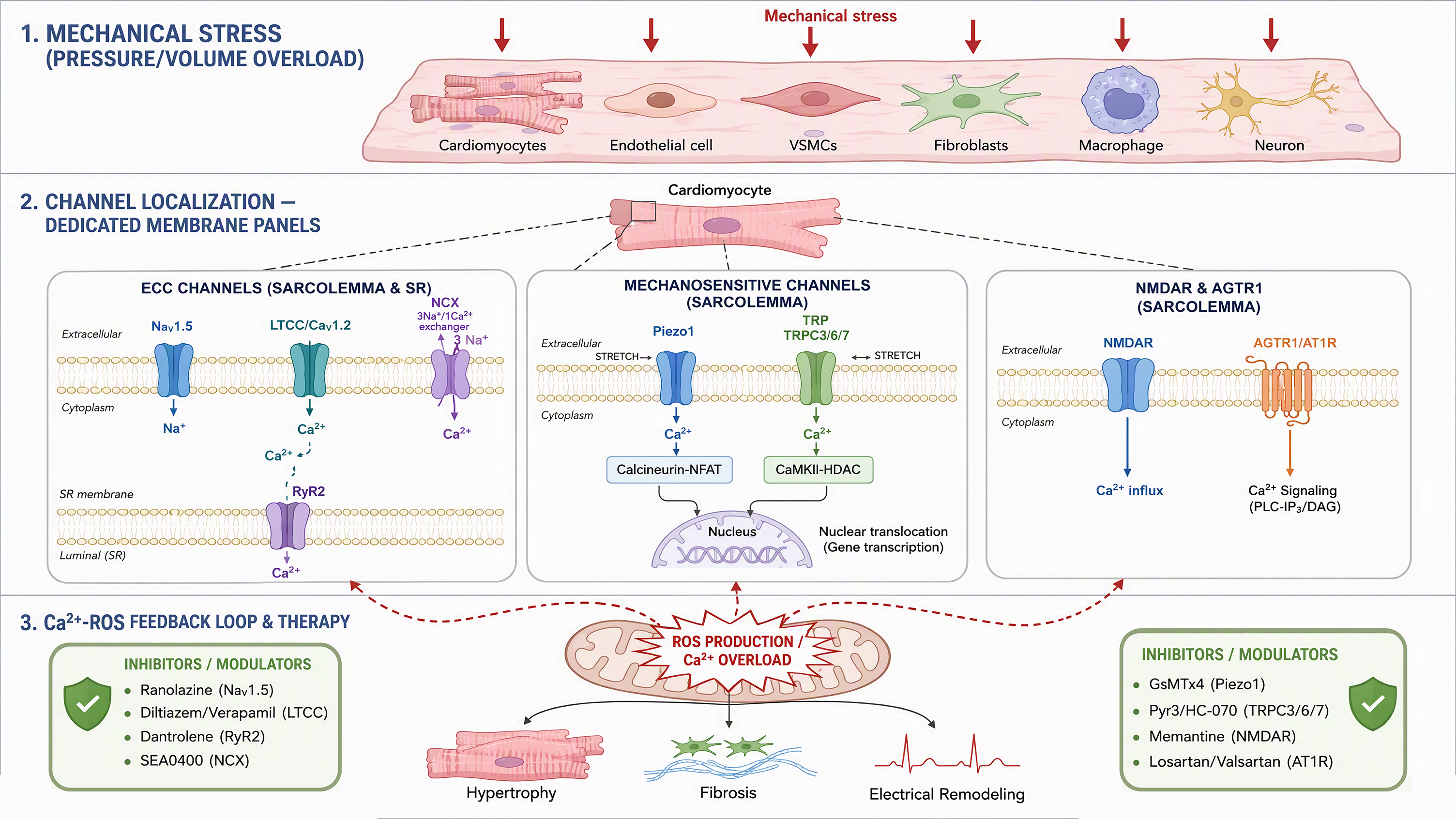
Figure 2. Cell-type-specific ion channel mechanisms of mechanical stress-induced cardiac remodeling. Pressure and volume overload generate mechanical stress that activates distinct ion-channel repertoires in cardiomyocytes, endothelial cells, vascular smooth muscle cells, cardiac fibroblasts, and perivascular sensory neurons and macrophages. Channel-mediated Ca2+/Na+ influx engages downstream signaling (calcineurin/NFAT, CaMKII/HDAC/MEF2, EDH-dependent vasodilation, myogenic tone, TGF-β/Smad-dependent fibrogenesis, sympathetic afferent activation, and innate immune activation). Mitochondrial Ca2+ overload generates ROS, which feedback to enhance channel activity through CaMKII oxidation, RyR2 oxidation, TRP S-nitrosylation, TRPC3-Nox2 coupling, and Na+-dependent mitochondrial ROS, forming self-reinforcing loops that drive hypertrophy, fibrosis, electrical remodeling, and vascular/neuro-immune dysfunction; candidate channel-directed therapeutic strategies are indicated at the bottom. The figure was created with Matplotlib (Python) and Adobe Illustrator.
Cardiomyocytes
Cardiomyocytes express the full complement of ECC channels (Nav1.5, LTCC, RyR2, NCX1) as well as mechanosensitive channels including TRPC1/3/6, TRPV2, TRPM4, TRPM7, Piezo1, and NMDAR. In these cells, mechanical stress activates Piezo1 and TRPC channels to produce sustained Ca2+ influx, mobilizing calcineurin/NFAT and CaMKII/HDAC signaling to drive hypertrophic growth, while channel-ROS feedback loops amplify this response and promote electrical instability (see Section "CONVENTIONAL SIGNALING PATHWAYS" and Sections "The TRP channel family"-"Non-canonical ion channels: the NMDAR" for details)[1,31,32,70].
Endothelial cells
Endothelial cells are the frontline sensors of hemodynamic forces. Endothelial TRPV4 channels generate basal Ca2+ influx events (TRPV4 sparklets) that activate intermediate- and small-conductance Ca2+-sensitive K+ channels, producing endothelium-dependent hyperpolarization and vasodilation; this mechanism is a major determinant of vascular resistance and blood pressure[65]. Endothelial Piezo1 senses shear stress and is essential for normal vascular development and the alignment of endothelial cells along the direction of blood flow; its disruption impairs vascular remodeling and angiogenesis[79,80]. Endothelial TRPC channels (TRPC1, TRPC3, TRPC5) also participate in angiogenesis, barrier function, and vasomotor regulation, and their dysfunction contributes to the microvascular abnormalities accompanying cardiac overload states[90,91].
Vascular smooth muscle cells (VSMCs)
In VSMCs, mechanosensitive channels regulate myogenic tone and structural adaptation of the vessel wall. TRPM4 is indispensable for pressure-induced myogenic constriction of cerebral and peripheral arteries, coupling membrane stretch to depolarization and Ca2+ influx through VGCCs[92]. Piezo1 is expressed in VSMCs and participates in hypertension-dependent arterial remodeling: smooth muscle Piezo1 activity promotes Ca2+-dependent transglutaminase activation and extracellular matrix cross-linking, thereby increasing arterial stiffness and amplifying cardiac afterload—an extracardiac mechanism by which mechanosensitive channels aggravate cardiac pressure overload[93]. VSMC TRPC channels (particularly TRPC6) also mediate agonist- and stretch-induced Ca2+ influx and vascular hyperreactivity in hypertension[90].
Cardiac fibroblasts
Cardiac fibroblasts lack contractile filaments but are rich in mechanosensitive channels that convert matrix mechanics into profibrotic signals. TRPM7-mediated Ca2+ signaling confers profibrotic properties on human atrial fibroblasts, and TRPM7 is upregulated in fibroblasts from patients with atrial fibrillation, linking channel activity to the fibrotic substrate of arrhythmias[33]. TRPV4 channels mediate the differentiation of cardiac fibroblasts into myofibroblasts by integrating mechanical and soluble (e.g., TGF-β) signals, promoting extracellular matrix deposition and myocardial stiffness[34]. Piezo1 in human atrial fibroblasts regulates cellular mechanical properties and matrix stiffness sensing and stimulates p38 MAPK-dependent interleukin-6 secretion, coupling mechanical stress to paracrine inflammatory amplification in the remodeling heart[35,94].
Perivascular sensory neurons and macrophages
Perivascular sensory neurons expressing TRPV1 and other TRP channels innervate the heart and coronary vessels, sense mechanical and chemical stimuli, and release neuropeptides such as calcitonin gene-related peptide (CGRP). In heart failure, enhanced cardiac sympathetic afferent activity—partly dependent on TRPV1-expressing afferent fibers—sustains sympathetic excitation; targeted ablation of these TRPV1-expressing cardiac sympathetic afferent fibers with resiniferatoxin attenuates cardiac remodeling and improves cardiovascular dysfunction in rats with heart failure, revealing a neurogenic contribution to adverse remodeling[68,69]. Cardiac macrophages, in turn, express Piezo1, which senses cyclic hydrostatic pressure and is essential for innate immune activation; Piezo1-mediated mechanosensing in myeloid cells couples tissue mechanics to inflammatory gene expression, thereby modulating the inflammatory phase of cardiac remodeling[95].
THERAPEUTIC TARGETS AND ADVANCES IN PHARMACOLOGICAL RESEARCH
The mechanistic insights described above identify several nodes at which ion channel function can be therapeutically targeted. This section summarizes the major channel-targeting strategies and then stratifies them by disease context and evidence level [Table 4]. At the outset, note that, except for CCBs, most of these strategies remain at the preclinical stage. Preclinical evidence includes TRPC inhibition in ischemic heart disease and pressure-overload hypertrophy[96-98], TRPV4 blockade in heart failure[99], and Piezo1 modulators such as Yoda1, GsMTx4, and Dooku1[100-102].
Channel-targeted therapeutic strategies stratified by disease context and evidence level
| Disease context | Candidate strategies (targets) | Representative drugs/interventions | Rationale | Evidence level | References |
| Pressure overload (hypertension, aortic stenosis) | Piezo1 inhibition; TRPC3/6 blockade; TRPM4 inhibition; RyR2 stabilization; AGTR1 blockade (inverse agonists) | GsMTx4; larixyl acetate; SAR7334; dantrolene; ARBs | Block mechanosensitive Ca2+ influx and calcineurin/NFAT activation; stabilize SR Ca2+ release; inhibit stretch-induced AGTR1 signaling | Cellular and animal models; Piezo1 upregulation confirmed in human hypertrophic myocardium | [1,14,64,70,77,97,114] |
| Volume overload (valvular regurgitation) | TRPV4 modulation; ECC channel monitoring; mechanosensor profiling | TRPV4 antagonists (preclinical); (no validated channel therapy yet) | Diastolic stretch recruits TRPV4 and mechanosensitive pathways; ECC channel regulation understudied | Cellular and animal models; direct comparative data lacking | [10,11,15] |
| HFrEF | RyR2 stabilization; NCX/INa,L modulation; NMDAR antagonism (exploratory); CCBs contraindicated in overt HFrEF | Dantrolene; SEA0400; memantine (preclinical) | Correct SR Ca2+ leak, Na+-dependent mitochondrial ROS, and excitotoxic Ca2+ influx | Animal model and human tissue data; no positive clinical trials for novel targets yet | [27,58,105,112,114] |
| HFpEF | Fibroblast-targeted TRPM7/TRPV4/Piezo1 inhibition; ROS-targeted antioxidants; endothelial TRPV4 blockade | HC-067047/GSK2193874; SS-31; mitoTEMPO (preclinical) | Attenuate fibrosis, myocardial stiffness, microvascular edema, and oxidative stress | Cellular and animal models; correlational evidence for TRPM7 in human atrial fibrosis | [33,34,99,117] |
| Ischemic heart disease | NMDAR blockade; TRPC inhibition; NCX inhibition | Memantine; SKF-96365-class compounds; SEA0400 (preclinical) | Limit ischemia-induced excitotoxic Ca2+ influx, Nav1.5 downregulation, and reperfusion Ca2+ overload | Cellular and animal models; human relevance unconfirmed | [36,96,116] |
CCBs
CCBs share a common mechanism of action: they exert their therapeutic effects by reducing calcium influx across the membrane. They are broadly divided into two subclasses: dihydropyridines (DHPs) and non-dihydropyridines (non-DHPs). These two drug classes have comparable antihypertensive efficacy but differ markedly in their pharmacological properties. DHP CCBs generally have stronger vasodilatory effects than non-DHP agents, whereas non-dihydropyridines have stronger negative chronotropic and negative inotropic effects, making them more suitable for patients with concomitant arrhythmias or those requiring β-blocker therapy. Dihydropyridines are indicated for hypertension, chronic stable angina, and vasospastic angina; non-dihydropyridines are likewise indicated for atrial fibrillation or atrial flutter and paroxysmal supraventricular tachycardia, and possess additional antiarrhythmic properties. The two subclasses have similar blood-pressure-lowering capacity; however, non-DHPs may offer advantages in managing chronic kidney disease and diabetic nephropathy, including antiproteinuric effects not replicated by DHPs; these effects may slow the progression of kidney disease[103-105].
CCBs have broad antihypertensive efficacy unaffected by sex, race, age, or dietary sodium intake, and they are approved to treat hypertension, angina, arrhythmias, and other conditions. Although CCB monotherapy has declined with the advent of newer antihypertensive agents, they have now become foundational components of many fixed-dose combination regimens, with the potential to improve blood pressure control and reduce cardiovascular event risk. CCBs remain first-line agents for hypertension and, compared with other antihypertensive drug classes, reduce the risk of adverse cardiovascular events, stroke, cardiovascular death, and myocardial infarction. They are well tolerated, with relatively few side effects, and can be used safely in the management of hypertension and angina; however, their use is associated with an increased risk of heart failure. In addition, CCB toxicity warrants attention because it can cause substantial morbidity and mortality; specific antidotal treatments are available, including intravenous calcium, glucagon, and high-dose insulin/euglycemic glucose regimens[106-109]. Nevertheless, the negative inotropic effects of non-dihydropyridine CCBs render them contraindicated in established heart failure with reduced ejection fraction, so their role is largely limited to prevention rather than reversal of remodeling[105].
Mechanosensitive channel modulators: Piezo1 and TRP channels
Pharmacological tools targeting mechanosensitive channels are being developed rapidly, although none has yet entered clinical trials for cardiac remodeling. For Piezo1, the spider venom peptide GsMTx4 blocks cation-selective mechanosensitive channels and is widely used in studies of Piezo1 function; the small molecule Yoda1 is a selective Piezo1 agonist, and its analog Dooku1 antagonizes Yoda1-evoked activation—these compounds constitute lead pharmacophores for future Piezo1-targeted drug development[100-102]. For TRP channels, combined blockade of TRPC3/TRPC6—whether by genetic means or with selective small molecules—suppresses pathological cardiac hypertrophy in mice; the natural diterpene larixyl acetate (a TRPC6 inhibitor) attenuates pressure overload-induced heart failure; and endogenous natriuretic peptide signaling also exerts antihypertrophic effects by inhibiting TRPC6 channel activity[64,97,98]. Within the TRPV subfamily, the TRPV4 antagonist GSK2193874 prevents and reverses pulmonary edema in experimental heart failure by normalizing endothelial barrier function, demonstrating the translational potential of vascular TRPV4 blockade; TRPV1 antagonism, in turn, attenuates hypertrophy and heart failure in mice through mechanisms that may involve both cardiac and neuronal TRPV1 pools[68,99].
NMDAR antagonists
The NMDA receptor is a subtype of glutamate receptor that regulates synaptic plasticity and intracellular Ca2+ homeostasis. Its uncontrolled activation leads to Ca2+ overload, causing cell contraction and alterations in intracellular pH. Uncontrolled NMDAR activation is a key factor in synaptic dysfunction. Targeting NMDA receptors with antagonists is a promising therapeutic strategy for neurological diseases, and studies in a variety of brain disorders have demonstrated favorable therapeutic effects. Memantine is an NMDA receptor antagonist clinically approved for the treatment of moderate-to-severe Alzheimer's disease and is currently the only NMDA receptor antagonist marketed for this indication. It improves symptoms but cannot cure the disease or halt its progression. Multiple NMDA receptor antagonists (including ketamine and MK-801) have shown therapeutic potential in experimental models of stroke, epilepsy, Parkinson's disease, and other encephalopathies[110,111].
Beyond their roles in the CNS, NMDA receptor activation also contributes to myocardial disease pathogenesis. Memantine exerts cardioprotective effects in heart failure (HF) by attenuating cardiac remodeling, lipid peroxidation, and neutrophil infiltration. Memantine may also enhance the chemotherapeutic efficacy of doxorubicin while reducing doxorubicin-induced cardiac oxidative stress and inflammation[112,113]. However, the efficacy of NMDAR blockade appears to be context-dependent: in a thyroxine-induced model, memantine prevented hypertension but not the associated cardiac remodeling; and to date, no clinical trials of NMDAR antagonists for cardiac disease have been reported. These agents should therefore be regarded as hypothesis-generating candidate strategies whose utility will depend on disease drivers and require confirmation in human tissue and clinical studies[9,89].
RyR2 stabilizers
Because excessive RyR2 phosphorylation and oxidation increase SR Ca2+ leak in hypertrophy and heart failure, stabilizing RyR2 is a mechanistically rational strategy. Dantrolene, the classic RyR inhibitor used for malignant hyperthermia, stabilizes inter-domain interactions within RyR2 and markedly improves both failing cardiomyocyte and whole-heart function in animal models of heart failure, correcting Ca2+-handling defects without suppressing physiological Ca2+ release[114]. More recent work shows that pharmacological RyR2 stabilization attenuates cardiac hypertrophy in pressure overload models by downregulating the TNF-α/NF-κB/NLRP3 inflammatory axis via calcineurin inhibition, linking correction of SR Ca2+ leak to anti-inflammatory and antihypertrophic effects[115]. These findings position RyR2 stabilizers as candidate agents with both structural and arrhythmic endpoints, although their effects in human myocardium and clinical settings remain to be established.
Modulation of the NCX
NCX1 is upregulated in hypertrophy and heart failure and contributes to contractile dysfunction and arrhythmogenesis, making NCX a long-standing therapeutic candidate[58,59]. Selective NCX inhibitors such as SEA0400 preferentially block reverse-mode (Ca2+ influx) exchange, attenuate Ca2+ overload-related injury in experimental models, and show antiarrhythmic efficacy in the setting of triggered activity[59,116]. However, the bidirectionality of NCX flux means that the net effect of inhibitors depends on cellular Na+ load and membrane potential, and clinical translation has been limited by concerns about suppressing forward-mode Ca2+ extrusion in the failing heart[58]. Targeting intracellular Na+ homeostasis upstream of NCX—for example, by reducing INa,L—is a complementary strategy that can simultaneously reduce mitochondrial oxidative stress and Ca2+ overload[26,27].
ROS-targeted interventions
Given the channel-ROS mutual amplification loop described in Section "Ion channels and cardiac remodeling: the Ca2+-Na+-ROS signaling triad", blocking oxidative stress at its source is an attractive adjunctive strategy. Mitochondria-targeted antioxidants have shown the most consistent cardiac efficacy: the mitochondria-targeted peptide SS-31 (elamipretide) improves hypertensive cardiomyopathy in mice by preserving mitochondrial function, and scavenging mitochondrial superoxide with mitoTEMPO reduces vascular oxidative stress and blood pressure in experimental hypertension[117,118]. By lowering ambient ROS levels, such interventions are expected to secondarily normalize the redox state of RyR2, CaMKII, and TRP channels, thereby breaking the Ca2+-ROS feed-forward cycle[28-30]. It should be
Combined multi-node intervention
Intracellular calcium overload contributes to the pathogenesis of multiple cardiovascular diseases. Conventional CCBs reduce calcium influx by blocking L-type voltage-gated calcium channels (LTCCs) and are classic treatments for hypertension and angina. NMDARs are also expressed in the heart, and their overactivation mediates pathological calcium influx, triggering calcium overload. These two calcium influx systems form an interactive network under pathological conditions. CCBs and NMDAR antagonists target voltage-gated and ligand-gated calcium influx pathways, respectively, and their combined application could achieve multi-node blockade of calcium overload.
Because mechanical stress activates multiple Ca2+-permeable pathways in parallel, simultaneously modulating multiple nodes may in theory yield additive protection. In isolated rat heart preparations, combined application of verapamil and glutamatergic modulation affected cardiac dynamics, coronary flow, and oxidative stress indices, providing preliminary proof of concept for combined targeting of LTCCs and glutamate receptors[119]. Combined regimens of CCBs and NMDAR antagonists are strictly hypothesis-generating: no controlled animal or clinical study has tested this combination for cardiac remodeling, and both drug classes have hemodynamic adverse effects (hypotension, bradycardia) that may be additive. Any future evaluation should therefore begin with rigorously designed preclinical studies in well-defined overload models, explicitly assessing effects independent of blood pressure changes, before clinical translation can be considered[89,112].
Stratification by disease context and evidence level
The therapeutic relevance of individual channel targets varies with the underlying disease context—pressure overload, volume overload, ischemic heart disease, or heart failure with reduced (HFrEF) or preserved (HFpEF) ejection fraction—and with the evidence level (which we categorize as cellular, animal model, human tissue, or clinical evidence) [Table 4]. Several overall patterns emerge. First, the evidence base is strongest for pressure overload models: cardiomyocyte Piezo1, TRPC3/6, TRPM4, and RyR2 have all been validated genetically and pharmacologically in animals, and upregulation of Piezo1 and TRPM7 has been corroborated in human hypertrophic and fibrillating myocardium[1,33,70,77]. Second, for HFrEF, the channel-targeting candidates with the most advanced translational prospects are RyR2 stabilizers and NCX/INa,L modulation, both supported by animal and human tissue data, whereas CCBs are contraindicated in overt HFrEF but are suitable for upstream prevention of hypertensive hypertrophy[58,105,114]. Third, for HFpEF and volume overload states—contexts characterized by diastolic dysfunction, fibrosis, and microvascular dysfunction—endothelial TRPV4, fibroblast TRPM7/TRPV4/Piezo1, and ROS-targeted interventions are biologically plausible but are currently supported mainly by animal and cellular evidence, representing an important knowledge gap[15,34,99]. Fourth, in ischemic heart disease, NMDAR blockade and TRPC inhibition have shown benefit in experimental ischemia-reperfusion and arrhythmia models, but likewise lack clinical confirmation to date[36,96]. This explicit stratification by evidence level should guide target prioritization in translational development.
DISCUSSION
This review systematically integrates the contributions of multiple ion channel families—ranging from classical voltage-gated channels to mechanosensitive and ligand-gated non-canonical receptors—to the pathophysiology of mechanical stress-induced cardiac remodeling. The central theme emerging from this integration is that cardiac remodeling is not driven by any single ion channel or signaling pathway in isolation, but rather arises from the emergent properties of a complex, multilayered mechano-calcium signaling network in which multiple channels engage in mutual functional crosstalk.
The traditional understanding of cardiac remodeling has been anchored in the LTCC-RAAS axis, with neurohumoral activation and voltage-gated Ca2+ influx regarded as the principal drivers of pathological hypertrophy. Although this framework has yielded important mechanistic insights and therapeutic successes—most notably establishing CCBs as first-line agents for hypertension and angina—it is increasingly clear that this model captures only part of the relevant biology. Mechanical stress, whether manifested as pressure overload or volume overload, activates a far broader repertoire of ion channels than previously appreciated[1,15]. Pressure overload predominantly engages Piezo1 and TRPC1/6 as direct mechanosensors, converting elevated wall stress into intracellular Ca2+ signals; whereas it has been proposed that volume overload may preferentially engage TRPV4 through sustained axial stretch, and may also involve NMDAR, direct experimental evidence for channel regulation under pure volume overload remains limited[9,15]. These distinctions are not merely academic: they carry important therapeutic implications, because the selective engagement of different channel ensembles under different hemodynamic conditions suggests that a "one-size-fits-all" channel blockade strategy may not be optimal. Within the RAAS, AGTR1 adds a receptor-level dimension to this paradigm: it can directly sense mechanical stress and activate hypertrophic ERK signaling in an angiotensin II-independent manner, thereby coupling mechanical load to RAAS-dependent growth programs even in the absence of systemic hormone generation[14,23].
The Ca2+-Na+-ROS triad
One of the most important unifying principles emerging from this review is a tightly integrated Ca2+-Na+-ROS signaling triad downstream of mechanosensitive channel activation. Mechanosensitive channel opening triggers an initial Ca2+ influx that activates calcineurin/NFAT and CaMKII, driving hypertrophic gene expression. Concurrently, Na+ influx through mechanosensitive and voltage-gated channels depolarizes the membrane, amplifying Ca2+ signals via reverse-mode NCX and enhanced LTCC opening. This Na+-Ca2+ amplification loop is critical for the robustness of the hypertrophic response, but it also creates susceptibility to arrhythmogenesis. The third arm of the triad, ROS generation, is initiated by Ca2+-dependent NOX activation and, critically, feeds back to enhance mechanosensitive channel activity and downstream signaling through well-defined oxidative modifications, including CaMKII methionine oxidation, RyR2 redox modification, and TRP channel S-nitrosylation[28-30]. This establishes a self-perpetuating vicious cycle of "ROS generation-calcium overload-ROS generation" that drives not only cardiomyocyte hypertrophy but also fibroblast activation and myocardial fibrosis. Two additional molecular couplings further reinforce this loop: TRPC3 stabilizes Nox2 under mechanical stress to amplify ROS generation; and elevated cytosolic Na+ in failing cardiomyocytes impairs mitochondrial Ca2+ uptake and antioxidant capacity, directly linking Na+ channel remodeling to oxidative stress[27,31]. The therapeutic implication is clear: effective intervention will likely require simultaneous targeting of multiple nodes within this triad, rather than isolated blockade of any single component.
Reappraising L-type calcium channels
The evidence reviewed here calls for a more nuanced re-evaluation of LTCC function in cardiac remodeling. Classically regarded as the principal gateway for pathological Ca2+ influx, LTCCs are now recognized to participate in a more complex regulatory architecture. Reduced LTCC activity can paradoxically promote hypertrophy through compensatory RyR2 Ca2+ leak and calcineurin/NFAT activation—a finding that reveals a non-monotonic relationship between LTCC function and remodeling. Moreover, LTCCs are physically and functionally coupled to mitochondria via the cytoskeleton (F-actin, β-tubulin), and this coupling is aberrantly activated in hypertrophic cardiomyopathy models to drive mitochondrial hypermetabolism—a finding that introduces an entirely new dimension to LTCC biology. AID-variant peptides targeting the LTCC β2 subunit can dissociate this abnormal channel-cytoskeleton coupling and safely prevent the development of HCM without affecting Ca2+ current or blood pressure—representing a paradigm shift that suggests the structural scaffolding functions of LTCCs may be as pathologically important as their ionic conductance. Together, these findings argue for moving beyond simple LTCC blockade toward strategies that selectively modulate channel-protein interactions[12,42-45,47-49].
Type 2 ryanodine receptors
Ryanodine receptors are critical mediators of pathological remodeling, and the evidence reviewed here establishes RyR2 as an indispensable node in the hypertrophic signaling network. Gain-of-function mutations such as RyR2-R176Q cause diastolic SR Ca2+ leak and markedly accelerate pressure overload-induced hypertrophy and heart failure, whereas heterozygous RyR2 knockout attenuates the hypertrophic response by suppressing calcineurin, ERK, and Akt signaling. Circ-RYR2—a circular RNA derived from the RYR2 locus—is downregulated in hypertrophy and heart failure, and restoring its expression normalizes Ca2+ handling without altering RyR2 protein levels—a finding that introduces an exciting new layer of regulatory complexity. This discovery suggests that therapeutic strategies aimed at modulating non-coding RNA expression may offer a means of regulating RyR2 function more precisely than directly targeting the channel[54,56,57,114].
TRP channels
TRP channels, as multimodal sensors of cardiac stress, occupy a unique position at the interface of mechanical sensing and Ca2+ signaling. TRPC1/3/6 respond directly to stretch to activate CaMKII and drive hypertrophy, whereas TRPV4 integrates shear stress and osmotic signals to regulate vascular tone via the eNOS/NO pathway. TRPM4, a Ca2+-activated non-selective cation channel, is upregulated in pressure overload hypertrophy and promotes remodeling through membrane depolarization-dependent CaMKII activation. A recently discovered functional interaction between TRPV4 and Piezo1 further underscores the networked nature of mechanosensitive signaling. Perhaps most importantly, TRP channel-mediated Ca2+ influx initiates and sustains the fibrotic ROS-calcium vicious cycle, positioning TRP channels as strategic upstream targets for interrupting pathological remodeling[31,32,41,63].
Piezo1 channels
Among mechanosensitive channels, Piezo1 is the best-characterized direct mechanosensor in the cardiovascular system. Its trimeric propeller-like architecture enables it to be directly gated by lipid bilayer tension, independent of cytoskeletal involvement. Cardiomyocyte-specific Piezo1 knockout and overexpression studies have established its critical role in initiating the pressure overload hypertrophic response through the dual signaling axes of CaM/CaN/NFAT and CaMKII/HDAC4/MEF2[1,2]. Notably, Piezo1 also regulates TRPM4 expression, forming a feed-forward mechanism that amplifies hypertrophic signaling[1,70]. The ability of Piezo1 to integrate vascular architecture with physiological shear stress further highlights its systemic importance in cardiovascular homeostasis[79,80]. Independent studies have shown that Piezo1-mediated mechanotransduction promotes hypertrophy by impairing Ca2+ homeostasis and activating calpain/calcineurin signaling, that Piezo1 is upregulated in human hypertrophic myocardium, and that mice carrying systemic PIEZO1 gain-of-function mutations spontaneously develop cardiac hypertrophy and fibrosis—evidence that collectively elevates Piezo1 from a candidate mechanosensor to a validated disease-modifying node[77,78]. As the field moves toward Piezo1-selective inhibitors—building on first-generation pharmacological tools such as GsMTx4, Yoda1, and its antagonist analog Dooku1—the key challenge will be to prevent pathological cardiac remodeling while preserving the physiological functions of this channel in vascular adaptation[100-102].
NMDA receptors
The inclusion of NMDARs in the cardiac ion channel landscape is one of the more unexpected developments in this field. Beyond their classical identity as neuronal channels, NMDARs are also stably expressed in cardiomyocytes and the cardiac conduction system, where their high Ca2+ permeability and slow inactivation kinetics permit sustained Ca2+ influx that can trigger mitochondrial Ca2+ overload and ROS generation[9,81,86]. Pathological NMDAR activation generates an arrhythmogenic substrate through dual mechanisms—structural (promoting fibrosis) and electrophysiological (downregulating Nav1.5 via AKT1/TBX3, slowing conduction, and prolonging repolarization)[36,85]. The observation that memantine prevents thyroxine-induced hypertension but not cardiac remodeling suggests that NMDAR-mediated Ca2+ signaling is only one component of a broader hypertrophic mechanism, consistent with the network-level model proposed here[89]. Although memantine attenuates remodeling and oxidative stress in other experimental models of heart failure and cardiotoxicity, the evidence base remains preclinical and model-dependent; therefore, its therapeutic claims should be regarded as preliminary and hypothesis-generating, pending confirmation in human tissues and clinical studies[9,112,113].
Cell-type-specific integration
Because different channels regulate the diverse cell types of myocardial tissue, the net outcome of concerted actions across cell types strongly influences cardiac remodeling. Endothelial TRPV4 sparklets and Piezo1 regulate vascular tone and angiogenic remodeling; smooth muscle TRPM4 and Piezo1 determine myogenic tone and arterial stiffness, thereby modulating cardiac afterload; fibroblast TRPM7, TRPV4, and Piezo1 drive fibrogenesis and myocardial stiffening; and TRPV1-expressing perivascular sensory afferent nerves and Piezo1-expressing macrophages couple tissue mechanics to neurohumoral and inflammatory amplification loops[33,65,69,93,95]. This cellular diversity implies that systemic blockade of a broadly expressed mechanosensor may produce opposing effects in different cell types, and that mechanosensitive channel therapy may ultimately require cell-targeted delivery.
Therapeutic implications
The mechanistic framework described above carries direct translational implications, albeit with varying degrees of maturity. Among conventional targets, CCBs are of value primarily for preventing hypertensive hypertrophy, rather than for reversing established remodeling or treating HFrEF[105,106]. RyR2 stabilizers and Na+/NCX-targeted strategies extend this classical pharmacology toward correcting SR Ca2+ leak and Na+-dependent oxidative stress, supported by animal and human tissue evidence but without completed clinical trials[27,58,114]. Among emerging targets, Piezo1 and TRP channel modulators, NMDAR antagonists, and ROS-targeted interventions are supported mainly by cellular and animal evidence [Table 4], and their clinical utility will depend on disease context, cell-type targeting, and rigorous translational validation. It should be particularly emphasized that the proposal of combined CCB and NMDAR antagonist therapy should be regarded strictly as a rationale for future preclinical research, rather than as an evidence-supported treatment option (see Section "Combined multi-node intervention")[89,119].
Limitations and future directions
First, most evidence on the role of mechanosensitive channels in cardiac remodeling comes from rodent models, and species differences in channel expression and mechanotransduction properties may limit direct translation to human disease. Second, the quantitative contribution of Piezo1-, TRP-, and NMDAR-mediated Ca2+ influx relative to LTCC-mediated influx in human cardiomyocytes under physiological and pathological mechanical loading remains incompletely defined. Third, developing truly channel subtype-selective drugs remains a major challenge; most currently available TRP and Piezo1 modulators lack the specificity required for clinical application. Fourth, the long-term effects of chronic suppression of mechanosensitive channels on cardiac contractile function—which depends on appropriate Ca2+ handling—require careful evaluation. Finally, mechanistic evidence is heavily skewed toward pressure overload models; direct comparative studies of channel regulation under volume overload are scarce, and therefore our distinction between pressure and volume overload [Table 1] should be regarded as preliminary[15,16]. In addition, individualized therapeutic strategies based on the relative activation of different channel ensembles in individual patients—for example, reflecting pressure overload-dominant versus volume overload-dominant physiological states—represent an exciting but as yet unrealized opportunity.
In summary, this review proposes an integrative framework in which mechanical stress-induced cardiac remodeling arises from the coordinated activity of multiple ion channels operating as an interconnected mechano-calcium signaling network. The shift from single-pathway to network-level understanding opens new avenues for targeted intervention and demands a fundamental reappraisal of research priorities and therapeutic strategies in the management of cardiac hypertrophy and heart failure.
CONCLUSION
This review proposes an integrated conceptual framework in which mechanical stress-induced cardiac remodeling is understood as the emergent output of a complex, multichannel mechano-calcium signaling network. We have systematically reviewed the contributions of three major classes of ion channels—classical ECC channels (the voltage-gated Na+ channel Nav1.5, LTCC, the SR Ca2+ release channel RyR2, and the Na+/Ca2+ exchanger NCX1), mechanosensitive channels (TRPC, TRPV, TRPM, Piezo1), and the non-canonical ligand-gated channel NMDAR—to the pathophysiology of cardiomyocyte hypertrophy, fibrosis, and electrical remodeling. The central unifying principle emerging from this integration is that these channels do not operate in isolation, but rather form a functionally interconnected network through shared downstream signaling nodes (calcineurin/NFAT, CaMKII, MAPK), reciprocal regulation by ROS, and mechanical signal amplification via the cytoskeleton.
Recognizing that mechanosensitive and non-canonical channels complement classical LTCC-mediated ECC and act in synergy with it under specific mechanical and pathological contexts compels us to carefully re-evaluate research priorities and therapeutic strategies for cardiac remodeling. The shift from single-pathway to network-level understanding opens new avenues for future research—including the hypothesis that combining classical CCBs with NMDAR antagonists may achieve multi-node blockade of Ca2+ overload (a strategy not yet validated in controlled animal or clinical studies targeting cardiac remodeling), as well as the potential of Piezo1-selective inhibitors to prevent pathological remodeling without compromising normal cardiac function. As the field advances, success in translating these mechanistic insights into clinical practice will depend on embracing the complexity of ion channel crosstalk, developing more selective pharmacological tools, and designing clinical trials that capture the mechanistic heterogeneity underlying phenotypically similar cardiac hypertrophy and heart failure.
DECLARATIONS
Authors’ contributions
Conceptualization, investigation, writing-original, writing-review & editing: Shi W
Validation, visualization, investigation: Ma Z
Writing-review & editing, software: Liu C
Supervision, project administration, funding acquisition, writing-review & editing: Sun H
Availability of data and materials
Not applicable.
AI and AI-assisted tools statement
During revision of this manuscript, Kimi (version Kimi K3, released 2026-7-16) was used to assist with English translation and the production of a graphical abstract to improve language, readability of some sentences, and visual presentation. The authors further manually checked and revised the translated text and graphical abstract. The tool did not influence the study design, data collection, analysis, interpretation, or the scientific content of the work. All authors take full responsibility for the accuracy, integrity, and final content of the manuscript.
Financial support and sponsorship
This work was supported by Jilin Provincial Department of Science and Technology (YDZJ202501ZYTS071).
Conflicts of interest
All authors declared no conflicts of interest.
Ethical approval and consent to participate
Not applicable.
Consent for publication
Not applicable.
Copyright
© The Author(s) 2026.
REFERENCES
1. Yu Z, Gong H, Kesteven S, et al. Piezo1 is the cardiac mechanosensor that initiates the cardiomyocyte hypertrophic response to pressure overload in adult mice. Nat Cardiovasc Res. 2022;1:577-91.
2. Lim GB. Piezo1 senses pressure overload and initiates cardiac hypertrophy. Nat Rev Cardiol. 2022;19:503-503.
3. Nakamura M, Sadoshima J. Mechanisms of physiological and pathological cardiac hypertrophy. Nat Rev Cardiol. 2018;15:387-407.
5. Mackrill JJ, Shiels HA. Evolution of excitation-contraction coupling. In: Islam MS, editors. Calcium Signaling. Cham: Springer International Publishing; 2019. pp. 281-320.
6. Heineke J, Molkentin JD. Regulation of cardiac hypertrophy by intracellular signalling pathways. Nat Rev Mol Cell Biol. 2006;7:589-600.
7. Zhang M, Ma Y, Ye X, Zhang N, Pan L, Wang B. TRP (transient receptor potential) ion channel family: structures, biological functions and therapeutic interventions for diseases. Sig Transduct Target Ther. 2023;8:261.
8. Jin C, Su S, Yu S, et al. Essential roles of PIEZO1 in mammalian cardiovascular system: from development to diseases. Cells. 2024;13:1422.
9. Soda T, Brunetti V, Berra-Romani R, Moccia F. The emerging role of N-methyl-D-aspartate (NMDA) receptors in the cardiovascular system: physiological implications, pathological consequences, and therapeutic perspectives. Int J Mol Sci. 2023;24:3914.
10. Rossi MA, Carillo SV. Cardiac hypertrophy due to pressure and volume overload: distinctly different biological phenomena? Int J Cardiol. 1991;31:133-41.
11. Papapostolou S, Kearns J, Costello BT, et al. Comparison of pressure vs volume overload ventricular wall stress in patients with valvular heart disease. J Am Coll Cardiol. 2024;84:635-44.
12. Goonasekera SA, Hammer K, Auger-Messier M, et al. Decreased cardiac L-type Ca2+ channel activity induces hypertrophy and heart failure in mice. J Clin Investig. 2012;122:280-90.
13. Ruwhof C. Mechanical stress-induced cardiac hypertrophy: mechanisms and signal transduction pathways. Cardiovasc Res. 2000;47:23-37.
14. Zou Y, Akazawa H, Qin Y, et al. Mechanical stress activates angiotensin II type 1 receptor without the involvement of angiotensin II. Nat Cell Biol. 2004;6:499-506.
15. Veteto AB, Peana D, Lambert MD, Mcdonald KS, Domeier TL. Transient receptor potential vanilloid-4 contributes to stretch-induced hypercontractility and time-dependent dysfunction in the aged heart. Cardiovasc Res. 2020;116:1887-96.
16. Izu LT, Kohl P, Boyden PA, et al. Mechano-electric and mechano-chemo-transduction in cardiomyocytes. J Physiol. 2020;598:1285-305.
17. Davis MJ, Earley S, Li Y, Chien S. Vascular mechanotransduction. Physiol Rev. 2023;103:1247-421.
18. Nourse JL, Pathak MM. How cells channel their stress: Interplay between Piezo1 and the cytoskeleton. Semin Cell Dev Biol. 2017;71:3-12.
19. Uray IP, Uray K. Mechanotransduction at the plasma membrane-cytoskeleton interface. Int J Mol Sci. 2021;22:11566.
20. Casarella S, Ferla F, Di Francesco D, Canciani E, Rizzi M, Boccafoschi F. Focal adhesion’s role in cardiomyocytes function: from cardiomyogenesis to mechanotransduction. Cells. 2024;13:664.
21. Chuang Y, Chen C. Force from filaments: the role of the cytoskeleton and extracellular matrix in the gating of mechanosensitive channels. Front Cell Dev Biol. 2022;10:886048.
22. Viola HM, Hool LC. The L-type Ca2+ channel: a mediator of hypertrophic cardiomyopathy. Channels. 2016;11:5-7.
23. Tang W, Strachan RT, Lefkowitz RJ, Rockman HA. Allosteric modulation of β-arrestin-biased angiotensin II type 1 receptor signaling by membrane stretch. J Biol Chem. 2014;289:28271-83.
24. Brown DI, Griendling KK. Regulation of signal transduction by reactive oxygen species in the cardiovascular system. Circ Res. 2015;116:531-49.
25. Elias-Llumbet A, Sharmin R, Berg-Sorensen K, Schirhagl R, Mzyk A. The interplay between mechanoregulation and ROS in heart physiology, disease, and regeneration. Adv Healthc Mater. 2024;13:2400952.
26. Wan E, Abrams J, Weinberg RL, et al. Aberrant sodium influx causes cardiomyopathy and atrial fibrillation in mice. J Clin Investig. 2015;126:112-22.
27. Kohlhaas M, Liu T, Knopp A, et al. Elevated cytosolic Na+ increases mitochondrial formation of reactive oxygen species in failing cardiac myocytes. Circulation. 2010;121:1606-13.
28. Erickson JR, Joiner MA, Guan X, et al. A dynamic pathway for calcium-independent activation of CaMKII by methionine oxidation. Cell. 2008;133:462-74.
29. Terentyev D, Györke I, Belevych AE, et al. Redox modification of ryanodine receptors contributes to sarcoplasmic reticulum Ca2+ leak in chronic heart failure. Circ Res. 2008;103:1466-72.
30. Yoshida T, Inoue R, Morii T, et al. Nitric oxide activates TRP channels by cysteine S-nitrosylation. Nat Chem Biol. 2006;2:596-607.
31. Kitajima N, Numaga-Tomita T, Watanabe M, et al. TRPC3 positively regulates reactive oxygen species driving maladaptive cardiac remodeling. Sci Rep. 2016;6:37001.
32. Kuwahara K, Wang Y, Mcanally J, et al. TRPC6 fulfills a calcineurin signaling circuit during pathologic cardiac remodeling. J Clin Investig. 2006;116:3114-26.
33. Du J, Xie J, Zhang Z, et al. TRPM7-mediated Ca2+ signals confer fibrogenesis in human atrial fibrillation. Circ Res. 2010;106:992-1003.
34. Adapala RK, Thoppil RJ, Luther DJ, et al. TRPV4 channels mediate cardiac fibroblast differentiation by integrating mechanical and soluble signals. J Mol Cell Cardiol. 2013;54:45-52.
35. Emig R, Knodt W, Krussig MJ, et al. Piezo1 channels contribute to the regulation of human atrial fibroblast mechanical properties and matrix stiffness sensing. Cells. 2021;10:663.
36. He Y, Zhang H, Zhang Q, et al. NMDA-type glutamate receptor activation promotes ischemic arrhythmias by targeting the AKT1-TBX3-Nav1.5 axis. Acta Physiol. 2025;241:e70085.
37. Ramirez RJ, Sah R, Liu J, Rose RA, Backx PH. Intracellular [Na+] modulates synergy between Na+/Ca2+ exchanger and L-type Ca2+ current in cardiac excitation-contraction coupling during action potentials. Basic Res Cardiol. 2011;106:967-77.
38. Hofmann F, Flockerzi V, Kahl S, Wegener JW. L-type CaV1.2 calcium channels: from in vitro findings to in vivo function. Physiol Rev. 2014;94:303-26.
39. Treinys R, Jurevičius J. L-type Ca2+ channels in the heart: Structure and regulation. Medicina. 2008;44:491.
40. Pallien T, Klussmann E. New aspects in cardiac L-type Ca2+ channel regulation. Biochem Soc Trans. 2020;48:39-49.
41. Gao H, Wang F, Wang W, et al. Ca2+ influx through L-type Ca2+ channels and transient receptor potential channels activates pathological hypertrophy signaling. J Mol Cell Cardiol. 2012;53:657-67.
42. Tonegawa K, Otsuka W, Kumagai S, et al. Caveolae-specific activation loop between CaMKII and L-type Ca2+ channel aggravates cardiac hypertrophy in α1-adrenergic stimulation. Am J Physiol Heart Circ Physiol. 2017;312:H501-14.
43. Tandan S, Wang Y, Wang TT, et al. Physical and functional interaction between calcineurin and the cardiac L-type Ca2+ channel. Circ Res. 2009;105:51-60.
44. Pedrozo Z, Criollo A, Battiprolu PK, et al. Polycystin-1 is a cardiomyocyte mechanosensor that governs L-type Ca2+ channel protein stability. Circulation. 2015;131:2131-42.
45. Manning JR, Yin G, Kaminski CN, et al. Rad GTPase deletion increases L-type calcium channel current leading to increased cardiac contraction. J Am Heart Assoc. 2013;2:e000459.
46. Li M, He H, Gong H, et al. NFATc4 and myocardin synergistically up-regulate the expression of LTCC α1C in ET-1-induced cardiomyocyte hypertrophy. Life Sci. 2016;155:11-20.
47. Ahern BM, Levitan BM, Veeranki S, et al. Myocardial-restricted ablation of the GTPase RAD results in a pro-adaptive heart response in mice. J Biol Chem. 2019;294:10913-27.
48. Viola H, Johnstone V, Cserne Szappanos H, et al. The L-type Ca2+ channel facilitates abnormal metabolic activity in the cTnI-G203S mouse model of hypertrophic cardiomyopathy. J Physiol. 2016;594:4051-70.
49. Er TS, Reinke D, Francis AJ, et al. Novel peptide therapeutics targeting the L-type calcium channel prevent hypertrophic cardiomyopathy by decreasing mitochondrial energetics. JACC Basic Transl Sci. 2025;10:101292.
50. Kobayashi T, Kurebayashi N, Murayama T. The ryanodine receptor as a sensor for intracellular environments in muscles. Int J Mol Sci. 2021;22:10795.
51. Fabiato A. Calcium-induced release of calcium from the cardiac sarcoplasmic reticulum. Am J Physiol Cell Physiol. 1983;245:C1-C14.
52. Waddell HM, Mereacre V, Alvarado FJ, Munro ML. Clustering properties of the cardiac ryanodine receptor in health and heart failure. J Mol Cell Cardiol. 2023;185:38-49.
53. Wang S, Song L, Lakatta EG, Cheng H. Ca2+ signalling between single L-type Ca2+ channels and ryanodine receptors in heart cells. Nature. 2001;410:592-6.
54. Pan W, Hunkler HJ, Chatterjee S, et al. A circular RNA derived from the ryanodine receptor 2 locus controls cardiac hypertrophy and calcium handling. Cell Mol Life Sci. 2025;82:359.
55. Gez LS, Hagalili Y, Shainberg A, Atlas D. Voltage-driven Ca2+ binding at the L-type Ca2+ channel triggers cardiac excitation-contraction coupling prior to Ca2+ influx. Biochemistry. 2012;51:9658-66.
56. Zou Y, Liang Y, Gong H, et al. Ryanodine receptor type 2 is required for the development of pressure overload-induced cardiac hypertrophy. Hypertension. 2011;58:1099-110.
57. Van Oort RJ, Respress JL, Li N, et al. Accelerated development of pressure overload-induced cardiac hypertrophy and dysfunction in an RyR2-R176Q knockin mouse model. Hypertension. 2010;55:932-8.
58. Pogwizd SM, Schlotthauer K, Li L, Yuan W, Bers DM. Arrhythmogenesis and contractile dysfunction in heart failure: roles of sodium-calcium exchange, inward rectifier potassium current, and residual β-adrenergic responsiveness. Circ Res. 2001;88:1159-67.
59. Shigekawa M, Iwamoto T. Cardiac Na+-Ca2+ exchange: molecular and pharmacological aspects. Circ Res. 2001;88:864-76.
60. Fleig A, Penner R. The TRPM ion channel subfamily: molecular, biophysical and functional features. Trends Pharmacol Sci. 2004;25:633-9.
61. Nishida M, Kurose H. Roles of TRP channels in the development of cardiac hypertrophy. Naunyn Schmiedebergs Arch Pharmacol. 2008;378:395-406.
62. Grimm M, Brown JH. β-adrenergic receptor signaling in the heart: role of CaMKII. J Mol Cell Cardiol. 2010;48:322-30.
63. Ohba T, Watanabe H, Murakami M, et al. Upregulation of TRPC1 in the development of cardiac hypertrophy. J Mol Cell Cardiol. 2007;42:498-507.
64. Seo K, Rainer PP, Shalkey Hahn V, et al. Combined TRPC3 and TRPC6 blockade by selective small-molecule or genetic deletion inhibits pathological cardiac hypertrophy. Proc Natl Acad Sci USA. 2014;111:1551-6.
65. Sonkusare SK, Bonev AD, Ledoux J, et al. Elementary Ca2+ signals through endothelial TRPV4 channels regulate vascular function. Science. 2012;336:597-601.
66. Katanosaka Y, Iwasaki K, Ujihara Y, et al. TRPV2 is critical for the maintenance of cardiac structure and function in mice. Nat Commun. 2014;5:3932.
67. Zhang Q, Qi H, Cao Y, et al. Activation of transient receptor potential vanilloid 3 channel (TRPV3) aggravated pathological cardiac hypertrophy via calcineurin/NFATc3 pathway in rats. J Cell Mol Med. 2018;22:6055-67.
68. Horton JS, Buckley CL, Stokes AJ. Successful TRPV1 antagonist treatment for cardiac hypertrophy and heart failure in mice. Channels. 2014;7:17-22.
69. Wang H, Wang W, Cornish KG, Rozanski GJ, Zucker IH. Cardiac sympathetic afferent denervation attenuates cardiac remodeling and improves cardiovascular dysfunction in rats with heart failure. Hypertension. 2014;64:745-55.
70. Guo Y, Yu Z, Wu J, et al. The Ca2+-activated cation channel TRPM4 is a positive regulator of pressure overload-induced cardiac hypertrophy. eLife. 2021;10:e66582.
71. Sah R, Mesirca P, Mason X, et al. Timing of myocardial Trpm7 deletion during cardiogenesis variably disrupts adult ventricular function, conduction, and repolarization. Circulation. 2013;128:101-14.
72. Chubanov V, Köttgen M, Touyz RM, Gudermann T. TRPM channels in health and disease. Nat Rev Nephrol. 2023;20:175-87.
73. Coste B, Mathur J, Schmidt M, et al. Piezo1 and Piezo2 are essential components of distinct mechanically activated cation channels. Science. 2010;330:55-60.
74. Cox CD, Bae C, Ziegler L, et al. Removal of the mechanoprotective influence of the cytoskeleton reveals PIEZO1 is gated by bilayer tension. Nat Commun. 2016;7:10366.
75. Wu J, Lewis AH, Grandl J. Touch, tension, and transduction - the function and regulation of Piezo ion channels. Trends Biochem Sci. 2017;42:57-71.
76. Jiang F, Yin K, Wu K, et al. The mechanosensitive Piezo1 channel mediates heart mechano-chemo transduction. Nat Commun. 2021;12:869.
77. Zhang Y, Su S, Li W, et al. Piezo1-mediated mechanotransduction promotes cardiac hypertrophy by impairing calcium homeostasis to activate calpain/calcineurin signaling. Hypertension. 2021;78:647-60.
78. Bartoli F, Evans EL, Blythe NM, et al. Global PIEZO1 gain-of-function mutation causes cardiac hypertrophy and fibrosis in mice. Cells. 2022;11:1199.
79. Li J, Hou B, Tumova S, et al. Piezo1 integration of vascular architecture with physiological force. Nature. 2014;515:279-82.
80. Ranade SS, Qiu Z, Woo S, et al. Piezo1, a mechanically activated ion channel, is required for vascular development in mice. Proc Natl Acad Sci USA. 2014;111:10347-52.
81. Gill SS, Pulido OM, Mueller RW, Mcguire PF. Molecular and immunochemical characterization of the ionotropic glutamate receptors in the rat heart. Brain Res Bull. 1998;46:429-34.
82. Zarei MM, Dani JA. Ionic permeability characteristics of the N-methyl-D-aspartate receptor channel. J Gen Physiol. 1994;103:231-48.
83. Tu Y, Yang Y, Kuo C. Modulation of NMDA channel gating by Ca2+ and Cd2+ binding to the external pore mouth. Sci Rep. 2016;6:37029.
84. Amin JB, Leng X, Gochman A, Zhou H, Wollmuth LP. A conserved glycine harboring disease-associated mutations permits NMDA receptor slow deactivation and high Ca2+ permeability. Nat Commun. 2018;9:3748.
85. Shi S, Liu T, Li Y, et al. Chronic N-methyl-d-aspartate receptor activation induces cardiac electrical remodeling and increases susceptibility to ventricular arrhythmias. Pacing Clin Electrophysiol. 2014;37:1367-77.
86. Gao X, Xu X, Pang J, et al. NMDA receptor activation induces mitochondrial dysfunction, oxidative stress and apoptosis in cultured neonatal rat cardiomyocytes. Physiol Res. ;2007:559-69.
87. Santulli G, Xie W, Reiken SR, Marks AR. Mitochondrial calcium overload is a key determinant in heart failure. Proc Natl Acad Sci USA. 2015;112:11389-94.
88. Moshal K, Metreveli N, Frank I, Tyagi S. Mitochondrial MMP activation, dysfunction and arrhythmogenesis in hyperhomocysteinemia. Curr Vasc Pharmacol. 2008;6:84-92.
89. Repas SJ, Saad NS, Janssen PML, Elnakish MT. Memantine, an NMDA receptor antagonist, prevents thyroxin-induced hypertension, but not cardiac remodeling. J Cardiovasc Pharmacol. 2017;70:305-13.
90. Earley S, Brayden JE. Transient receptor potential channels in the vasculature. Physiol Rev. 2015;95:645-90.
91. Solano AS, Lavanderos B, Metwally E, Earley S. Transient receptor potential channels in vascular mechanotransduction. Am J Hypertens. 2025;38:151-60.
92. Earley S, Waldron BJ, Brayden JE. Critical role for transient receptor potential channel TRPM4 in myogenic constriction of cerebral arteries. Circ Res. 2004;95:922-9.
93. Retailleau K, Duprat F, Arhatte M, et al. Piezo1 in smooth muscle cells is involved in hypertension-dependent arterial remodeling. Cell Rep. 2015;13:1161-71.
94. Blythe NM, Muraki K, Ludlow MJ, et al. Mechanically activated Piezo1 channels of cardiac fibroblasts stimulate p38 mitogen-activated protein kinase activity and interleukin-6 secretion. J Biol Chem. 2019;294:17395-408.
95. Solis AG, Bielecki P, Steach HR, et al. Mechanosensation of cyclical force by PIEZO1 is essential for innate immunity. Nature. 2019;573:69-74.
96. Falcón D, Galeano-Otero I, Martín-Bórnez M, et al. TRPC channels: dysregulation and Ca2+ mishandling in ischemic heart disease. Cells. 2020;9:173.
97. Jia M, Liu W, Zhang K, et al. Larixyl acetate, a TRPC6 inhibitor, attenuates pressure overload‑induced heart failure in mice. Mol Med Rep. 2024;29:49.
98. Kinoshita H, Kuwahara K, Nishida M, et al. Inhibition of TRPC6 channel activity contributes to the antihypertrophic effects of natriuretic peptides-guanylyl cyclase-A signaling in the heart. Circ Res. 2010;106:1849-60.
99. Thorneloe KS, Cheung M, Bao W, et al. An orally active TRPV4 channel blocker prevents and resolves pulmonary edema induced by heart failure. Sci Transl Med. 2012;4:159ra148.
100. Syeda R, Xu J, Dubin AE, et al. Chemical activation of the mechanotransduction channel Piezo1. eLife. 2015;4:e07369.
101. Suchyna TM, Johnson JH, Hamer K, et al. Identification of a peptide toxin from Grammostola spatulata spider venom that blocks cation-selective stretch-activated channels. J Gen Physiol. 2000;115:583-98.
102. Evans EL, Cuthbertson K, Endesh N, et al. Yoda1 analogue (D ooku1) which antagonizesY oda1-evoked activation ofP iezo1 and aortic relaxation. Br J Pharmacol. 2018;175:1744-59.
103. Elliott WJ, Ram CVS. Calcium channel blockers: calcium channel blockers. J Clin Hypertens. 2011;13:687-9.
104. Frishman WH. Calcium channel blockers: differences between subclasses. Am J Cardiovasc Drugs. 2007;7:17-23.
105. Godfraind T. Calcium channel blockers in cardiovascular pharmacotherapy. J Cardiovasc Pharmacol Ther. 2014;19:501-15.
106. Weir MR. Calcium Channel blockers: their pharmacologic and therapeutic role in hypertension. Am J Cardiovasc Drugs. 2007;7:5-15.
107. Zhu J, Chen N, Zhou M, et al. Calcium channel blockers versus other classes of drugs for hypertension. Cochrane Database Syst Rev. 2022:2022.
108. Eisenberg MJ, Brox A, Bestawros AN. Calcium channel blockers: an update. Am J Med. 2004;116:35-43.
110. Rafe MR, Saha P, Bello ST. Targeting NMDA receptors with an antagonist is a promising therapeutic strategy for treating neurological disorders. Behav Brain Res. 2024;472:115173.
111. Zambrano P, Suwalsky M, Jemiola-Rzeminska M, Strzalka K. Studies on the interaction of NMDA receptor antagonist memantine with cell membranes: a mini-review. Chem Biol Interact. 2018;283:47-50.
112. Abbaszadeh S, Javidmehr A, Askari B, Janssen PM, Soraya H. Memantine, an NMDA receptor antagonist, attenuates cardiac remodeling, lipid peroxidation and neutrophil recruitment in heart failure: a cardioprotective agent? Biomed Pharmacother. 2018;108:1237-43.
113. Ghafouri-Asbagh A, Tabatabaei K, Vaez H, et al. Memantine, an NMDA receptor antagonist, attenuates doxorubicin-induced cardiac oxidative stress and inflammation in mouse 4T1 breast cancer model. Breast Cancer. 2025;19:11782234251393421.
114. Kobayashi S, Yano M, Suetomi T, et al. Dantrolene, a therapeutic agent for malignant hyperthermia, markedly improves the function of failing cardiomyocytes by stabilizing interdomain interactions within the ryanodine receptor. J Am Coll Cardiol. 2009;53:1993-2005.
115. Gao Y, Li S, Liu X, et al. RyR2 stabilizer attenuates cardiac hypertrophy by downregulating TNF-α/NF-κB/NLRP3 signaling pathway through inhibiting calcineurin. J Cardiovasc Transl Res. 2024;17:481-95.
116. Tanaka H, Nishimaru K, Aikawa T, Hirayama W, Tanaka Y, Shigenobu K. Effect of SEA0400, a novel inhibitor of sodium-calcium exchanger, on myocardial ionic currents. Br J Pharmacol. 2009;135:1096-100.
117. Dai D, Chen T, Szeto H, et al. Mitochondrial targeted antioxidant peptide ameliorates hypertensive cardiomyopathy. J Am Coll Cardiol. 2011;58:73-82.
118. Dikalova AE, Bikineyeva AT, Budzyn K, et al. Therapeutic targeting of mitochondrial superoxide in hypertension. Circ Res. 2010;107:106-16.
Cite This Article
 Open Access
How to Cite
Shi W, Ma Z, Liu C, Sun H. Key ion channels in mechanical stress-induced cardiac remodeling: from mechanotransduction to therapeutic targets. Vessel Plus. 2026;10:53. https://dx.doi.org/10.20517/2574-1209.2026.50
Download Citation
If you have the appropriate software installed, you can download article citation data to the citation manager of your choice. Simply select your manager software from the list below and click on download.
Export Citation File
Type of Import
Tips on Downloading Citation
Citation Manager File Format
Type of Import
Direct Import: When the Direct Import option is selected (the default state), a dialogue box will give you the option to Save or Open the downloaded citation data. Choosing Open will either launch your citation manager or give you a choice of applications with which to use the metadata. The Save option saves the file locally for later use.
Indirect Import: When the Indirect Import option is selected, the metadata is displayed and may be copied and pasted as needed.
About This Article
Special Topic
Copyright

Data & Comments
Data
0









Comments
Comments must be written in English. Spam, offensive content, impersonation, and private information will not be permitted. If any comment is reported and identified as inappropriate content by OAE staff, the comment will be removed without notice. If you have any queries or need any help, please contact us at [email protected].