

fig5
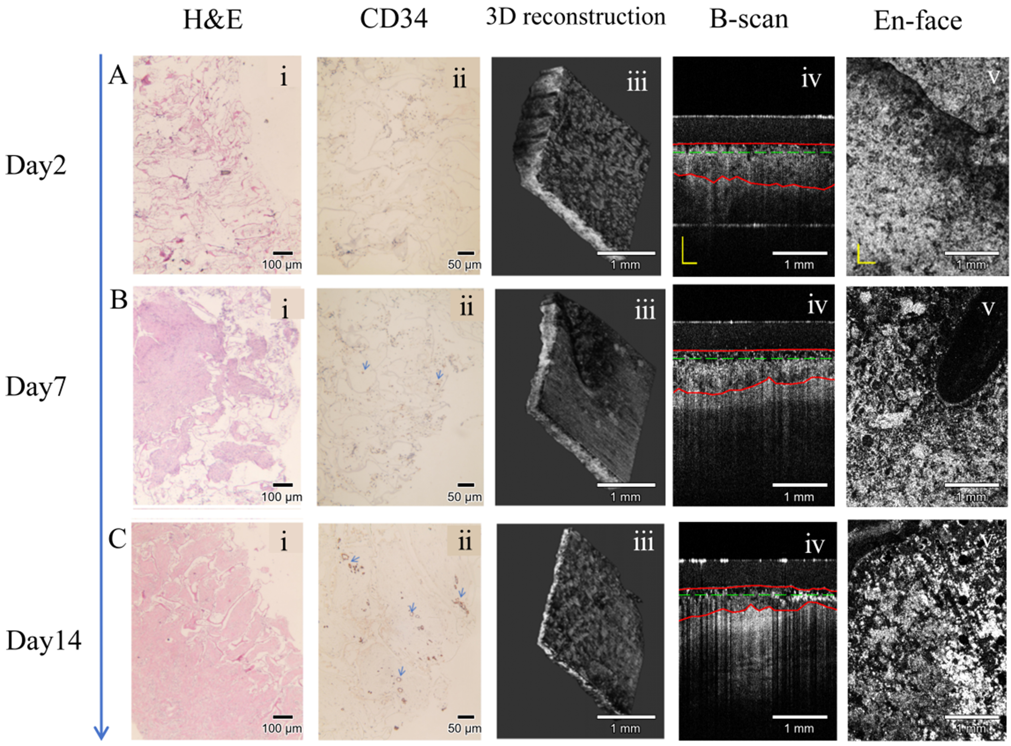
Figure 5. Representative clinical validation images obtained on postoperative days 2 (A), 7 (B), and 14 (C): (i) H&E-stained histological sections; (ii) CD34 immunohistochemistry; (iii) three-dimensional reconstructions of the collagen sponge layer; (iv) B-scan OCT images; and (v) en face OCT images. H&E: Hematoxylin and eosin; CD34: cluster of differentiation 34; OCT: optical coherence tomography; B-scan: brightness scan.