

fig4
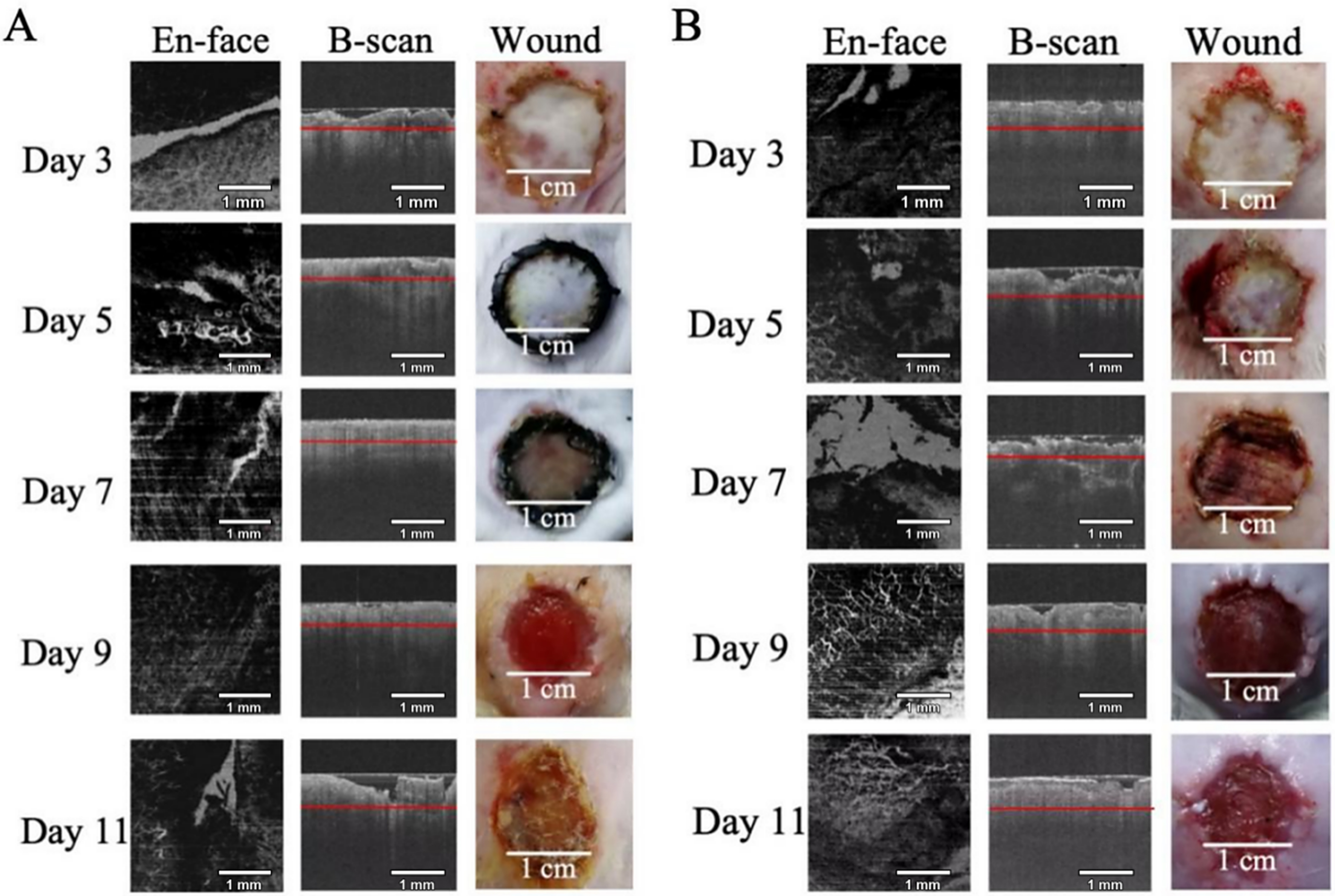
Figure 4. En face OCT images, corresponding B-scans, and macroscopic wound photographs on postoperative days 3, 5, 7, 9, and 11. (A) Wounds treated with autologous skin grafts and Beifuji (recombinant bovine basic fibroblast growth factor); (B) Wounds treated with autologous skin grafts alone (control group). Red lines on the B-scans indicate the depth at which the en face images were generated. B-scan: Brightness scan; OCT: optical coherence tomography.