

fig1
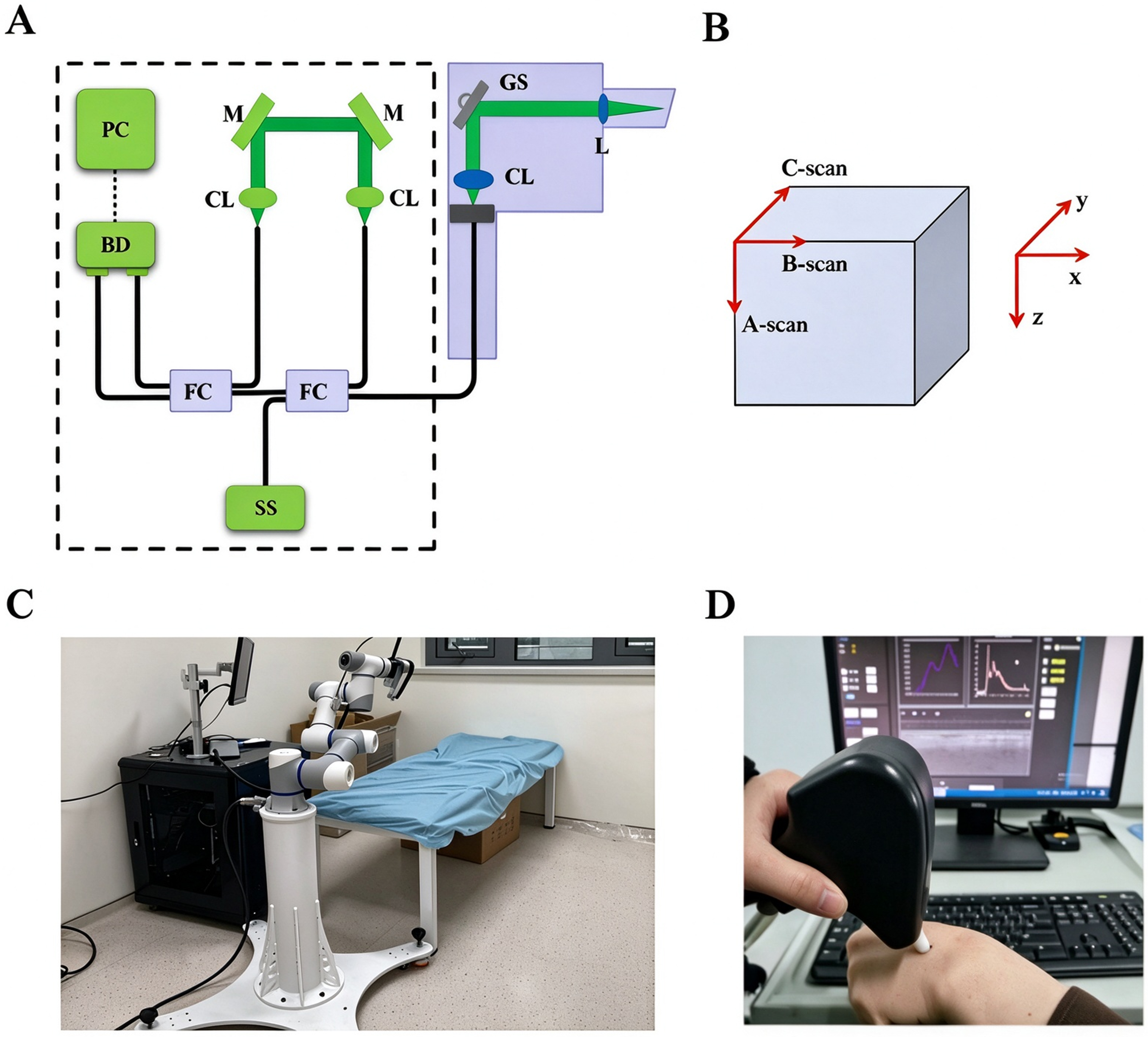
Figure 1. Schematic and setup of the OCT system. (A) Schematic illustrating the basic principles of OCT; (B) OCT scanning modes. A single scan along the depth direction (z-axis) is defined as an A-scan. A series of A-scans acquired along the lateral direction (x-axis) forms a two-dimensional cross-sectional B-scan; (C) Overview of the OCT instrument; (D) Photograph of the OCT system in operation. (C and D) are original photographs captured by the authors. OCT: Optical coherence tomography; A-scan: amplitude scan; B-scan: brightness scan.