

An autonomous regenerative bioelectronic dressing powered by wound exudate for high-quality skin repair
 0
0 Graphical Abstract
Abstract
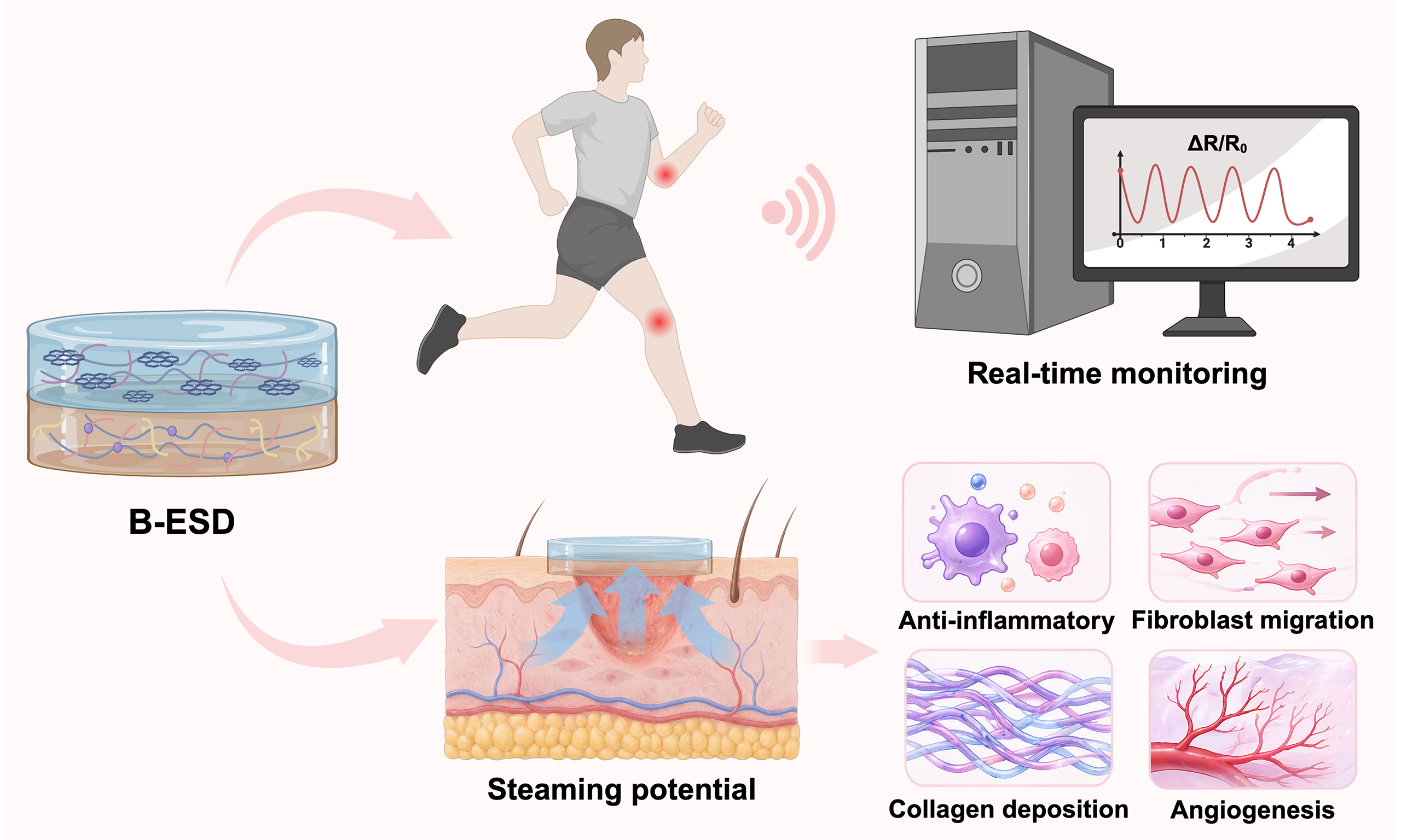
Endogenous electric field (EF) plays a crucial role in the wound healing process, yet conventional electrical stimulation strategies remain constrained by their dependence on bulky external power supplies. Herein, we report a bilayer electrogenerative smart dressing (B-ESD) that generates a self-sustained EF through a robust streaming potential induced by unidirectional pumping of wound exudate. This biofluid-driven mechanism successfully amplified the attenuated wound potential from 343.8 to 921.0 mV without requiring any external power source. Comprehensive in vitro and in vivo evaluations demonstrated that this self-sustained EF mitigated acute inflammation, accelerated fibroblast migration, and promoted highly organized collagen deposition. Immunohistochemical analysis further revealed that the B-ESD regulated the expression of Type I collagen, CD31, and transforming growth factor-beta 1 at the wound site, improving the quality of wound repair. Consequently, the B-ESD achieves a 98.72% wound closure rate within 14 days, with reduced scar formation and the regrowth of mature skin appendages. Moreover, the B-ESD serves as a high-fidelity sensor to monitor physiological motions, offering a promising integrated platform that combines bioelectric wound therapy with real-time biomechanical monitoring for advanced wound management in the future.
Keywords


 Download PDF
INTRODUCTION
As the primary barrier of the human body, the skin shields internal tissues from external physical, chemical, and biological threats[1]. However, skin is highly susceptible to injury, and large open wounds remain a significant global public health challenge, affecting millions of individuals annually[2]. Cutaneous wound healing proceeds through four overlapping yet distinct stages: hemostasis, inflammation, proliferation, and remodeling[3,4]. When any of these stages is disturbed, healing tends to be delayed, inflammation is often prolonged, and pathological scars may form[5]. Dressings that can both accelerate tissue repair and limit scarring have therefore become a major goal in wound care research[6].
Multiple factors influence the wound healing process, including biochemical signals[7,8], inflammatory responses[9,10], and biophysical stimuli[11]. Among these, electrical stimulation (ES) is of particular interest because it can mimic or strengthen the endogenous electric field (EF)[12,13]. Upon skin injury, the transepithelial potential (TEP) collapses at the wound site, and the resulting lateral voltage gradient gives rise to the wound endogenous EF[14]. This endogenous EF is essential for healing, as it directs fibroblast proliferation and migration and thus supports re-epithelialization and angiogenesis[15]. However, conventional ES devices depend on external power supplies and wired electrodes, and their bulky hardware, limited portability, and potential battery leakage restrict clinical use[16-18]. Moreover, most contemporary dressings function solely as passive therapeutic barriers, lacking the diagnostic capability to provide real-time physiological feedback[19]. To overcome these obstacles, there is an urgent need for smart dressings capable of generating self-sustained bioelectric stimulation without external power while simultaneously serving as a reliable real-time monitor.
In this study, we developed a bilayer electrogenerative smart dressing (B-ESD) that integrates endogenous EF reconstruction with high-fidelity physiological sensing for smart wound management. The B-ESD exhibits exceptional biocompatibility, balanced stretchability (138.2%), compressive resistance (225.1 kPa), high conductivity (1.31 S/m), tissue adhesiveness (7.5 kPa) and exudate management. The engineered wettability gradient induces a capillary-driven unidirectional exudate transport. This flow interacts with the carboxylate matrix, generating a continuous streaming potential. This biofluid-driven mechanism successfully enhanced the attenuated wound EF to 184.2 V/m without requiring any external power source. In vivo evaluations in a full-thickness rat model demonstrate that this enhanced EF accelerated the resolution of inflammation, directed fibroblast migration and proliferation to promote re-epithelialization, and supported highly organized collagen deposition. Notably, the B-ESD advanced the remodeling period by restoring a mature epithelial microstructure, ultimately improving repair quality and inhibiting scar formation. Furthermore, the B-ESD accurately monitors multi-scale physiological activities and joint kinematics in real time through deformation-induced resistance variations. This multifunctional hydrogel platform provides a self-driven strategy for concurrent wound therapy and personalized biomechanical monitoring.
EXPERIMENTAL
Materials
Acrylamide (AM, ≥ 99%), sodium alginate (SA, low viscosity), and sodium carboxymethyl cellulose (CMC, M.W. 250,000) were purchased from Sigma-Aldrich (St. Louis, MO, USA). Dopamine hydrochloride (DA, 98%) was obtained from Aladdin Biochemical Technology Co., Ltd. (Shanghai, China). Anhydrous calcium chloride (CaCl2 ≥ 96%), N,N-methylenebisacrylamide (MBAA), ammonium persulfate (APS), and N,N,N',N'-tetramethylethylenediamine (TEMED) were supplied by Sinopharm Chemical Reagent Co., Ltd. (Shanghai, China). Reduced graphene oxide (rGO) dispersion was acquired from Nanjing XFNANO Materials Tech Co., Ltd. (Nanjing, China).
For biological assays, human foreskin fibroblasts (HFFs) were purchased from the Cell Bank of the Chinese Academy of Sciences. Dulbecco’s modified Eagle’s medium (DMEM), fetal bovine serum (FBS), and penicillin/streptomycin were purchased from Gibco (Grand Island, NY, USA). The Cell Counting Kit-8 (CCK-8) and Live/Dead Viability/Cytotoxicity Kit were obtained from Beyotime Biotechnology (Shanghai, China). Primary antibodies against CD31, transforming growth factor-beta 1 (TGF-β1), Type I collagen (Col I), and corresponding secondary antibodies were purchased from Abcam (Cambridge, UK). Phosphate-buffered saline (PBS), 4% paraformaldehyde, hematoxylin and eosin (H&E) staining kit, and Masson’s Trichrome staining kit were obtained from Servicebio Technology (Wuhan, China). Sodium hydroxide (NaOH) and normal saline (0.9% NaCl) were supplied by Sigma-Aldrich (St. Louis, MO, USA). Tegaderm transparent film dressing was purchased from 3M (St. Paul, MN, USA). Fresh porcine skin used for the adhesion tests was purchased from a local market in Shanghai, China, and used within 6 h after collection. All other chemicals were of analytical grade and used as received without further purification. Deionized (DI) water (18.2 MΩ·cm) purified by a Milli-Q system (Millipore, USA) was used throughout the experiments.
Fabrication of the PCCD layer
The PCCD hydrogel [polyacrylamide/sodium carboxymethyl cellulose/calcium chloride/dopamine (PAM/CMC/CaCl2/DA, PCCD)] served as the bottom layer and was synthesized via a one-pot free radical polymerization method. Briefly, DA powder was dissolved in an alkaline aqueous solution (pH = 11) and vigorously stirred under ambient air for 20 min to form polydopamine (PDA). Concurrently, CMC and CaCl2 were dissolved in DI water at 60 °C and stirred for 2 h to ensure thorough metal chelation. Then, the AM monomer was dissolved into the mixture, followed by the addition of the crosslinker MBAA (0.04% w/v relative to AM) and the initiator APS (1% w/v relative to AM). The pre-gel solution was degassed to remove air bubbles, injected into a mold, and polymerized at room temperature to obtain the PCCD hydrogel. The optimized formulation, denoted as PC0.15C0.10D (containing 0.15% w/v CMC and 0.10 M CaCl2), was used for subsequent experiments unless otherwise specified.
Fabrication of the B-ESD
To construct B-ESD, the top PSG layer [polyacrylamide (PAM)/SA/rGO] was cast directly onto the pre-formed PCCD layer to ensure robust interfacial adhesion. First, SA (1.0% w/v relative to AM) and a defined amount of rGO were dispersed in DI water via ultrasonication, followed by the dissolution of AM, MBAA, APS and TEMED to form the PSG pre-gel solution. This solution was then carefully poured onto the surface of the semi-cured PCCD hydrogel in the mold. The final B-ESD was peeled off and stored in a humidified container for further characterization. To investigate the effect of rGO content on the electrical properties of B-ESD, PSG pre-gel solutions containing different rGO contents were prepared under otherwise identical conditions. Specifically, rGO contents of 0, 0.5, 1.0, and 2.0 wt% relative to AM were used and the corresponding samples were denoted as rGO0, rGO0.5, rGO1.0, and rGO2.0, respectively. The rGO1.0 formulation was used as the optimized formulation in the main study.
Characterizations
The chemical structures of the hydrogels were analyzed using Fourier transform infrared spectroscopy (FTIR, Nicolet iS20, Thermo Fisher, USA) in the range of 4,000-300 cm-1. The surface and cross-sectional morphologies were observed by scanning electron microscopy (SEM, SU8100, Hitachi, Japan) after freeze-drying and gold sputtering.
Mechanical and adhesive property tests
A universal testing machine (Instron 5967, USA) was used for both tensile and compressive measurements. Adhesion was evaluated by the lap shear method on several substrates, including porcine skin, glass, and metal. Fresh porcine skin from a local market (Shanghai, China) was stripped of subcutaneous fat and residual hair, rinsed with DI water and PBS, and tested within 6 h. The hydrogel was sandwiched between two substrate pieces with a contact area of 20 × 20 mm2. The assembly was pulled at a speed of 10 mm/min until separation, and the adhesion strength was calculated by dividing the maximum load by the contact area.
Swelling and water contact angle tests
The swelling ratio (SR) was measured gravimetrically. Dried hydrogels were weighed (Wd) and immersed in PBS (pH 7.4) at 37 °C. At predetermined time intervals, the samples were removed, surface water was gently blotted, and the wet weight (Ws) was recorded. The SR was calculated as SR (%) = (Ws - Wd)/Wd × 100%. The surface hydrophilicity was evaluated using a contact angle goniometer (JY-82C, Chengde Dingsheng, China). A 5 μL droplet of DI water was placed on the surface of the PSG and PCCD layers, respectively. The dynamic change of the contact angle was recorded to analyze the water absorption rate.
Biocompatibility evaluation
Cell Culture: HFFs were purchased from the Cell Bank of the Chinese Academy of Sciences. The cells were authenticated by the supplier and confirmed to be free of mycoplasma, bacterial, and fungal contamination before use. HFFs were cultured in DMEM supplemented with 10% FBS and 1% penicillin/streptomycin at
Live/Dead Assay: Hydrogel extracts were prepared by soaking samples in culture medium for 24 h. HFFs were seeded in 24-well plates and incubated with the extracts. After 1, 2, and 3 days, cells were stained with Calcein-AM (live) and Propidium Iodide (dead) and imaged using a fluorescence microscope (Nikon, Badhoevedorp, the Netherlands).
CCK-8 Assay: Cell proliferation was quantified using the CCK-8. HFFs were incubated with hydrogel extracts for 4, 8, 24 and 48 h. The optical density (OD) at 450 nm was measured using a microplate reader (Multiskan FC, Thermo Fisher Scientific, China).
Hemolysis Test: Fresh Sprague-Dawley (SD) rat blood was obtained from orbital venous blood collection of healthy SPF-grade SD rats maintained in our animal facility. Blood collection was performed under deep anesthesia to minimize pain and stress. The donor rats used for blood collection were separate from those used in the wound-healing experiments. The collected blood was diluted with normal saline before use. Hydrogel samples were incubated with diluted blood at 37 °C for 1 h. PBS and deionized water (ddH2O) served as negative and positive controls, respectively. The supernatant absorbance at 545 nm was measured to calculate the hemolysis ratio.
In vitro cell migration assay
To independently evaluate the effect of ES generated by the B-ESD on cell migration, a modified non-contact scratch wound-healing assay was performed. Briefly, HFFs were seeded into the lower chamber of a 6-well plate and cultured to achieve a 100% confluent monolayer. To decouple cell migration from proliferation, the cells were subjected to serum starvation for 12 h prior to the assay. A standardized linear scratch was subsequently created across the cell monolayer using a sterile 200 μL pipette tip. The functional samples were then sterilized and strategically placed in the upper Transwell inserts. The co-culture system was then incubated under standard physiological conditions. The progression of wound closure was monitored, and images were captured at 0, 12 and 24 h. The cell migration ratio was calculated according to:
Cell Migration Ratio (%) =
Electrical performance measurements
For the in situ electrical measurements, a separate cohort of rats was used. Rats were randomly divided into four groups: Control (untreated), PSG, PCCD, and B-ESD (n = 3 per group). The open-circuit voltage of the hydrogel samples was measured using a digital source meter (Keithley 2450, Tektronix, USA) with a current source output of 0 μA. To evaluate the wound EF, the potential difference was measured between the wound center and the epidermis at the wound edge, with the wound-edge epidermis used as the reference point. For the 10 mm circular wound model, the center-to-edge distance was taken as 5 mm, and the local EF was calculated as E = ΔV/d, where ΔV is the measured potential difference and d is the measurement distance. The reported values are presented as mean ± standard deviation from multiple wounds in each group.
Ex vivo electrogenerative output characterization
The simulated exudate (PBS, pH = 7.4) was perfused through the B-ESD at controlled flow rates (0, 5, 10 and 20 μL/min) using a syringe pump, and the open-circuit voltage (OCV) and short-circuit current (Isc) were recorded with a Keithley 2450 source meter. Polarity-reversal controls were performed by inverting the device orientation or reversing the flow direction. Long-term output stability was evaluated under continuous perfusion, with the OCV recorded at 0, 1, 3, 6, 12, 24 and 48 h.
Water-retention and encapsulation test
Fully hydrated samples were weighed (W0) and maintained at 32 °C to simulate the skin-surface temperature. The sample weight (Wt) was recorded at 0, 1, 3, 6, 12, 24 and 48 h, and the water retention was calculated as Wt/W0 × 100%. For the encapsulated group (E-B-ESD), the hydrated B-ESD was covered with a Tegaderm 3M transparent film before the test. The OCV retention of encapsulated and non-encapsulated samples was recorded in parallel over 48 h.
In vivo wound healing assessment
Animal Model: All animal procedures were approved by the Institutional Animal Care and Use Committee (IACUC) of Fudan University (Approval No. YSAPL20260269). Adult male SD rats (200-250 g) were housed in an SPF-grade facility under controlled temperature (22-25 °C), humidity (50%-60%), and a 12 h light/dark cycle, with free access to food and water. All rats were acclimatized for 7 days before surgery. Before wound modeling, rats were anesthetized by intraperitoneal injection of 1 mL of 1.25% tribromoethanol (Avertin), and a full-thickness circular excisional wound (10 mm diameter) was created on the dorsal skin under sterile conditions.
Animal Grouping: Rats were randomly divided into four groups: Control (untreated), Tegaderm 3M, PCCD, and B-ESD. For longitudinal wound-closure monitoring, 5 rats per group were followed until day 14. These same animals were euthanized on day 14 for histological and immunohistochemical analyses. To assess intermediate histological changes, an additional independent cohort of 3 rats per group was euthanized on day 7 for tissue collection. Thus, the wound-healing and histological/immunohistochemistry (IHC) experiments used 8 rats per group in total, and no animal was sampled at more than one terminal time point.
Treatment: After wound creation, the wound surface was gently rinsed with sterile normal saline and covered with the assigned dressing. No topical or systemic antibiotics were routinely applied. Dressings were changed every 2 days, and wound healing and signs of infection were monitored daily. Humane endpoints included body-weight loss greater than 20%, persistent inability to eat or drink, severe wound infection or bleeding, abnormal vital signs, or irreversible motor dysfunction. No animal reached the humane endpoints during the study.
Evaluation: Photographs of the wounds were taken on days 0, 3, 7, 10, and 14, and wound areas were measured by investigators blinded to the group allocation. The wound healing ratio was calculated as:
Wound Healing Ratio (%) =
Histological and immunohistochemical analysis
Wound tissues were collected from 3 rats per group on day 7 and from 5 rats per group on day 14. Normal skin samples were collected from unwounded dorsal skin regions of the same batch of SD rats at least 1 cm away from the wound edge. Samples were fixed in 4% paraformaldehyde, embedded in paraffin, and sectioned (5 μm). Sections were stained with H&E for morphological analysis and Masson’s Trichrome for collagen assessment. For IHC, sections were incubated with primary antibodies against CD31, Col I and TGF-β1, followed by secondary antibodies. Semi-quantitative analysis of immunohistochemical images was performed using ImageJ. The expression levels of Col I and TGF-β1 were quantified using the integrated optical density (IOD), which was calculated by multiplying the positive stained area by the mean optical density (IOD = Area × Mean OD). For CD31-stained sections, angiogenesis was evaluated by counting CD31-positive (brown-stained) vessels per unit area, reported as vessel density (vessels/mm2).
Sensing performance evaluation
To systematically evaluate the electromechanical responsiveness, the B-ESD was connected to a programmable universal testing machine (Instron 5967, USA) to undergo precisely controlled cyclic stretching, compression, and bending deformations. Concurrently, the real-time electrical signals were continuously acquired utilizing a Keithley 2450 source meter. For in vivo motion monitoring, the B-ESD was conformally attached to specific anatomical sites of healthy volunteers, including the throat for swallowing and deep breathing, finger joints for finger bending, the wrist for wrist bending, and the elbow joint for elbow bending. Swallowing and deep breathing were each repeated four times, whereas finger, wrist, and elbow bending were each repeated five times under the same testing conditions. A total of three healthy volunteers participated in the non-invasive motion-monitoring experiments. All experiments involving healthy were approved by Ethics Committee of the School of Life Sciences, Fudan University (Approval No.H2026078).
Statistical analysis
Data are presented as mean ± standard deviation unless otherwise stated. Statistical analyses were performed using ImageJ and Origin 2021. Schematic illustrations were created with BioRender.com under the appropriate license, as indicated in the corresponding figure captions. For the longitudinal wound-closure data, two-way repeated-measures analysis of variance (ANOVA) was used, with group as the between-subject factor and time as the within-subject factor. Between-group comparisons at each time point were then performed using one-way ANOVA followed by Tukey’s post hoc test. For all other datasets, statistical significance was assessed using one-way ANOVA followed by Tukey’s post hoc test. The significance of differences was indicated in the graphs by P value or by (*P < 0.05, **P < 0.01, ***P < 0.001). All experiments were performed with at least three independent replicates. Sample sizes (n) for each experiment are provided in the corresponding figure captions (n ≥ 3).
RESULTS AND DISCUSSION
Fabrication and characterizations of B-ESD
To meet the complex requirements of the wound microenvironment, the B-ESD was bioinspired to feature a bilayer architecture. It consists of a top PSG composite layer and a bottom PCCD composite layer. The PCCD layer was engineered as a tissue-adhesive, porous substrate with skin-like resilience, while the PSG layer provided a robust conductive backing [Figure 1]. Within the PCCD layer, CMC was incorporated to enhance strength via hydrogen bonding and DA was incorporated to facilitate robust interfacial bonding through synergistic reversible non-covalent and irreversible covalent interactions. Biologically relevant calcium ions have been proven to play important physiological functions, ranging from intracellular signal transduction to the regulation of biological tissue mechanics via chelation[20]. Inspired by this, Ca2+ ions were introduced to regulate inter-chain interactions through metal-chelate bonding with CMC. Within the PSG layer, PAM and SA were entangled to form a rigid double network, and the incorporation of rGO nanosheets increased the conductivity. Beyond these mechanical and adhesive functions, the bilayer design is essential for electrogeneration. The porous PCCD layer promotes wound-exudate uptake and directional ion transport through the carboxylate-rich hydrogel network, whereas the rGO-containing PSG layer serves as a conductive charge-collecting and charge-spreading layer. The coupled liquid transport and interfacial charge redistribution generate a streaming potential, thereby providing a self-sustained electrical cue at the wound interface [Figure 1]. The B-ESD was fabricated via a sequential casting strategy, where the PSG precursor was cast directly onto the pre-polymerized PCCD substrate [Figure 2A]. Unlike simple physical lamination, this method ensures robust interfacial adhesion through interpenetrating polymer networks and covalent/non-covalent bonding at the junction [Figure 2B].
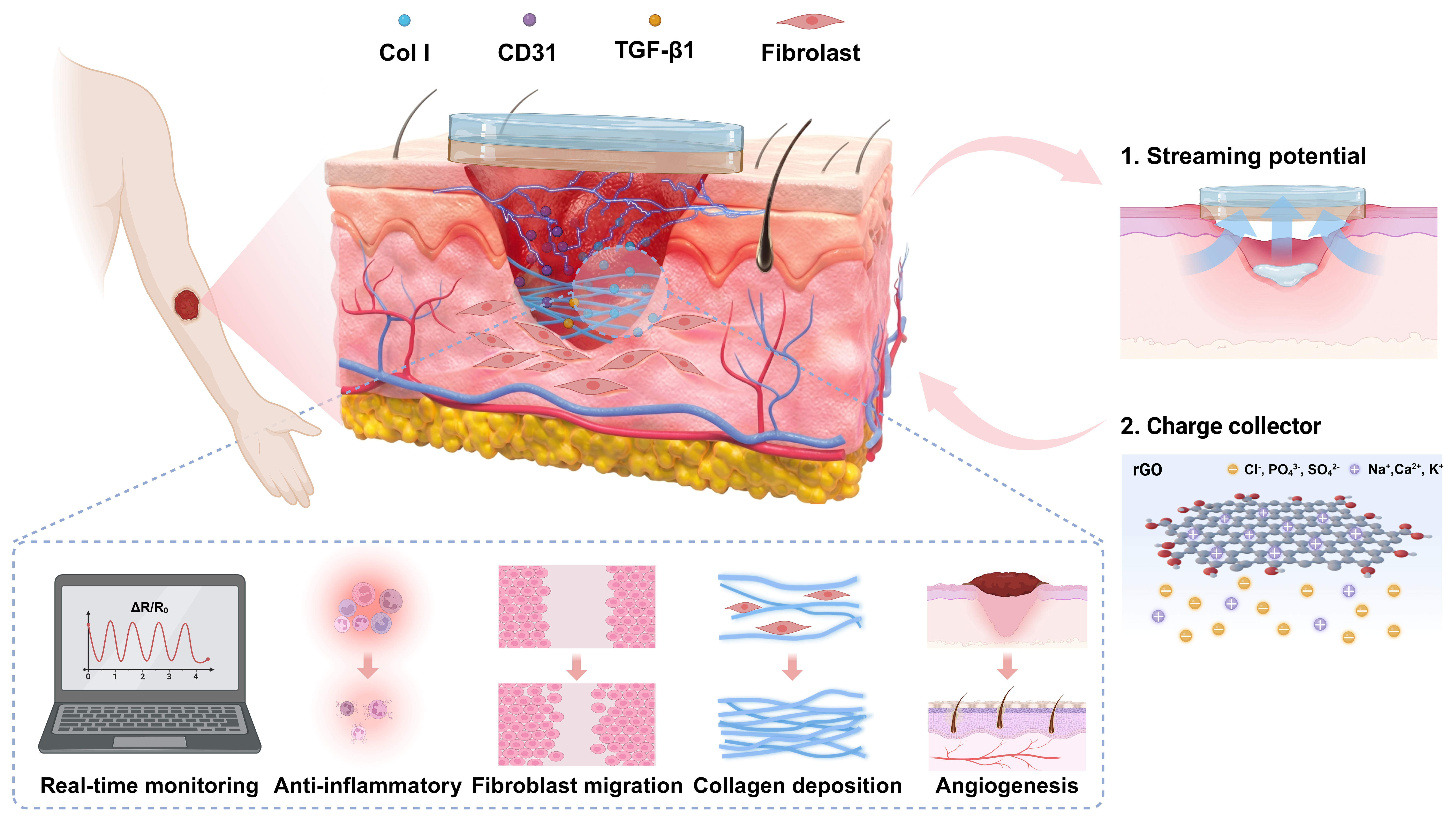
Figure 1. Design and working mechanism of the B-ESD for reduced scar formation and real-time monitoring. Created in BioRender. Du, X. (2026) https://BioRender.com/p4rb6uu. B-ESD: Bilayer electrogenerative smart dressing; Col I: Type I collagen; TGF-β1: transforming growth factor-beta 1; rGO: reduced graphene oxide.
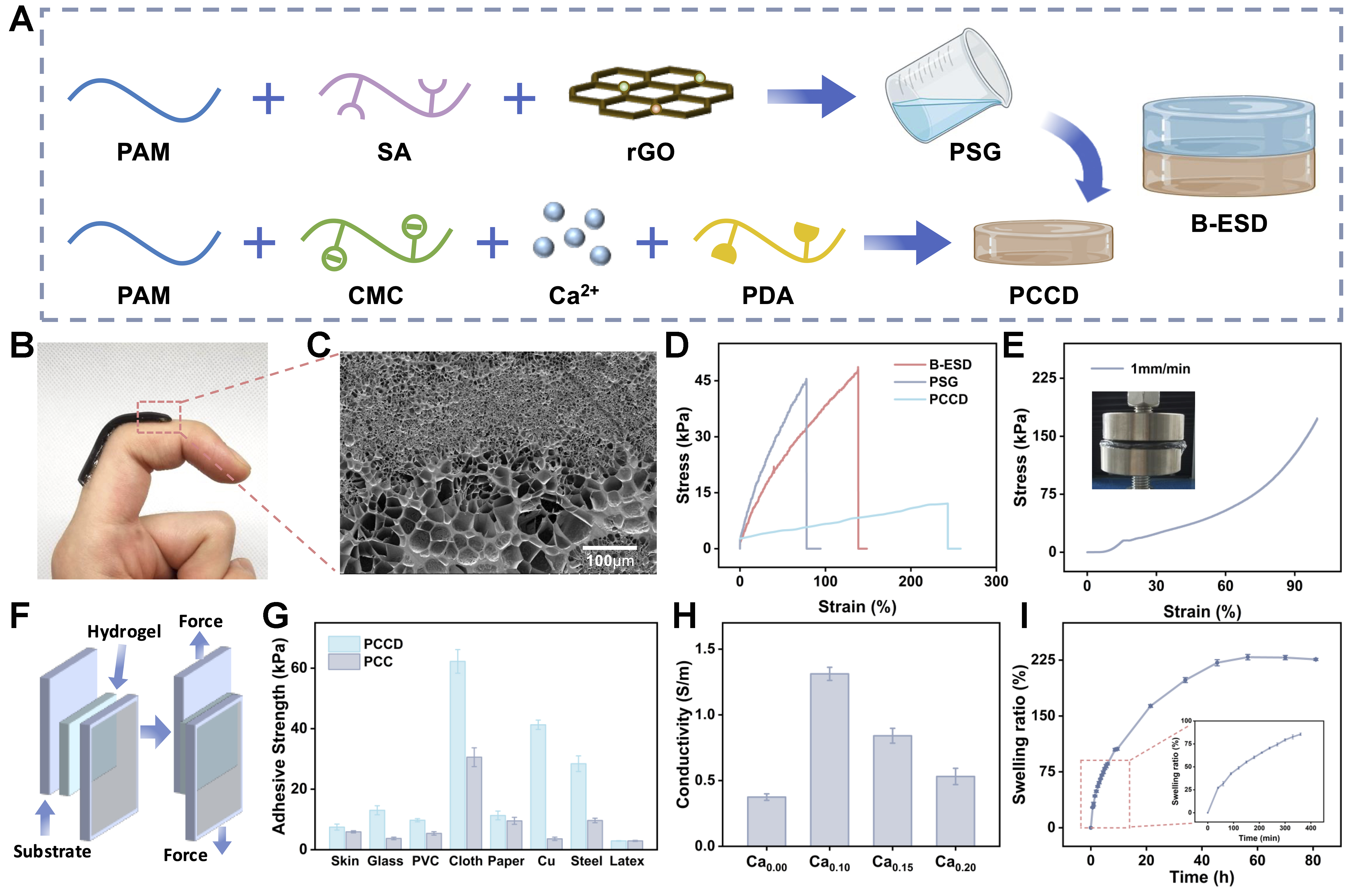
Figure 2. Fabrication and characterization of the B-ESD. (A) Schematic of the sequential casting strategy for fabricating the B-ESD; (B) Digital photograph of the B-ESD applied on the finger joint taken by the authors; (C) SEM image of the B-ESD cross-section showing a seamless interface and pore-size gradient; (D) Tensile stress-strain curves of PSG, PCCD, and B-ESD; (E) Compression stress-strain curve of B-ESD. The inset displays a photograph of the experimental setup during the compression test; (F) Schematic of lap shear test; (G) Adhesive strength of PCCD and PCC on various substrates; (H) Conductivity of the B-ESD at different Ca2+ concentrations (subscripts denote concentration in mol/L); (I) SR curve of B-ESD. Data are expressed as mean ± standard deviation (n = 3). B-ESD: Bilayer electrogenerative smart dressing; SEM: scanning electron microscopy; PSG: polyacrylamide/sodium alginate/reduced graphene oxide (PAM/SA/rGO); PCCD: polyacrylamide/sodium carboxymethyl cellulose/calcium chloride/dopamine; PCC: polyacrylamide/sodium carboxymethyl cellulose/calcium chloride; SR: swelling ratio; PAM: polyacrylamide; SA: sodium alginate; rGO: reduced graphene oxide; CMC: sodium carboxymethyl cellulose; PDA: polydopamine; PVC: polyvinyl chloride.
The structural and chemical integration of the bilayer network was systematically verified. FTIR analysis confirmed the successful polymerization and interactions among components [Supplementary Figure 1]. Specifically, the significant broadening of the O–H/N–H stretching band around 3,400 cm-1 indicates the formation of a robust hydrogen-bonding network within both hydrogel layers. Furthermore, the distinct appearance and shifts of the carboxylate peaks (at around 1,600 and 1,400 cm-1) explicitly confirm the successful integration of SA and the metal-chelate bonding between Ca2+ ions and CMC. Morphologically, the SEM image of the B-ESD cross-section revealed a seamless interface without obvious delamination, guaranteeing structural integrity during subsequent mechanical deformations [Figure 2C].
Mechanically, this dual-layer reinforcement strategy addressed the common trade-off between adhesion and cohesion. The PCCD layer achieved a maximum tensile strain of 243.2% and a stress of 12.1 kPa at the optimized Ca2+ and CMC concentrations [Supplementary Figure 2]. The PSG layer exhibited high tensile stress of 45.5 kPa but limited extensibility. The B-ESD, however, combined the advantages of both components, achieving a high tensile stress of 48.7 kPa and a balanced elongation at break of 138.2% [Figure 2D]. The elastic modulus of B-ESD, calculated from the initial linear region (0.5%-15% strain) of the tensile stress–strain curve, was determined to be 61.2 kPa, indicating a compliant mechanical profile suitable for intimate contact with soft skin tissue [Supplementary Figure 3][21]. Furthermore, the B-ESD exhibited high compressive resistance, withstanding over 225.1 kPa of stress at 99.7% strain without structural failure [Figure 2E], making it suitable for applications on high-mobility body parts.
To leverage mussel-inspired adhesive strategies, DA was incorporated into the PCCD matrix. This superior adhesive performance is primarily ascribed to the catechol group of DA, which exhibits potent binding affinities across diverse substrates[22]. Lap shear tests [Figure 2F] demonstrated that PCCD significantly outperformed its DA-free counterpart (PCC), with an enhancement of 26.5% on the adhesion strength of porcine skin, reaching 7.5 kPa [Figure 2G]. This moderate adhesion was functionally required for the intended wound-dressing application. It enables conformal fixation on curved and high-mobility body regions, reduces interfacial displacement during joint motion, and helps maintain stable skin–hydrogel contact for reliable electrical recording, while avoiding excessive adhesion that may cause secondary trauma during dressing replacement [Figure 2B].
An ideal conductivity is crucial for maintaining the endogenous bioelectric environment of the wound site and ensures multiple applications of B-ESD as a real-time physiological sensor and monitor. The PCCD layer provides ionic pathways facilitated by mobile Na+, Ca2+, and Cl- ions, while the rGO nanosheets in the PSG layer create an electronically conductive network[23-26]. The conductivity of the hydrogel reached a maximum of 1.31 S/m at the optimized Ca2+ concentration [Figure 2H]. Notably, this value is highly compatible with the physiological conductivity of human tissues, which typically ranges from 0.1 to 1.5 S/m. Such electrical matching minimizes the contact impedance at the hydrogel-tissue interface[27,28].
Favorable SRs and exudate management are also crucial properties for wound healing applications. The B-ESD exhibited rapid swelling during the initial 7 h, reaching an equilibrium SR of 221.4% after 45.5 h [Figure 2I]. The pronounced water absorption capability and swelling properties of the B-ESD enable effective absorption of wound exudate, especially during the first few days of wound healing, thereby reducing infection risk and providing an ideal moist environment for recovery[29].
Biocompatibility and biosafety of B-ESD
Fibroblasts are critical for wound healing[30]. To evaluate biocompatibility and biosafety of PCCD, PSG and B-ESD, a series of experiments were conducted to systematically assess the potential effects of the hydrogels on the proliferation, migration, and cellular functions of HFFs.
First, live/dead fluorescence staining was performed by co-culturing HFF cells with extracts of the three hydrogels. Live cells were distinguished by green fluorescence, and dead cells by red fluorescence. Cells in all hydrogel groups and the control group exhibited continuous proliferation, with a progressive, time-dependent increase in cell number over the 3-day culture period in all groups [Figure 3A]. Quantitative statistics of cell viability further confirmed this conclusion [Figure 3B]. At each time point, no significant differences were observed between the hydrogel-treated groups and the control group (P > 0.05). CCK-8 assays were also conducted separately for the three hydrogels to further evaluate the cytocompatibility. After 48 h of co-culture with HFF cells, the cell viability in all groups exceeded 95% (P > 0.05), with the B-ESD group reaching 101%, indicating that none of the hydrogels exhibited noticeable cytotoxicity [Figure 3C-E].
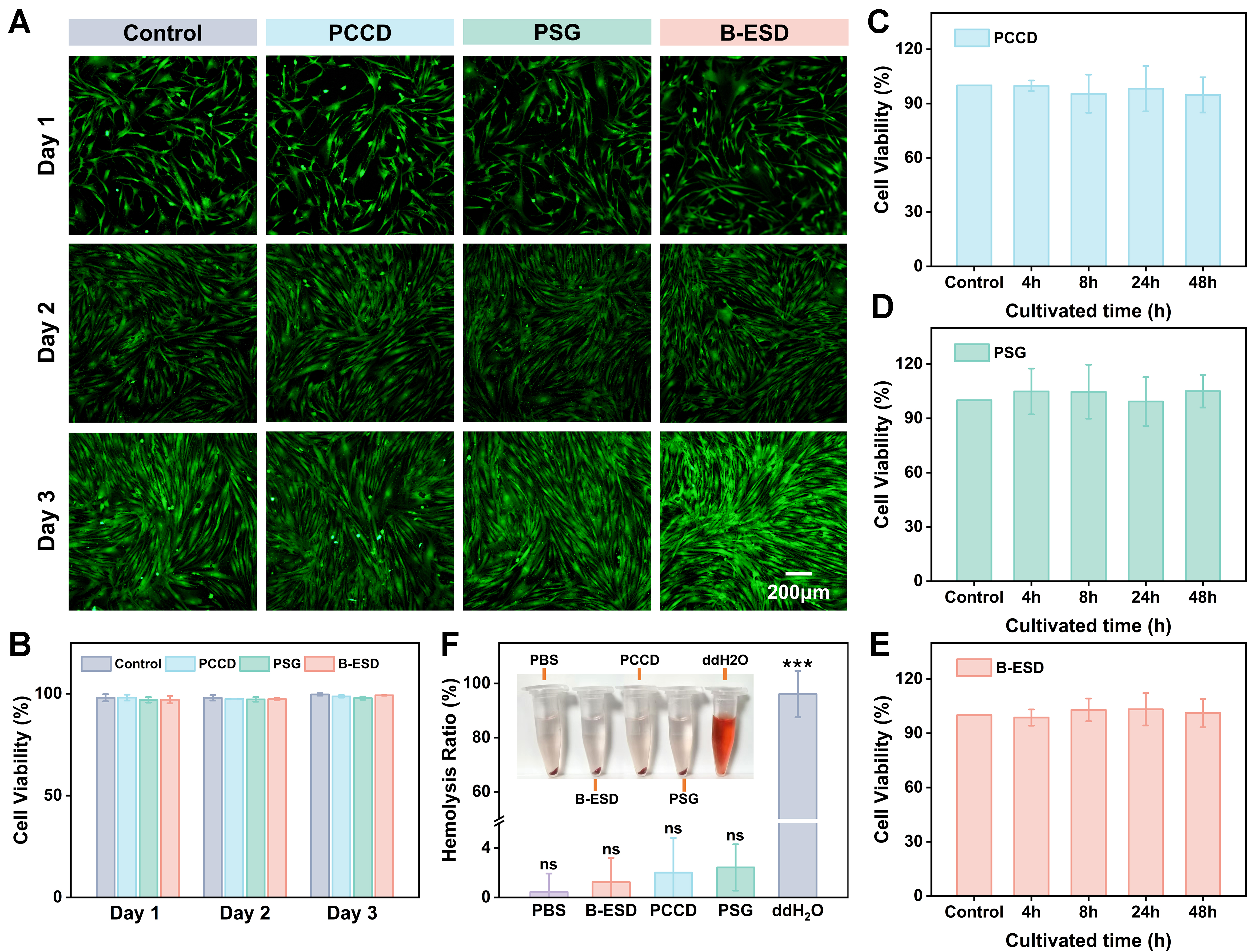
Figure 3. Biocompatibility and biosafety evaluations. (A) Live/dead fluorescence staining images of HFF cells co-cultured with different hydrogel extracts at day 1, 2, and 3; (B) Quantitative analysis of cell viability based on live/dead staining; (C-E) Cell viability of HFF cells evaluated by CCK-8 assay over 48 h of co-culture with extracts of (C) PCCD, (D) PSG, and (E) B-ESD; (F) Hemolysis assay of the hydrogel extracts. Data are expressed as mean ± standard deviation (n = 3, ***P < 0.001, ns: not significant). HFF: Human foreskin fibroblast; CCK-8: Cell Counting Kit-8; PCCD: polyacrylamide/sodium carboxymethyl cellulose/calcium chloride/dopamine; PSG: polyacrylamide/sodium alginate/reduced graphene oxide (PAM/SA/rGO); B-ESD: bilayer electrogenerative smart dressing; PBS: phosphate-buffered saline; ddH2O: double-distilled water.
Furthermore, hemolysis assays showed that none of the hydrogel extracts induced red blood cell rupture upon contact. After centrifugation, the supernatants appeared colorless and transparent, with no significant difference compared to the PBS control group [Figure 3F]. The hemolysis rates of all hydrogel groups remained below the 5% threshold established by the American Society for Testing and Materials standard practice (ASTM: F 756-00), indicating superior hemocompatibility. These experimental results further validate the biosafety of B-ESD for biomedical applications. In addition, the hydrogels positively influence fibroblast proliferation and viability, suggesting their potential in promoting cell growth and tissue repair.
Effect of B-ESD on wound endogenous EFs
EFs play a pivotal regulatory role in wound healing processes by orchestrating cell migration, promoting angiogenesis, and modulating inflammatory responses. However, large-open wounds are frequently characterized by significantly attenuated EFs, primarily attributable to ionic gradient disruption and impaired charge conduction resulting from tissue damage[31]. Herein, the asymmetric B-ESD successfully restored and amplified this endogenous EF through a self-sustained electrokinetic mechanism [Figure 4A].
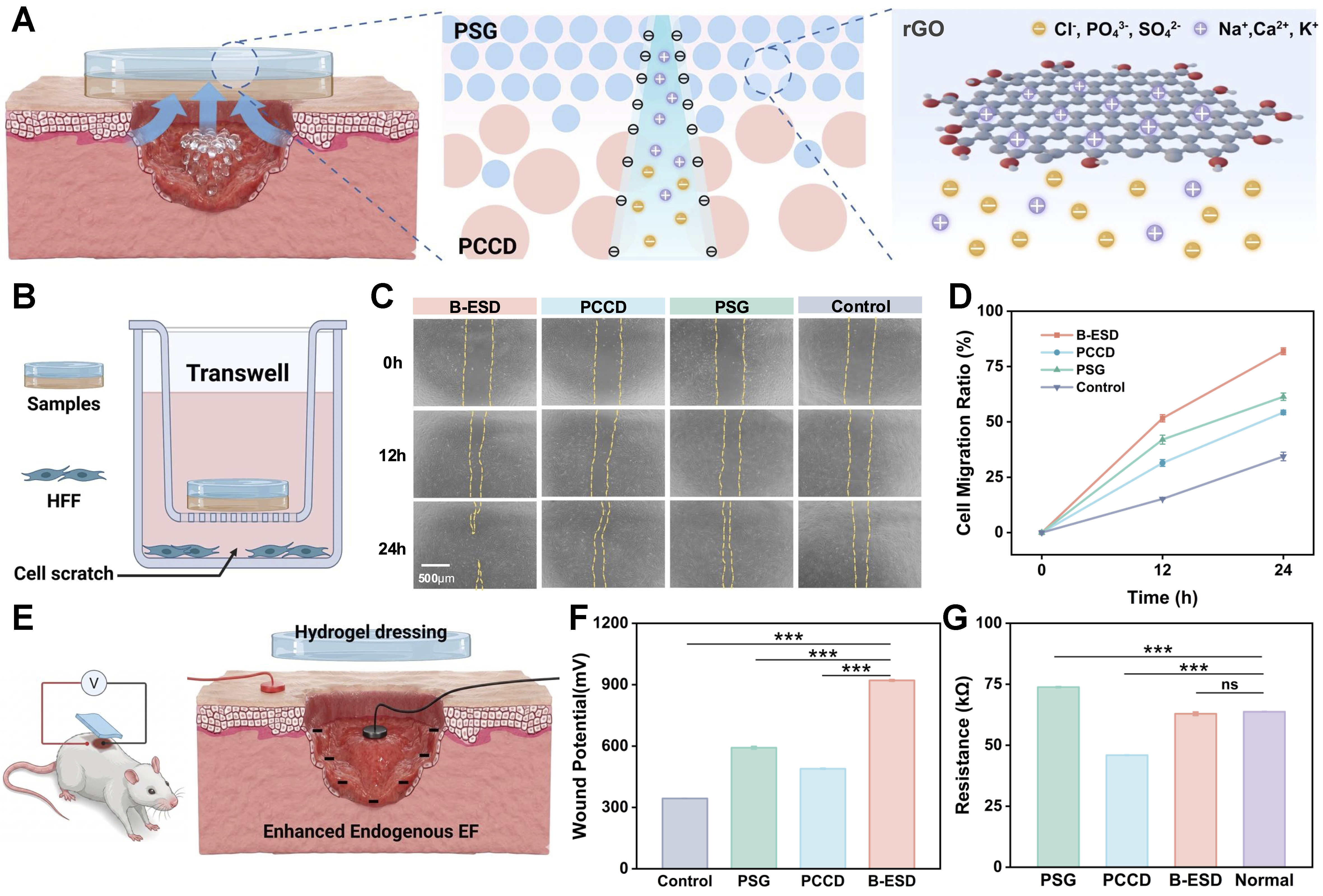
Figure 4. Mechanism of endogenous EF amplification and subsequent in vitro and in vivo evaluations. (A) Schematic illustration of streaming potential generation driven by unidirectional exudate pumping and rGO charge collection; (B) Setup of the modified Transwell cell scratch healing test; (C) Representative images and (D) quantitative analysis of HFF cell migration ratios at 0, 12, and 24 h; (E) Schematic of in situ potential measurement on a rat wound model; (F) Wound potential and (G) resistance tested under different treatments. Data are expressed as mean ± standard deviation (n = 3, ***P < 0.001, ns: not significant). (A, B, and E) were created with BioRender. Du, X. (2026) https://BioRender.com/6b3d3he. EF: Electric field; rGO: reduced graphene oxide; HFF: human foreskin fibroblast; PSG: polyacrylamide/sodium alginate/reduced graphene oxide (PAM/SA/rGO); PCCD: polyacrylamide/sodium carboxymethyl cellulose/calcium chloride/dopamine; B-ESD: bilayer electrogenerative smart dressing.
The B-ESD possessed a wettability gradient, with the bottom PCCD layer absorbing exudate rapidly (3 s) compared to the top PSG layer (9 s) [Supplementary Figure 4]. Coupled with an engineered pore size gradient, this asymmetric structure supports preferential bottom-to-top fluid transport[32]. As the exudate flows through the hydrogel network, migrating cations (e.g., Na+, Ca2+) interact with the dense, negatively charged carboxylate groups (-COO-) of the CMC and SA matrix. This convective ion transport generates a robust streaming potential, inducing vertical charge separation. Crucially, the B-ESD overcomes the limitations of single-layer hydrogels through a unique bilayer structure, in which rGO is integrated into the top layer. By acting as a robust charge collector, the integrated rGO stabilizes arriving ions through cation–π interactions, thereby preventing premature EF attenuation [Figure 4A][33]. To further validate the electrical contribution of rGO, an rGO-free bilayer hydrogel was prepared by removing rGO from the top PSG layer while maintaining the same PCCD bottom layer and bilayer configuration. Compared with the rGO-containing B-ESD, the rGO-free bilayer exhibited lower electrical conductivity, higher resistance, weaker voltage output, and faster voltage attenuation in the PBS-hydrated state [Supplementary Figures 5 and 6]. These results indicate that rGO functions as a conductive charge-collecting and charge-retention component, thereby contributing to the maintenance of the streaming-potential-derived electrical output. Under controlled perfusion of simulated wound exudate, the isolated B-ESD generated an open-circuit voltage of approximately 135 mV and a short-circuit current of approximately 90 nA. The output increased with the flow rate and saturated above 10 μL/min, and its polarity reversed upon inverting the device orientation or reversing the flow direction, confirming the flow-driven electrokinetic origin of the electrical output [Supplementary Figure 7].
A modified cell scratch healing test was further performed to evaluate whether fibroblast migration is enhanced in the presence of the electrogenerative hydrogel dressings. The experimental setup is shown in Figure 4B, where the HFFs were seeded in the lower chamber, while the samples were placed in the upper Transwell chamber. A standardized linear scratch was subsequently created across the cell monolayer using a sterile 200 μL pipette tip. The results showed that cells treated with the B-ESD exhibited a markedly faster migration rate than those in the other groups [Figure 4C]. Quantitative analysis revealed that the cell migration ratio in the B-ESD group reached 81.9% after 24 h, while the single-layer PSG and PCCD groups only reached 61.4% and 54.3%, respectively [Figure 4D].
To validate the bioelectrical performance of the dressing in a physiological environment, in situ voltage and electrical resistance were measured on full-thickness wounds in SD rats. A rat full-thickness skin defect model (5 mm in radius) was established, and the potential difference (ΔV) between the wound center and the epidermis edge was measured [Figure 4E and Supplementary Figure 8]. The corresponding electric field (E) was estimated as E = ΔV/r, where r = 5 mm in this model. The untreated wound exhibited a baseline potential of 343.8 mV, corresponding to a natural endogenous EF of 68.8 V/m. Upon application, the B-ESD-covered wound exhibited a markedly elevated potential of 921.0 mV, which represents the combined readout of the endogenous wound potential and the streaming-potential output of the dressing. The wound potential of B-ESD group was significantly higher than those of the PSG (592.0 mV) and PCCD (489.2 mV) groups [Figure 4F and Supplementary Figure 9]. The average wound EF intensity in the B-ESD group was 184.2 V/m, which was 1.6 and 1.9 times that of the PSG and PCCD groups, respectively [Supplementary Figure 10]. To evaluate whether this electrical cue can persist across the multi-day healing period, we further monitored the temporal evolution of the B-ESD output under simulated wound-exudate conditions. The B-ESD maintained a measurable potential over 48 h, retaining approximately 63.5% of its initial output at 48 h, whereas the rGO-free bilayer retained only 23.6% [Supplementary Figures 6 and 11]. The results confirmed that the B-ESD successfully reshaped and amplified the endogenous EF, providing a favorable and sustainable electrical microenvironment for accelerated tissue regeneration[34,35]. Furthermore, the B-ESD possessed a resistance of 62.9 kΩ, which was remarkably close to that of the surrounding normal skin
B-ESD accelerated wound repair in large full-thickness wounds
To further validate the therapeutic efficacy of B-ESD in accelerating wound healing, large-scale full-thickness skin defect models (10 mm in diameter) were established on the dorsal area of SD rats. The wound self-healing model covered with commercially available Tegaderm 3M was used as the positive control, while untreated group served as the negative control. A single-layer PCCD hydrogel group was employed as a control to isolate the inherent biological effects of the hydrogel components [Figure 5A and B].
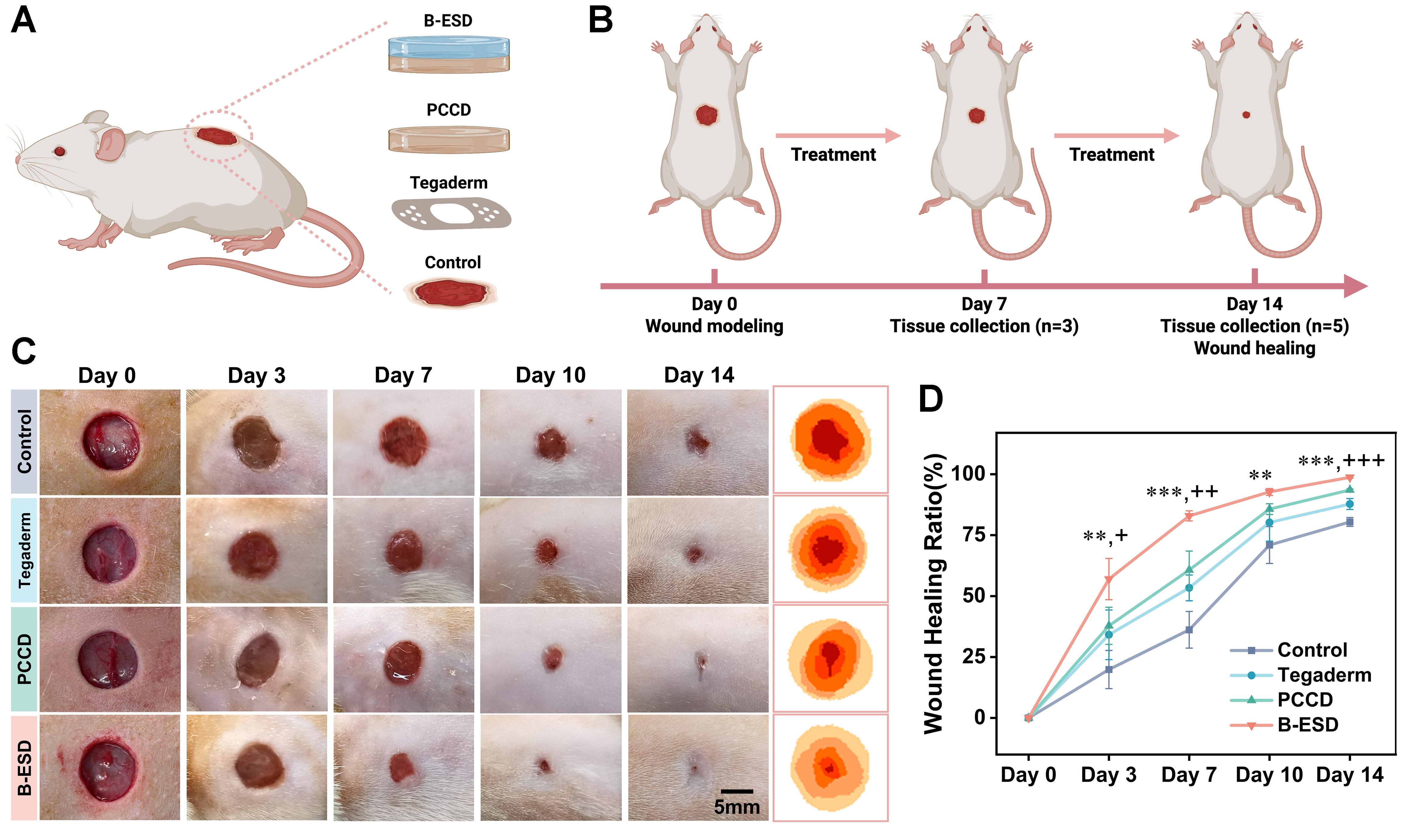
Figure 5. In vivo therapeutic efficacy of B-ESD in a rat full-thickness wound model. (A) Schematic of the rat full-thickness wound model and treatment mode in different groups; (B) Treatment procedure and timeline; (C) Representative optical photographs of wound closure under different treatment modes over 14 days; (D) Quantitative analysis of the wound-healing ratios over 14 days with statistical annotations at the indicated time points. Data are expressed as mean ± standard deviation (n = 5, **P < 0.01, ***P < 0.001 for B-ESD vs. Control, and +P < 0.05, ++P < 0.01, +++P < 0.001 for B-ESD vs. Tegaderm). (A and B) were created with BioRender. Du, X. (2026) https://BioRender.com/yfllnny. B-ESD: Bilayer electrogenerative smart dressing; PCCD: polyacrylamide/sodium carboxymethyl cellulose/calcium chloride/dopamine.
Wound healing progression was monitored via serial photographic documentation and quantitative analysis of rat wounds on days 0, 3, 7, 10 and 14 [Figure 5C and D]. Throughout the observation period, both PCCD and B-ESD hydrogels accelerated wound healing compared with other groups, with B-ESD showing the most pronounced improvement, suggesting that the bilayer electrogenerative architecture offered benefits beyond the hydrated and adhesive hydrogel matrix [Figure 5C]. In the early transition stage from inflammation to proliferation (day 3), the B-ESD group achieved a wound healing ratio of 56.99%, which was 1.67 and 2.87 times that of the Tegaderm group (34.14%) and the control group (19.87%), respectively. As healing progressed to day 10, the wound healing ratio in the B-ESD group rose sharply to 92.75%, retaining a clear advantage over other groups, indicating a sustained acceleration of tissue repair rather than a transient early-stage effect. By the final stage (day 14), the wounds in the B-ESD group were nearly completely healed, achieving a high closure rate of 98.72%, which surpassed that of the control group (80.43%), the Tegaderm group (87.80%), and the PCCD group (93.52%) [Figure 5D]. Two-way repeated-measures ANOVA of the longitudinal wound-closure data confirmed significant main effects of group (P < 0.01) and time (P < 0.001), together with a significant group × time interaction (P < 0.001), demonstrating that the B-ESD significantly altered the healing trajectory rather than merely the endpoint outcome. The superior closure performance of B-ESD highlights the synergistic contribution of moisture retention, exudate drainage, and self-sustained ES.
Histologic evaluation of wound tissue
Wound healing generally contains four stages: hemostasis, inflammation, proliferation and remodeling[3]. To elucidate the effect of the reshaped endogenous EF on the kinetics of wound repair, histological assessments of wound tissue were performed on day 7 and day 14 using H&E and Masson staining.
Given that the resolution of the inflammatory phase is a prerequisite for the onset of proliferation, H&E staining was performed on day 7 to evaluate the density of inflammatory cells, predominantly neutrophils, at the wound margin [Figure 6A]. While accumulations of neutrophils were observed in the wound sites across all groups, the B-ESD group exhibited a remarkably low neutrophil density of only 27.64 pieces/mm2, followed by the PCCD group with 41.46 pieces/mm2. In sharp contrast, the Tegaderm and control groups showed significantly higher densities, reaching an average of 49.75 pieces/mm2 and 73.70 pieces/mm2, respectively [Figure 6B]. These results demonstrated that B-ESD effectively mitigates acute inflammation triggered by injury, thereby facilitating a faster transition to the subsequent healing phases. The low inflammation level observed in the B-ESD group was primarily attributed to the DA-mediated ROS-scavenging properties, as well as the excellent absorption and moisturizing properties of hydrogels.
Figure 6. Histological evaluation of wound tissues. (A) Representative H&E and Masson staining images of wound tissues on day 7; (B) Density of neutrophils on day 7; (C-E) Quantitative analysis of (C) re-epithelialization length, (D) epidermal thickness, and (E) collagen content; (F) Representative H&E and Masson staining images of wound tissues on day 14. Data are expressed as mean ± standard deviation (n = 3 for day-7 histological analysis; n = 5 for day-14 histological analysis, *P < 0.05, **P < 0.01, ***P < 0.001, ns: not significant). H&E: Hematoxylin and eosin; PCCD: polyacrylamide/sodium carboxymethyl cellulose/calcium chloride/dopamine; B-ESD: bilayer electrogenerative smart dressing.
The proliferative phase, as characterized by the formation and re-epithelialization of new granulation tissue, is a key stage of wound repair[4]. As shown in Figure 6A, compared with other groups, B-ESD demonstrated a superior capacity to accelerate wound closure, consistent with the ability of bioelectric cues to guide epithelial and fibroblast migration toward the wound center. The magnified images highlighted the newly formed epithelial layer (outlined by black dashed lines). Quantitative analysis demonstrated that the B-ESD group achieved the maximum epithelialization length (512.5 μm) and thickness (104.8 μm), significantly surpassing the other groups, indicating accelerated re-epithelialization during the proliferative phase [Figure 6C and D]. Regenerated granulation tissue consists primarily of proliferating fibroblasts and the extracellular matrix (ECM) they secrete, so Masson staining was utilized to visualize and quantify collagen deposition. While the Control and Tegaderm groups were covered with thick scabs, the B-ESD group showed minimal scabbing and the highest ratio of collagen deposition [Figure 6A]. The statistical analysis of collagen content [Figure 6E] revealed that the B-ESD group had the highest collagen content (44.5%), followed by the PCCD (35.8%), Tegaderm (19.2%), and control groups (15.4%). In the hydrogel-treated groups, especially in the B-ESD group, collagen fibers appeared more organized and were accompanied by enhanced angiogenesis, indicating that the electrogenerative hydrogel promoted faster growth of granulation tissue and orderly fibroblast alignment.
By day 14, both the B-ESD group and PCCD group achieved complete epithelial coverage. The B-ESD group displayed a well-stratified epidermal structure with high tissue integrity, whereas the Tegaderm and control groups exhibited inflammatory infiltration, residual scabs and immature, irregularly thickened epidermal layers [Figure 6F]. Statistical results showed that the B-ESD group (43.3 μm) was closest to that of normal native skin (42.0 μm), indicating normalized epidermal remodeling rather than hyperplastic repair [Figure 6D]. This demonstrated that the B-ESD accelerates re-epithelialization length without inducing abnormal epithelial thickening. Crucially, the B-ESD group featured an abundance of regenerated skin appendages, including hair follicles and sebaceous glands, alongside with mature blood vessels [Figure 6F]. Masson’s staining at day 14 confirmed the maturation of the dermal matrix. The collagen fibers in the B-ESD group were densely packed and arranged in a regular, basket-weave-like pattern, closely resembling healthy skin. The collagen content in the B-ESD group reached 86.6%, significantly surpassing the Tegaderm group (55.1%) and the control group (44.9%) [Figure 6E]. The above results indicated that B-ESD significantly accelerated the entire wound healing process through a stage-coordinated mechanism: early stabilization of the inflammatory microenvironment, enhanced re-epithelialization and granulation tissue formation during proliferation, and organized collagen remodeling during the remodeling phase. This coordinated response is consistent with the reconstructed endogenous EF acting as a bioelectric cue to guide cell migration and matrix organization.
Immunohistochemical analysis of wound tissue
Repair of full-thickness skin wounds frequently proceeds imperfectly, giving rise to hypertrophic scarring and the disappearance of skin appendages. Such scars compromise not only appearance but also patients’ quality of life, owing to associated pruritus, pain, and impaired function[37]. Given that excessive Col I deposition is a fundamental hallmark of hypertrophic ECM, we evaluated Col I expression via IHC analysis on day 14[38]. In the control and Tegaderm groups, the regenerated dermal layers were characterized by densely packed, thickened, and rigidly parallel-aligned Col I bundles [Figure 7A], with semi-quantitative analysis confirming the high expression levels [Figure 7B]. Although the PCCD group exhibited lower Col I expression, localized fibrous aggregations were observed within the wound bed. On the contrary, the deposited collagen fibers in the B-ESD group exhibited a highly mature, basket-weave-like pattern with a moderate expression level relevant to normal skin [Figure 7B]. These results indicated that B-ESD successfully attenuated excessive collagen deposition during the late remodeling phase, thereby inhibiting scar formation.
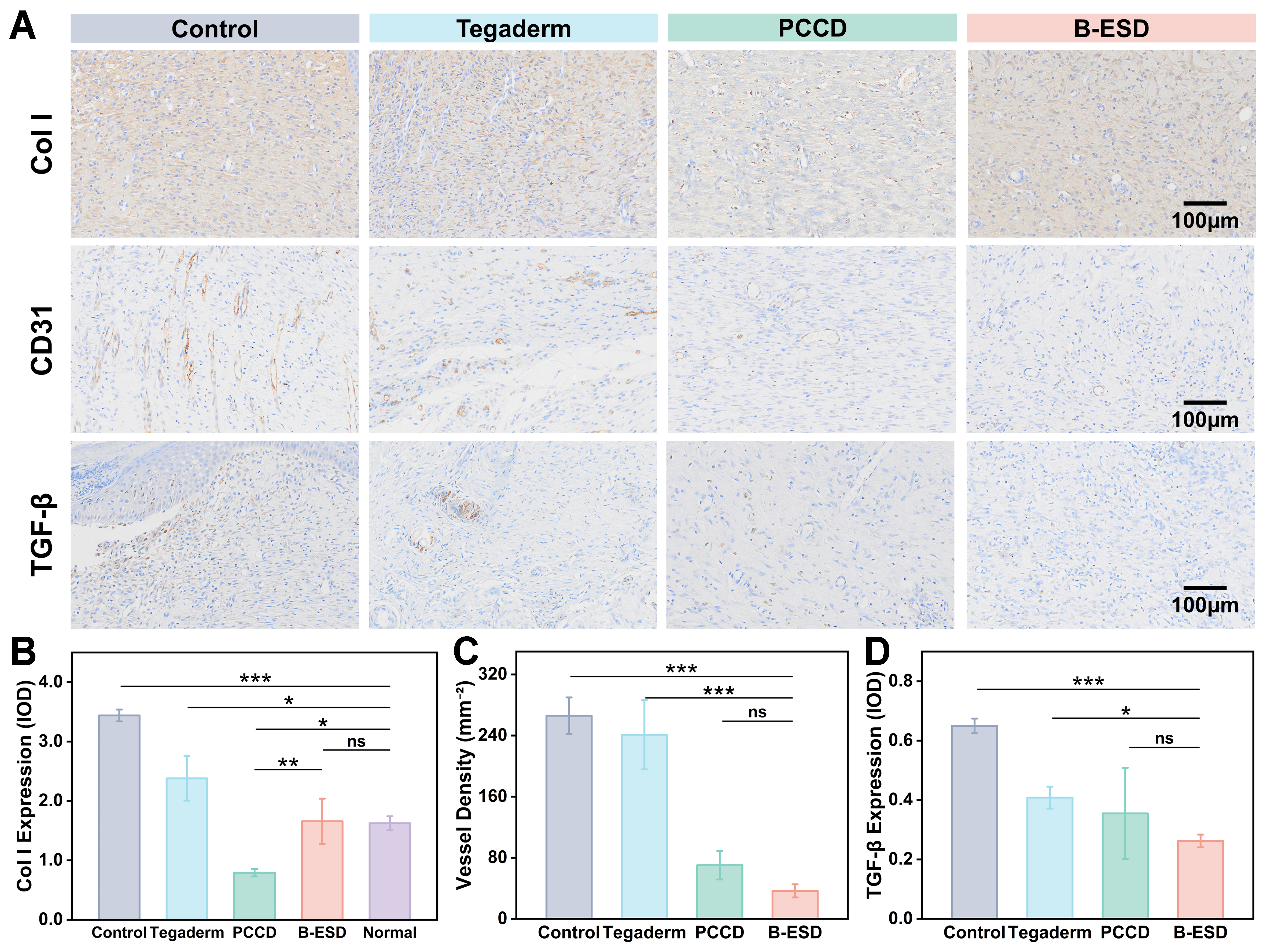
Figure 7. IHC analysis of ECM remodeling and angiogenesis. (A) Representative IHC images of Col I, CD31, and TGF-β1 expression in wound sections on day 14; (B and D) Semi-quantitative analysis (IOD) of Col I expression and TGF-β1 expression levels across different groups; (C) Quantitative analysis of angiogenesis expressed as vessel density (number of CD31-positive vessels per mm2). Data are expressed as mean ± standard deviation (n = 5, *P < 0.05, **P < 0.01, ***P < 0.001, ns: not significant). IHC: Immunohistochemistry; ECM: extracellular matrix; Col I: Type I collagen; TGF-β1: transforming growth factor-beta 1; IOD: integrated optical density; PCCD: polyacrylamide/sodium carboxymethyl cellulose/calcium chloride/dopamine; B-ESD: bilayer electrogenerative smart dressing.
During wound healing, angiogenesis ensures the delivery of nutrients and oxygen throughout the inflammation and proliferation phases, whereas uncontrolled angiogenesis in the remodeling phase may lead to scar formation, excessive fibrosis and even pathological hyperplasia[39]. Therefore, CD31, a characteristic endothelial cell marker, was used to identify newly formed blood vessels on day 14. In the control and Tegaderm groups, the regenerated tissue contained abundant CD31-positive vessels with a disorganized, morphologically immature architecture, a pattern typical of scar-associated angiogenesis [Figure 7A], and quantitative analysis confirmed the high vessel density [Figure 7C]. In the B-ESD group, the vessel density was markedly lower, and the vessels displayed a mature, well-stabilized topology [Figure 7A and C]. These results indicated that the wound has transitioned into the stage of vascular maturation[40].
TGF-β1 is also regarded as an important factor in cutaneous wound repair. It orchestrates the initial phases of healing by recruiting inflammatory cells and upregulating ECM-related genes to drive fibroblast proliferation and migration[41]. However, the overexpression of TGF-β1 exacerbates fibrotic responses and promotes scar formation[42]. IHC results showed that the expression of TGF-β1 remained high in the control and Tegaderm group on day 14, particularly localizing within the scar tissue. In contrast, the B-ESD group showed a suppressed TGF-β1 expression of 0.26 [Figure 7A and D]. The above findings validated that B-ESD effectively facilitated the remodeling of the ECM, promoted vascular maturation and inhibited scar formation.
B-ESD as a high-performance sensor for real-time monitoring
Early evaluation of wound healing efficacy remains challenging, especially in cases of extensive or large-area wounds[43]. Traditional clinical wound management heavily relies on periodic visual inspections, which necessitate frequent dressing changes. This method not only disturbs the healing process and even causes secondary damage, but also exposes the vulnerable wound bed to external pathogens[44]. Integrating real-time sensing capabilities into advanced hydrogel dressings fundamentally overcomes these limitations[45,46]. Beyond its therapeutic efficacy, the B-ESD integrates excellent mechanical robustness with high conductivity, enabling its application as a high-performance iontronic sensor. Upon mechanical deformation, the geometrical variations of the hydrogel and the subsequent alterations in its internal ion-conducting pathways lead to precisely detectable fluctuations in electrical resistance[47].
To systematically quantify the electromechanical responsiveness of B-ESD, relative resistance changes (ΔR/R0) of the hydrogel under different mechanical modes were investigated. As illustrated in Figure 8A, the ΔR/R0 exhibited a monotonic increase with applied tensile strain. The calculated gauge factor (GF) of 1.31 indicates reliable sensitivity for capturing subtle stretching [Figure 8B]. The resistance signal was stable during five cycles, indicating the sensing steadiness of B-ESD. Furthermore, the B-ESD demonstrated good sensitivity to compression stress with a pressure sensitivity of 0.38%·kPa-1 within the pressure range of 0.5-10.0 kPa [Figure 8C and D]. Moreover, B-ESD could precisely differentiate bending angles ranging from 30° to 180° [Supplementary Figure 13].
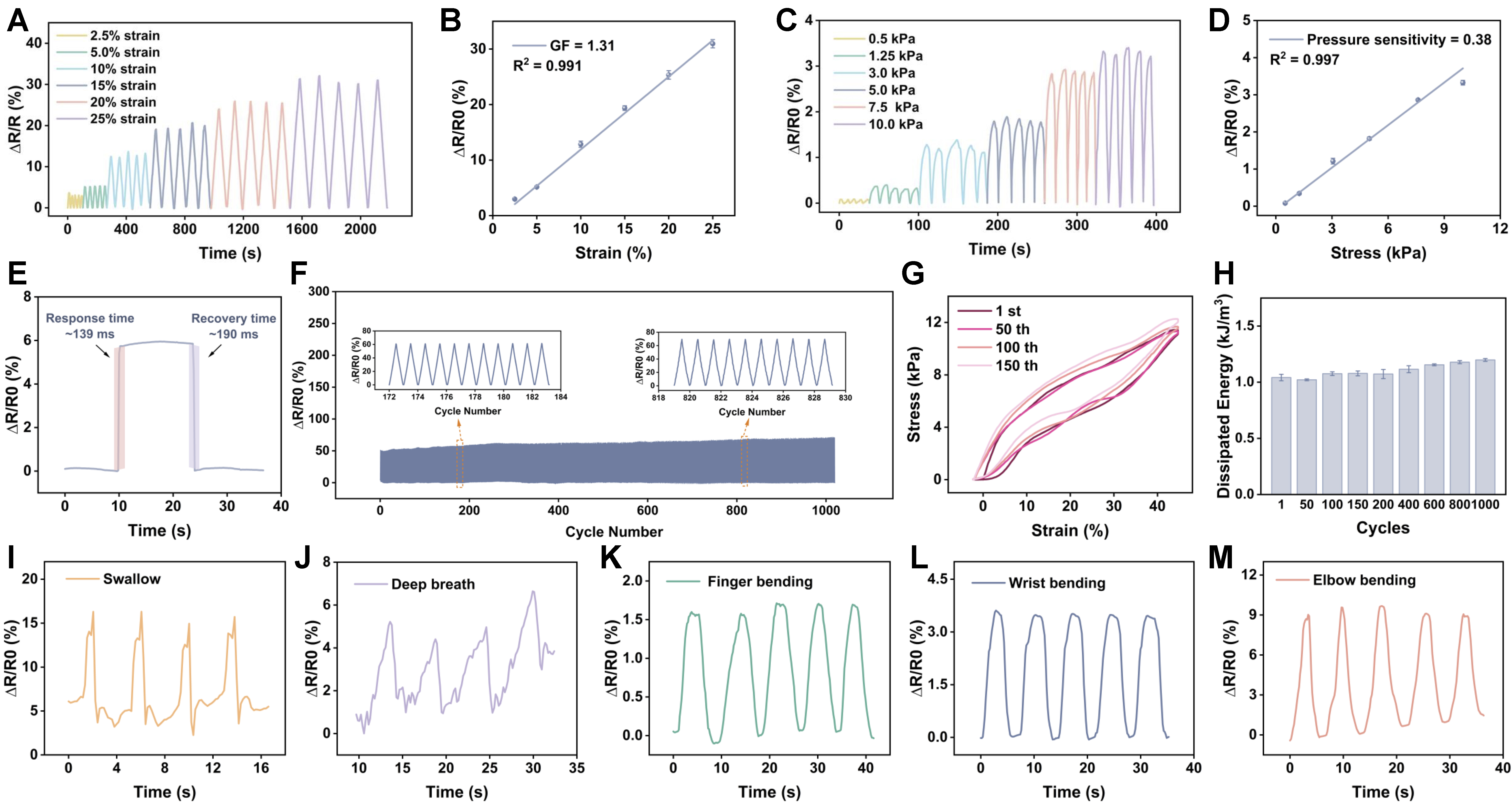
Figure 8. Sensing properties and real-time monitoring capabilities of the B-ESD. (A and B) Relative resistance changes (ΔR/R0) and corresponding GFs under different tensile strains; (C and D) Relative resistance changes (ΔR/R0) and corresponding pressure sensitivity under different compression stresses; (E) Response and recovery time evaluation; (F) Long-term durability test of B-ESD with 45% tensile strain for 1,000 cycles; (G) Cyclic tensile stress-strain curves and (H) calculated dissipated energy over 1,000 cycles; (I and J) Real-time monitoring of subtle physiological signals: (I) swallowing and (J) deep breathing; (K-M) Real-time monitoring of large-scale joint kinematics: (K) finger, (L) wrist, and (M) elbow bending. B-ESD: Bilayer electrogenerative smart dressing; GFs: gauge factors.
The B-ESD also exhibited prompt electromechanical responsiveness during the rapid loading–unloading process. When deformation occurred, the resistance responded instantaneously with a response time of
Reliability and fatigue resistance are paramount for practical application of flexible electronics. The B-ESD featured remarkable long-term durability, maintaining consistent ΔR/R0 outputs with only a slight baseline drift and no appreciable signal degradation over 1,000 continuous stretching-releasing cycles at 45% strain [Figure 8F]. This structural stability was further corroborated by the cyclic mechanical hysteresis loops. The stress-strain curves of the B-ESD under repeated stretching (150 cycles) highly overlapped [Figure 8G], and the calculated dissipated energy exhibited only a negligible increase after 1,000 cycles [Figure 8H]. These results verified that the sacrificial bonds within the hydrogel network can effectively dissipate energy while maintaining network integrity, ensuring excellent elasticity and fatigue resistance of B-ESD and robust performance during prolonged use[48]. These hysteresis and energy-dissipation results also indicate favorable viscoelastic compatibility for wound-contact applications, thereby reducing sustained interfacial stress transfer to fragile granulation tissue and supporting stable conformal contact with skin[49]. Although a slight baseline drift in ΔR/R0 was observed due to inevitable water evaporation, this can be effectively mitigated by encapsulation in practical clinical scenarios. To quantify this effect, we evaluated the water-retention behavior of B-ESD with and without encapsulation over a 48 h wear-relevant period. Encapsulation markedly reduced water loss and preserved the hydrated state of the hydrogel. After 48 h at 32 °C, the encapsulated B-ESD retained approximately 88.8% of its initial hydrated weight, compared with 62.0% for the non-encapsulated sample [Supplementary Figure 14]. Consistently, the encapsulated B-ESD retained more stable streaming-potential output than the non-encapsulated sample [Supplementary Figure 15]. These results demonstrate the effectiveness of encapsulation for maintaining both therapeutic electrical output and sensing reliability during practical use.
Encouraged by its outstanding sensitivity and robust stability, the B-ESD was mounted onto various body parts as a wearable skin-mountable device for rehabilitation monitoring. The sensor successfully detected subtle physiological signals, such as swallowing and deep breathing, presenting distinct and characteristic signal peaks [Figure 8I and J]. Furthermore, it accurately recorded large-scale joint movements. When attached to the finger [Figure 8K], wrist [Figure 8L] and elbow [Figure 8M], the B-ESD output repeatable and consistent waveforms corresponding to the flexion motions. Notably, due to the varying range of motion (ROM) and induced strains at different joints, B-ESD generated electrical signals with distinct and distinguishable amplitudes, enabling the precise clarification of specific body parts. Importantly, this sensing capability is highly relevant to wound management rather than merely rehabilitation monitoring, as wounds located at mobile regions are continuously exposed to bending, stretching and compression during daily activities. By recording the amplitude, frequency, and duration of local movements, B-ESD may help evaluate whether patient’s activity exceeds the mechanical tolerance of the healing wound, thereby assisting rehabilitation training and enabling dynamic assessment of healing progression[50,51]. Such biomechanical feedback may also help reduce secondary tissue damage caused by excessive movement and limit mechanically induced scar formation[52]. Therefore, B-ESD provides not only self-sustained bioelectric therapy but also real-time biomechanical information for more informed wound management.
CONCLUSIONS
Main conclusions
In summary, we successfully developed a B-ESD that serves as a multifunctional platform for accelerated wound healing and real-time monitoring. Driven by an engineered hydrophilicity and pore-size gradient, the unidirectional fluid pumping induces a robust vertical streaming potential across the B-ESD. More importantly, the incorporation of rGO on the top layer of B-ESD prevents premature field attenuation and sustains a persistent electrical stimulus. By restoring and amplifying the disrupted wound EF, B-ESD provided a sustained bioelectric cue that coordinated inflammation resolution, fibroblast migration, re-epithelialization, angiogenesis, and collagen remodeling. Consequently, the B-ESD markedly shortened the early-stage inflammatory stage, accelerated re-epithelialization and facilitated a 98.72% wound closure rate within 14 days. Notably, the B-ESD inhibited scar formation while orchestrating the regeneration of skin appendages and modulating collagen deposition, significantly improving the quality of wound repair. Concurrently, the dynamic cross-linking network endows the B-ESD with exceptional stretchability, fatigue resistance, and high sensitivity, enabling the precise, long-term real-time monitoring of human motions and subtle physiological signals. The B-ESD strategy not only presents a multifunctional platform for the clinical management of large-scale wounds on highly mobile body parts but also broadens the horizons for the design of next-generation, self-powered wearable bioelectronics.
Limitations and future perspectives
Although the B-ESD demonstrates an integrated strategy for exudate management, flow-coupled electrogeneration, accelerated wound repair, and biomechanical monitoring, several aspects deserve further investigation to support its translation toward practical wound care. The electrogenerative performance was systematically characterized under controlled ex vivo perfusion and further evaluated by in situ wound-potential measurements. Future studies incorporating protein-containing artificial wound fluid and long-term on-wound electrical monitoring would provide a more comprehensive understanding of how wound-fluid composition and secretion kinetics affect the streaming-potential output during actual wear.
The therapeutic benefit of B-ESD likely arises from the combined effects of exudate drainage, moisture retention, tissue adhesion, impedance matching, and bioelectric modulation. More refined electrical-on/off designs and real-time mapping of the wound bioelectric microenvironment would help clarify the relative contribution of the electrokinetic cue. Evaluation in chronic, infected, diabetic, or large-animal wound models would further assess the robustness of this dressing under clinically challenging conditions.
The sensing function of B-ESD was demonstrated in healthy volunteers, supporting its potential for biomechanical feedback in mobile wound regions. Future studies in wound-bearing subjects are needed to establish clinically relevant motion thresholds and validate long-term signal stability during repeated dressing use.
DECLARATIONS
Acknowledgments
The graphical abstract was created with BioRender.com [Created in BioRender. Du, X. (2026) https://BioRender.com/osoz171].
Authors’ contributions
Conceived the central idea, designed the project, and acquired funding: Guo, R.; Dai, H.; Wang, L. (Le Wang)
Synthesized the hydrogel materials, designed and performed the majority of the experiments, and wrote the original draft: Du, X.
Assisted with the in vivo animal models, histological analysis, and immunohistochemical (IHC) quantification: Shi, C.
Assisted in conducting the in vitro cell assays: Wang, L. (Le Wang)
Contributed to the material characterizations: Ye, Q.; Wang, L. (Liwei Wang)
Contributed to the data visualization: Zhang, W.; Xiao, R.
Comprehensively reviewed and edited the manuscript: Guo, R.; Dai, H.; Wang, L. (Le Wang)
Jointly supervised this work: Guo, R.; Dai, H.; Wang, L. (Le Wang)
All authors discussed the results, commented on the manuscript, and approved the final version.
Availability of data and materials
The data supporting the findings of this study are available from the corresponding authors upon reasonable request.
AI and AI-assisted tools statement
Not applicable.
Financial support and sponsorship
This work was supported by the National Key Research and Development Program of China (2024YFE0204600), National Natural Science Foundation of China (62305068 and 62074044), Shanghai Post-doctoral Excellence Program (2021016), Shanghai Rising-Star program (22YF1402000), Jilin Provincial Scientific and Technological Development Program (YDZJ202201ZYTS402), and Shanghai 2025 “Pioneer Plan” biological hybrid robot theme (the third batch) project (25XF3201000).
Conflicts of interest
All authors declared that there are no conflicts of interest.
Ethical approval and consent to participate
All animal procedures were approved by the Institutional Animal Care and Use Committee of Fudan University (Approval No. YSAPL20260269). The study was approved by the School of Life Sciences, Fudan University (Approval No. H2026078). For human motion-monitoring experiments, informed consent was obtained from all volunteers prior to participation.
Consent for publication
Not applicable.
Copyright
© The Author(s) 2026.
Supplementary Materials
REFERENCES
2. Sen, C. K. Human wounds and its burden: an updated compendium of estimates. Adv. Wound. Care. 2019, 8, 39-48.
4. Strecker-McGraw, M. K.; Jones, T. R.; Baer, D. G. Soft tissue wounds and principles of healing. Emerg. Med. Clin. North. Am. 2007, 25, 1-22.
5. Cao, X.; Wu, X.; Zhang, Y.; Qian, X.; Sun, W.; Zhao, Y. Emerging biomedical technologies for scarless wound healing. Bioact. Mater. 2024, 42, 449-77.
6. Jonidi Shariatzadeh, F.; Currie, S.; Logsetty, S.; Spiwak, R.; Liu, S. Enhancing wound healing and minimizing scarring: a comprehensive review of nanofiber technology in wound dressings. Prog. Mater. Sci. 2025, 147, 101350.
7. Schultz, G. S.; Davidson, J. M.; Kirsner, R. S.; Bornstein, P.; Herman, I. M. Dynamic reciprocity in the wound microenvironment. Wound. Repair. Regen. 2011, 19, 134-48.
8. Barrientos, S.; Stojadinovic, O.; Golinko, M. S.; Brem, H.; Tomic-Canic, M. Growth factors and cytokines in wound healing. Wound. Repair. Regen. 2008, 16, 585-601.
10. Eming, S. A.; Krieg, T.; Davidson, J. M. Inflammation in wound repair: molecular and cellular mechanisms. J. Invest. Dermatol. 2007, 127, 514-25.
11. Li, J.; Xie, Y.; Liu, G.; et al. Bioelectret materials and their bioelectric effects for tissue repair: a review. ACS. Appl. Mater. Interfaces. 2024, 16, 38852-79.
12. Zhou, Y.; Ma, X.; Yu, C.; et al. A wearable self-charging electroceutical device for bacteria-infected wound healing. ACS. Nano. 2024, 18, 15681-94.
13. Lv, Y.; Li, Y.; Pan, Y.; et al. Progress in the application of conductive hydrogels in wound healing: a review. Nanoscale. Adv. 2026, 8, 1490-507.
14. Luo, R.; Dai, J.; Zhang, J.; Li, Z. Accelerated skin wound healing by electrical stimulation. Adv. Healthc. Mater. 2021, 10, e2100557.
15. Zhao, M.; Song, B.; Pu, J.; et al. Electrical signals control wound healing through phosphatidylinositol-3-OH kinase-gamma and PTEN. Nature 2006, 442, 457-60.
16. Wu, Y.; Lu, Y.; Wu, C.; et al. Conductive dual hydrogen bonding hydrogels for the electrical stimulation of infected chronic wounds. J. Mater. Chem. B. 2021, 9, 8138-46.
17. Liu, Z.; Wei, W.; Tremblay, P. L.; Zhang, T. Electrostimulation of fibroblast proliferation by an electrospun poly (lactide-co-glycolide)/polydopamine/chitosan membrane in a humid environment. Colloids. Surf. B. Biointerfaces. 2022, 220, 112902.
18. Li, P.; Xu, J.; Shi, Q.; et al. Pulse capacitive coupling electric field regulates cell migration, proliferation, polarization, and vascularization to accelerate wound healing. Adv. Wound. Care. 2023, 12, 498-512.
19. Wang, E.; Xu, L.; Luo, L.; et al. Tissue-fluid-driven symbiotic electronic textile accelerates and guides tissue self-healing. Cell. Biomater. 2026.
20. Kläning, E.; Christensen, B.; Sørensen, E. S.; Vorup-Jensen, T.; Jensen, J. K. Osteopontin binds multiple calcium ions with high affinity and independently of phosphorylation status. Bone 2014, 66, 90-5.
21. Pailler-Mattei, C.; Bec, S.; Zahouani, H. In vivo measurements of the elastic mechanical properties of human skin by indentation tests. Med. Eng. Phys. 2008, 30, 599-606.
22. Lee, H.; Scherer, N. F.; Messersmith, P. B. Single-molecule mechanics of mussel adhesion. Proc. Natl. Acad. Sci. U. S. A. 2006, 103, 12999-3003.
23. Tang, P.; Han, L.; Li, P.; et al. Mussel-inspired electroactive and antioxidative scaffolds with incorporation of polydopamine-reduced graphene oxide for enhancing skin wound healing. ACS. Appl. Mater. Interfaces. 2019, 11, 7703-14.
24. Kim, B.; Park, J.; Lee, J. Y. Conductive double-network hydrogel composed of sodium alginate, polyacrylamide, and reduced graphene oxide. Korean. J. Chem. Eng. 2023, 40, 352-60.
25. Wang, G.; Yang, F.; Zhou, W.; Xiao, N.; Luo, M.; Tang, Z. The initiation of oxidative stress and therapeutic strategies in wound healing. Biomed. Pharmacother. 2023, 157, 114004.
26. Sánchez-Abella, L.; Ruiz, V.; Pérez-San Vicente, A.; Grande, H.; Loinaz, I.; Dupin, D. Reactive oxygen species (ROS)-responsive biocompatible polyethylene glycol nanocomposite hydrogels with different graphene derivatives. J. Mater. Sci. 2021, 56, 10041-52.
27. Cong, Y.; Fu, J. Hydrogel-tissue interface interactions for implantable flexible bioelectronics. Langmuir 2022, 38, 11503-13.
28. Tang, H.; Li, Y.; Liao, S.; Liu, H.; Qiao, Y.; Zhou, J. Multifunctional conductive hydrogel interface for bioelectronic recording and stimulation. Adv. Healthc. Mater. 2024, 13, e2400562.
29. Xu, Z.; Fan, J.; Tian, W.; et al. Cellulose‐based pH‐responsive Janus dressing with unidirectional moisture drainage for exudate management and diabetic wounds healing. Adv. Funct. Mater. 2024, 34, 2307449.
30. Yu, R.; Zhang, H.; Guo, B. Conductive biomaterials as bioactive wound dressing for wound healing and skin tissue engineering. Nanomicro. Lett. 2021, 14, 1.
31. Martin-Granados, C.; McCaig, C. D. Harnessing the electric spark of life to cure skin wounds. Adv. Wound. Care. 2014, 3, 127-38.
32. Qin, Y.; Wang, Y.; Sun, X.; et al. Constant electricity generation in nanostructured silicon by evaporation-driven water flow. Angew. Chem. Int. Ed. Engl. 2020, 59, 10619-25.
33. Zhao, G.; Zhu, H. Cation-π interactions in graphene-containing systems for water treatment and beyond. Adv. Mater. 2020, 32, e1905756.
34. Wang, K.; Zhang, J. Electric field regulation in wound healing: from endogenous mechanisms to self-powered wearable device applications. J. Sci. Adv. Mater. Devices. 2026, 11, 101177.
35. Fan, L.; Dai, Z.; Bai, X.; et al. (K,Na)NbO3-based lead-free ceramic/polyvinylidene fluoride piezoelectric composite for biomimetic wound healing via endogenous electric field simulation. Biomater. Adv. 2026, 186, 214909.
36. Zhou, Q.; Dai, H.; Yan, Y.; et al. From short circuit to completed circuit: conductive hydrogel facilitating oral wound healing. Adv. Healthc. Mater. 2024, 13, e2303143.
37. Finnerty, C. C.; Jeschke, M. G.; Branski, L. K.; Barret, J. P.; Dziewulski, P.; Herndon, D. N. Hypertrophic scarring: the greatest unmet challenge after burn injury. Lancet 2016, 388, 1427-36.
38. Xue, M.; Jackson, C. J. Extracellular matrix reorganization during wound healing and its impact on abnormal scarring. Adv. Wound. Care. 2015, 4, 119-36.
39. Korntner, S.; Lehner, C.; Gehwolf, R.; et al. Limiting angiogenesis to modulate scar formation. Adv. Drug. Deliv. Rev. 2019, 146, 170-89.
40. Veith, A. P.; Henderson, K.; Spencer, A.; Sligar, A. D.; Baker, A. B. Therapeutic strategies for enhancing angiogenesis in wound healing. Adv. Drug. Deliv. Rev. 2019, 146, 97-125.
41. Shi, A.; Li, J.; Qiu, X.; et al. TGF-β loaded exosome enhances ischemic wound healing in vitro and in vivo. Theranostics 2021, 11, 6616-31.
42. Zhang, J.; Zheng, Y.; Lee, J.; et al. A pulsatile release platform based on photo-induced imine-crosslinking hydrogel promotes scarless wound healing. Nat. Commun. 2021, 12, 1670.
43. Chen, F.; Wu, M.; Dong, Q.; et al. Arbitrarily shapeable and conductive hydrogel with “Magic Cube” like structure for real-time monitoring and promoting wound healing. Compos. Part. B. Eng. 2022, 238, 109903.
44. Kekonen, A.; Bergelin, M.; Eriksson, J. E.; Vaalasti, A.; Ylänen, H.; Viik, J. Bioimpedance measurement based evaluation of wound healing. Physiol. Meas. 2017, 38, 1373-83.
45. Hao, F.; Wang, L.; Chen, B.; Qiu, L.; Nie, J.; Ma, G. Bifunctional smart hydrogel dressing with strain sensitivity and NIR-responsive performance. ACS. Appl. Mater. Interfaces. 2021, 13, 46938-50.
46. Zhao, Y.; Li, Z.; Song, S.; et al. Skin‐inspired antibacterial conductive hydrogels for epidermal sensors and diabetic foot wound dressings. Adv. Funct. Mater. 2019, 29, 1901474.
47. Jiang, J.; Zhao, L. Halometallate ionic liquid dynamically regulates zwitterionic hydrogels by synergistic multiple‐bond networks. Adv. Funct. Mater. 2025, 35, 2417688.
48. Shi, Y.; Guan, Y.; Liu, M.; et al. Tough, antifreezing, and piezoelectric organohydrogel as a flexible wearable sensor for human-machine interaction. ACS. Nano. 2024, 18, 3720-32.
49. Chen, Q.; Li, S.; Li, K.; Zhao, W.; Zhao, C. A skin stress shielding platform based on body temperature-induced shrinking of hydrogel for promoting scar-less wound healing. Adv. Sci. 2024, 11, e2306018.
50. Li, M.; Zhang, Y.; Lian, L.; et al. Flexible accelerated‐wound‐healing antibacterial MXene‐based epidermic sensor for intelligent wearable human‐machine interaction. Adv. Funct. Mater. 2022, 32, 2208141.
51. Koller, T. Mechanosensitive aspects of cell biology in manual scar therapy for deep dermal defects. Int. J. Mol. Sci. 2020, 21, 2055.
Cite This Article
 Open Access
How to Cite
Du, X.; Shi, C.; Ye, Q.; Wang, L.; Xiao, R.; Zhang, W.; Wang, L.; Guo, R.; Dai, H. An autonomous regenerative bioelectronic dressing powered by wound exudate for high-quality skin repair. Soft Sci. 2026, 6, 90. https://dx.doi.org/10.20517/ss.2026.153
Download Citation
If you have the appropriate software installed, you can download article citation data to the citation manager of your choice. Simply select your manager software from the list below and click on download.
Export Citation File
Type of Import
Tips on Downloading Citation
Citation Manager File Format
Type of Import
Direct Import: When the Direct Import option is selected (the default state), a dialogue box will give you the option to Save or Open the downloaded citation data. Choosing Open will either launch your citation manager or give you a choice of applications with which to use the metadata. The Save option saves the file locally for later use.
Indirect Import: When the Indirect Import option is selected, the metadata is displayed and may be copied and pasted as needed.
About This Article
Copyright

Data & Comments
Data
0












Comments
Comments must be written in English. Spam, offensive content, impersonation, and private information will not be permitted. If any comment is reported and identified as inappropriate content by OAE staff, the comment will be removed without notice. If you have any queries or need any help, please contact us at [email protected].