

Clinical translation of findings from rare disease animal models to veterinary patients: practical reality or elusive dream
 0
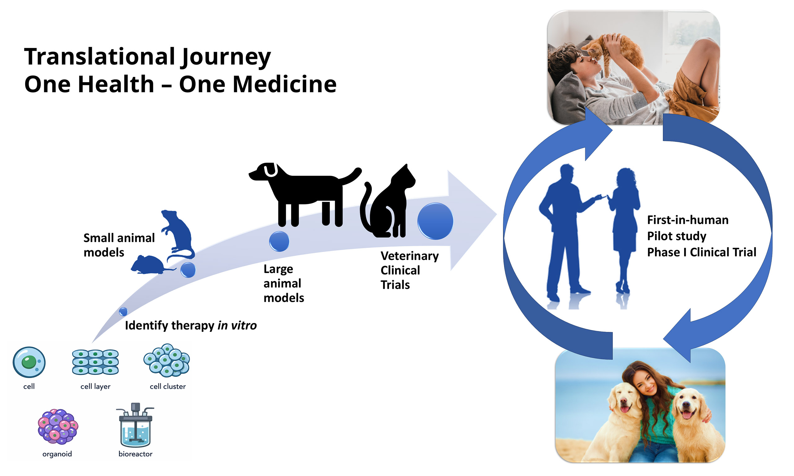
Graphical Abstract
Abstract
Animal research plays a critical role in developing disease treatments. Most studies are done in mice but larger species, phylogenetically closer to humans, are sometimes used. In the genomic age, numerous rare inherited diseases and attendant treatment challenges have been characterized. The US Food and Drug Administration and similar international agencies have enacted laws like the Orphan Drug Act for humans and the Minor Use and Minor Species Animal Health Act for animals to incentivize therapy development for these rare conditions. The remarkable curative potential of new cell and gene therapies (CGTs) has drawn attention to the drug approval process. Questions have been raised about whether animal findings adequately translate to humans. One justification for using animals in research has been the potential for them to ultimately benefit. This review highlights the bidirectional interplay of human and animal studies in keeping with the One Health - One Medicine concept. Examples of innovative drugs, sophisticated surgeries, and CGTs now being used in companion animals with rare diseases are provided. Related issues discussed in the paper include potential reasons for inconsistent translation of experimental animal findings in human clinical trials, barriers to using specialized treatments more broadly, ethical concerns associated with rare disease treatments, and prospects for developing alternatives that would benefit animals and reduce their use in research. The goal of this review is to encourage broader use of advanced therapies for rare companion animal diseases while helping to ensure ongoing research is done responsibly.
Keywords


 Download PDF
INTRODUCTION
Animal models have been used since antiquity to define the basis of human health and disease[1,2]. This work has been especially critical for rare conditions whose underlying causes often defy definition[3]. While most studies have been done in mice[4], larger species, including nonhuman primates[5], pigs[6], sheep[7], dogs[8], and cats[9], have been used [Figure 1]. These larger species are generally considered more like humans phylogenetically than rodents[10-13], with comparable size[14], anatomy[15], physiology[15,16], and immunity[17]. Dogs and cats have an added benefit of sharing the home environment with humans, exposing them to the same infectious agents and potential toxins[18]. Moreover, pets are routinely assessed in veterinary practice with sophisticated diagnostic tests that serve as outcome parameters in human clinical trials[19]. Use of these same tests in preclinical research allows results to be directly compared to those from humans. Finally, the relatively condensed life spans of dogs and cats have made them valuable sentinels for predicting disease effects[20].
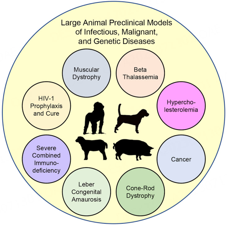
Figure 1. Schematic of several large animal species used in gene editing studies for mostly rare diseases. Reproduced without modification from Maynard et al.[13]. © 2021 The Authors. Published by Elsevier, under a CC BY-NC-ND license.
Critical studies of normal function and development in companion animals include early landmark research such as Pavlov’s use of dogs to define conditioning behavior[21], Sir Charles Sherrington’s canine spinal cord injury experiments establishing key aspects of reflex function including phantom scratching seen in syringomyelia[22,23], and the seminal work done by Hubel and Wiesel in cats that revolutionized our understanding of visual processing[24]. The impact of these studies extends from defining general disease concepts to understanding the basis of rare conditions, including Urbach-Wiethe disease (Pavlovian fear conditioning)[25], acute flaccid myelitis (disruption of spinal reflex pathways)[26], and congenital cataracts (amblyopia)[27]. Specific rare genetic diseases influenced by this research extend across body systems in humans and companion animals. One example that involves fear conditioning and reflex function is hyperekplexia (startle disease) seen in dogs and people[28]. More broadly, naturally occurring canine and feline diseases have served as critical models for many rare human diseases[8,29,30]. This species interconnectedness and bidirectional translation of clinical and research findings serve as a pillar of the One Health - One Medicine concept benefiting both humans and animals[31].
In addition to their role as disease models, dogs and, to a much lesser degree, cats have been used in tandem with rodents through the US Food and Drug Administration’s (FDA) drug approval process in which two species are tested[32,33]. Using this approach, the dog has been particularly valuable in identifying adverse drug effects[34,35]. Nonetheless, recent papers have shown that animal preclinical findings do not consistently translate to humans[36-38]. Additional ethical concerns have been raised[39]. Largely because of these issues, the FDA Modernization Act 2.0 passed by the US Congress in 2022[40-42] and further clarified by the 3.0 Act now under consideration[43] has removed the requirement for animals in drug approval and stipulated that alternatives would be allowed. Use of so-called new approach methodologies/methods (NAMs), such as cell/tissue culture, computer modeling, and “organ on a chip” technologies, has been encouraged to assess drug toxicity[44] and efficacy[45]. These initiatives are occurring in concert with changes in the conduct of clinical trials, as greater focus is being placed on approving orphan drugs, often after accelerated review[46]. The speed with which this shift has occurred, and the associated changes in the drug approval process, have caused concerns that a loss of rigor could compromise patient care[47,48].
One long-held justification for using dogs and cats in research has been the potential for translating treatments to these same species as veterinary patients[49-51]. Encouragingly, we are now seeing progress at multiple levels as specialized therapies are making a difference in animals’ lives. Examples in this Special Issue extend across a range of innovative medical[52] and surgical[53,54] treatments that were once seen as experimental and have now become part of the standard of veterinary care. These approaches are being complemented by cell and gene therapies (CGTs)[55-57] therapies in which companion animal models were critically important to establish proof of concept prior to human use.
This review also addresses challenges for developing rare disease treatments and applying them more broadly, reasons for inconsistent translation of experimental animal findings in human clinical trials, and the potential for using alternatives that will both reduce experimental animal use and benefit dogs and cats with rare diseases. The collective goal of the review and associated papers in the Rare Disease and Orphan Drugs Journal (RDODJ) Special Issue is to encourage broader use of advanced therapies for rare companion animal diseases while helping to ensure ongoing research is done responsibly.
RARE DISEASE INCIDENCE CRITERIA AND DRUG DEVELOPMENT INCENTIVE PROGRAMS
While individual rare diseases occur infrequently, they collectively have a tremendous global impact, affecting ~30 M people in the US, ~29 million in the EU, and ~ 400 million worldwide[58] [Figure 2]. As a result, there is strong patient advocacy for these conditions through organizations such as the National Organization for Rare Disorders in the US[59]. This has facilitated passage of laws to enhance access and incentivize drug development for affected populations[60].
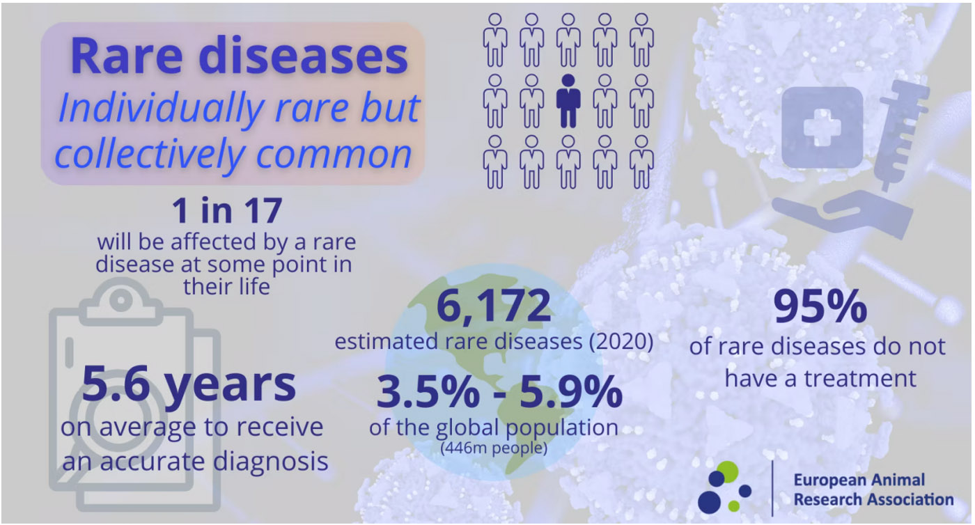
Figure 2. Schematic showing the collective impact of rare diseases on the global population. From the European Animal Research Association (EARA)[3]. Reprinted with permission.
To establish public policy and provide incentives to develop therapies, governments have established guidelines defining the incidence of rare diseases[61]. Projecting through the US 1983 Orphan Drug Act[62], the FDA defines rare diseases as those affecting fewer than 200,000 people (~1 in 1,750; ~60/100,000) in the US[47]. This act is intended to help address barriers for treatment development such as the limited patient population and cost by incentivizing companies. Notably, more common diseases (affecting > 200,000 people) may qualify for incentives if there is no reasonable expectation that a drug will be developed to treat affected individuals. Analogous legislation, with comparable disease incidence figures and incentives, was subsequently enacted in Japan[63], the European Union (EU)[64], and other countries to encourage drug development for rare diseases[65,66].
Relevant to this RDODJ Special Issue, the FDA also sought to incentivize development of legal medications for rare diseases in major animal species (dogs, cats, horses, etc.) and for any diseases in minor species (zoo animals, fish, etc.) through the Minor Use and Minor Species (MUMS) Animal Health Act of 2004[67]. This is particularly important in veterinary medicine as there are fewer funding sources and limited options for third-party payers (see discussion of challenges below).
Disease incidence rates to qualify for MUMS were recently updated to thresholds of 80,000 cases annually for dogs and 150,000 for cats in the US[68]. Using pet ownership data estimating that there are ~89.7 M pet dogs and ~73.8 M cats in the US[69], this translates to incidence figures of ~1 in 1,100 and 490, respectively. Several drugs to treat cancer in dogs, as an example Verdinexor (KPT-335) for lymphoma[70], have been approved through the MUMS law[71]. A related FDA process allows conditional approval under two circumstances, namely (1) if a drug “addresses a serious or life-threatening disease, or (2) addresses an unmet animal or human health need, for which demonstrating effectiveness would require a complex or particularly difficult study or studies[72]”. Felycin-CA1 for hypertrophic cardiomyopathy in cats was recently approved using these criteria[73]. In addition, approval is pending for an intravenous mesenchymal stromal cell (MSC) therapy targeting refractory feline chronic gingivostomatitis (FCGS)[74,75] (As discussed further below, the FDA has also created a special accommodation for use of GS-441524 to treat feline infectious peritonitis [FIP]).
Comparable incentives are available for animals in the EU through the Veterinary Medicinal Products Regulation (Regulation [EU] 2019/6)[76].
ONE HEALTH - ONE MEDICINE
Concept and application to rare diseases (see graphical abstract)
German physician and pathologist Rudolf Virchow was a leading advocate of comparative medicine. He conducted studies in the 19th century on a range of infectious diseases affecting both humans and animals[77]. Virchow coined the word zoonosis for the potential of interspecies transmission and famously said, “Between animal and human medicine there are no dividing lines, nor should there be. The object is different, but the experience obtained constitutes the basis of all medicine”[78].
Canadian physician William Osler and American veterinary epidemiologist Calvin Schwabe have each been credited with the term “One Medicine[79]”. Osler, considered to be the father of modern medicine, was an eminent advocate of comparative pathology at the dawn of the 20th century. He taught both medical and veterinary students in Montreal and later served on the Johns Hopkins faculty. Schwabe subsequently rekindled the concept of comparative medicine through his work on infectious diseases internationally and extensive writings. More recently, comparative medicine advocates have referred to this paradigm as One Health and incorporated environmental and ecological considerations[77,78,80].
This Special Issue follows in the spirit of the bidirectional flow of information between human and veterinary medicine, thus benefiting both people and animals[31]. The One Health - One Medicine approach is especially applicable to rare diseases, as experimental and clinical findings can be leveraged to overcome challenges caused by small patient numbers and costs that would otherwise impede treatment development[31,81,82].
Limitations in clinical translation
The discipline of comparative medicine leverages not just similarities but also differences (a component of biological variability) between and within species[83,84]. Importantly, while these differences may limit translation of experimental and clinical findings between animals and humans in some cases, they also provide insight into disease mechanisms. Major interspecies differences that can influence treatment response include the pharmacokinetics of drug metabolism and immunologic pathways. A classic example of pharmacokinetic differences relates to the anticonvulsant, diphenylhydantoin (Dilantin), which while being a mainstay of seizure control in humans, is ineffective in dogs due to its short half-life in that species[85]. Similarly, speaking to immunologic differences, the more reduced inflammatory response seen with immunotherapy in dogs will limit its effectiveness in managing cancer and diminish the ability of canine models to predict adverse effects to CGTs[86].
Further illustrating the impact of intraspecies differences, individuals with rare diseases can have markedly different clinical syndromes even though they have the same genetic mutation[87]. For instance, siblings with Gaucher disease due to BA1 gene mutations[88] and canine littermates with superoxide dismutase (SOD1) mutations causing degenerative myelopathy[89] may have widely different clinical features. This so-called variable expressivity and its associated “phenotypic variation” likely occur largely due to modifier genes and epigenetic effects[90,91]. From a drug development perspective, resulting differences in outcome parameters complicate demonstrating significance between treatment groups during clinical trials.
CHALLENGES FOR DEVELOPING VETERINARY RARE DISEASE TREATMENTS
The examples of specialized treatments now being used in companion animals with rare diseases discussed below and in other papers of this Special Issue are encouraging. However, paralleling the experience in people[92-94], challenges such as the interrelated issues of small patient populations and high treatment development costs will limit their broader use in companion animals. In fact, these issues are even more problematic in dogs and cats with rare diseases[95]. Extrapolating from humans, the low case numbers will limit collection of patient natural history data, making it difficult to develop mechanistic studies[93,96]. As a result, rare disease pathogenesis will continue to be poorly understood and treatment strategies and outcome parameters for clinical trials will be ill-conceived[97]. These deleterious effects have been further complicated by the lack of animal models[30,98], making it even more difficult to define disease pathogenesis and test treatments. And beyond these challenges, the pharmaceutical industry faces a substantial disincentive in rare diseases, with disproportionately high costs compared to the small number of patients who may benefit[94,99]. Development of CGTs has been especially challenging because each treatment can cost millions of dollars[100].
Maximizing data from small patient populations
The two cornerstones of recruiting individuals with rare diseases to clinical trials, whether human or animal, are strong patient advocacy and collaboration across centers[93,101]. An example for humans is the Rare Diseases Clinical Research Network established to facilitate data sharing among research groups and build trust between researchers and patients[102]. The recent COVID-19 pandemic illustrates this point. Upon recognizing the potential global impact of this infection on rare disease patients, the RDCRN has prioritized cross-consortia collaboration.
While less developed in veterinary medicine, collaborative networks also play an important role in promoting research to develop treatments for rare companion animal diseases. Innovative multi-center veterinary clinical networks include the Comparative Oncology Trials Consortium managed by the National Institutes of Health (NIH)[103,104] and the Clinical and Translational Science Awards One Health Alliance[105].
These networks work closely with advocacy groups and registries to raise awareness and recruit patients for clinical trials in both humans[101,106,107] and animals[108-110] with rare diseases.
Funding rare disease treatments
The markedly greater challenges faced in managing veterinary rare diseases are best illustrated by the disparity in available funding. Support for humans in the US comes principally from the NIH and private foundations. While only 0.1% of the NIH $39B 2019 fiscal year budget went towards rare disease research, it still amounted to $38 M spread over a range of diseases[111]. These federal grants have often included funding for canine and feline models to study comparative aspects of human disease. The fact that these studies have, at the same time, tremendously advanced our understanding of companion animal disease pathogenesis and treatment should be considered in the debate on preclinical research translation. In any case, cumulative support for animal models through the NIH is substantial with one group estimating current funding of 193 ongoing projects using dogs and cats as models totals $1.2B[112].
The Dog Genome Project is an example of an NIH-funded project that has played a critical role in advancing not just human but also canine health. Funding has allowed identification of genetic mutations contributing to rare diseases in both species[113]. Disease examples in dogs include retinitis pigmentosa[114] and certain cancers[115]. The NIH’s plan to deemphasize “animal-only” grants, choosing instead to focus on NAMs[116], will likely impact comparative medicine programs such as these. With that said, animal model funding will likely continue, at least in the short term, as the original announcement indicated traditional grants would still be awarded. Supporting this notion, a list of recently funded NIH grants reportedly included proposals calling for animal use[112].
Private disease foundations also provide critical support for rare disease research in humans[117]. For instance, the National Organization for Rare Disorders awards hundreds of thousands yearly[118]. Analogous companion animal advocacy groups such as the American Kennel Club (AKC) and its AKC Canine Health Foundation[119] and the EveryCat Health Foundation (formerly the Winn Feline Foundation)[120] allocate critical funding for dog and cat disease research. For context, the AKC Canine Health Foundation has provided a total of more than $80 M since 1995, with a current portfolio of $14.6 M spread over 155 grants to support canine research in ~20 countries worldwide[119,121]. While most of these funds have understandably focused on more common diseases, support has also gone to rare conditions, including hemophilia[122], osteosarcoma[123], and degenerative myelopathy[124] covered in papers of this Special Issue.
Comparative research programs such as the Canine Genome Project[113] discussed above emphasize the importance of One Health - One Medicine and its bidirectional research translation in advancing our understanding of rare diseases. Data from innovative CGT veterinary trials can support the use of similar protocols in humans[125], including investigational new drug (IND) applications[126]. And preclinical drug toxicity studies in animals, especially dogs, have laid the foundation for use of similar therapeutic approaches in pets[127]. Indeed, the mutual benefits to humans and animals are emphasized by the AKC Animal Health Foundation’s impact statement: “While CHF’s primary focus is improving the health of dogs, the One Health approach extends its benefits to all species, recognizing the broader implications of our work”[119].
The importance of One Health and its international reach in funding canine research was further emphasized by a recent study from the United Kingdom[128]. Looking at the period 2012-2022, sources of funding to support canine health and welfare research were classified across three groups: “wide scope” to include governmental councils and medical charities concerned generally with human health, funders specifically aligned with animals, and breed community organizations. Pointing to the critical nature of funding for comparative biomedical research, the wide-scope funders contributed £41.2M (71.2%) of the total $57.8M compared to £14.7 M (28.1%) for animal health organizations, and £370K (0.6%) from breed groups [Figure 3].
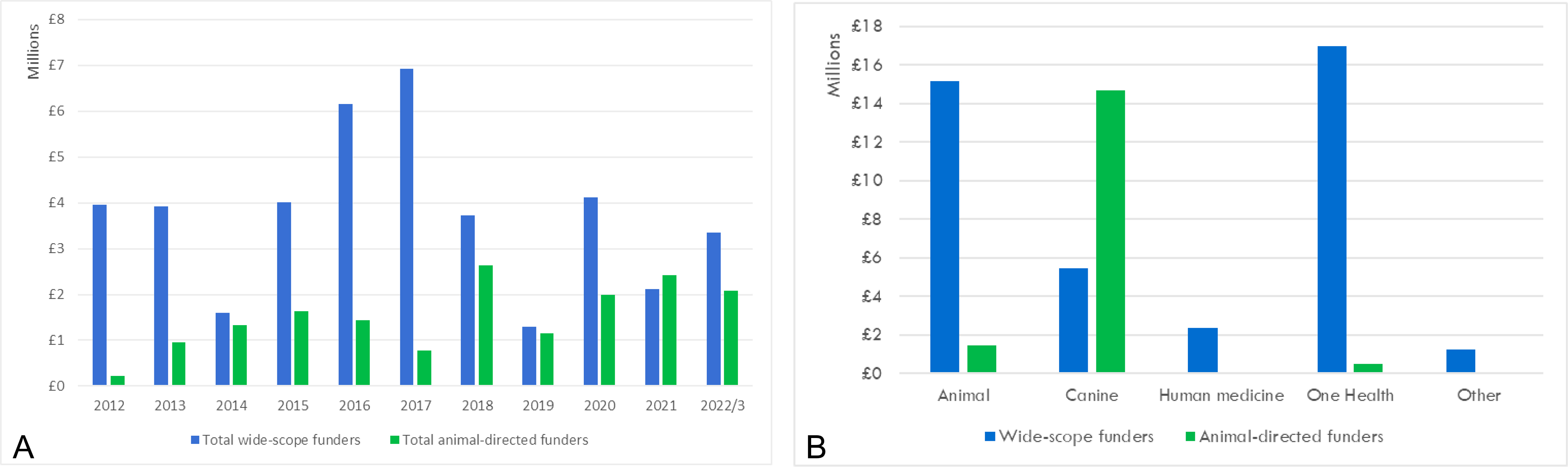
Figure 3. Histograms depicting proportional funding of canine health and welfare research in the United Kingdom (UK) for 2012-2022 supported by “wide scope” funders (UK government funding councils and medical charities concerned generally with human health) and organizations specifically aligned with animals. Funding is shown by year (A) and category (animal; canine; human medicine; One Health; and other) (B). As detailed in the paper, One Health grants generally were not focused on dogs alone but rather came from a broader human medical perspective. The 2017 peak included two sizable One Health grants on rabies and diabetes mellitus that accounted for almost half the total annual funding. The figure is reprinted with permission from[128]. Adhering to the CC BY 4.0 agreement.
Looking to future funding of companion animal rare disease research, the greatest challenge will be in using CGTs in affected animals. Here, the hemophiliac dog studies discussed by Doshi et al.[129] in this Special Issue are informative. Without funding from NIH, the ~ $3 M adeno-associated virus (AAV) per patient vector expense would have been far too costly for client-owned dogs. More encouragingly, companies like Scout Bio (recently acquired by Ceva Santé Animale)[130] and Rejuvenate Bio[131] are now providing gene therapy for common chronic companion animal diseases with much larger patient populations. Hopefully, over time, funding paradigms will make these treatments possible for rare diseases. In any case, managing costs of CGTs in rare diseases will undoubtedly be guided by experiences with human gene therapy for the same conditions[100]. These expenses will likely decline as original research and development investment costs are recouped, and manufacturing efficiencies are gained[132,133].
While similar challenges apply for using CGTs to treat companion animals with rare diseases, there may be greater opportunities for cell-based treatments. This includes the use of MSCs for neurological diseases[134] and immunotherapy for osteosarcoma[135] discussed in this Special Issue.
The role of insurance in covering rare disease treatment costs
Although policies differ and details are still evolving, insurance plays a major role worldwide in covering the costs of rare disease treatments for humans[136]. In contrast, veterinary (pet) insurance has a limited impact for affected animals. This disparity begins with the low rate of coverage among US pet owners as only about 4% of dogs (~ 4.3 M) and 1% of cats (~1.1 M) are insured at an approximate annual cost of $1,400 and $1,150, respectively[137]. Notably, coverage is much higher in some countries, amounting to around 40% in Sweden and 25% in the United Kingdom[138].
Key differences with human policies limit the usefulness of pet insurance in managing rare disease treatment[139,140]. Most veterinary policies exclude preexisting (genetic) conditions and experimental treatments. Furthermore, reimbursable costs may be capped annually, per condition, or for the animal’s lifespan. Accordingly, claims are mostly for routine health concerns such as treatable gastrointestinal and dermatologic diseases[141].
Moving forward, as owners increasingly think of their pets as family members[142], the US pet insurance market is expected to grow from $10.1 B to $38.3 B, a 14.5% annual growth rate, between 2023 and 2033[143]. Companies will almost assuredly seek to distinguish themselves. For instance, Pets Best and Trupanion have pioneered coverage for “rare procedures” such as bone marrow transplantation and stem cell therapy within the past 15 years[144,145]. Insurance for pre-existing conditions is also evolving, with some companies now paying for treatments of “curable” conditions[146]. Despite this progress, gene therapies are still seen as experimental and not covered.
RARE DISEASE THERAPIES
This section on treatments is particularly important as it speaks directly to advanced health care that is becoming available to animals with rare diseases. The discussion is divided into two complementary parts, first addressing general principles and approaches for treatment followed by specific therapies for inherited and acquired conditions. Other papers in the Special Issue are referenced to provide greater detail on certain diseases and treatments.
Emphasis is placed on approaches and/or diseases in which companion animals have played a major role in developing treatments and for which these approaches are now being employed in veterinary patients. Reflecting my own clinical and research experience and the papers that were ultimately submitted to the Special Issue, this review focuses on diseases of dogs and cats.
General principles and approaches
As discussed above, developing treatments for rare diseases is inherently more challenging. Also, since disease mechanisms are in many cases poorly defined, treatments that could be considered “experimental” entail carefully controlled clinical trials[147]. Given these factors, management of rare diseases is often both multimodal and multidisciplinary[148,149]. Here to facilitate discussion, therapies are categorized as either “conventional” (Western, mainstream, or allopathic medicine) approaches (drugs, radiation, and surgery) or more advanced methods that are typically grouped under CGTs.
Conventional therapy
Drugs/Small molecules. There is no definitive treatment for approximately 95% of rare diseases, meaning that patient care is usually palliative[150]. Bringing drugs to market is also costly and protracted[92]. Lafora disease, a rare fatal, autosomal recessive neurodegenerative disorder best characterized in humans[151] and dogs[152-155] provides a good example of the limitations of drug therapy and the role animal models can play.
People with Lafora disease have spontaneous mutations in either the EPM2A or NHLRC1 (EPM2B) genes[151], whereas only the NHLRC1 gene is mutated in dogs[152-155]. The two species develop analogous phenotypes, including progressive myoclonic epilepsy and classical intracytoplasmic inclusion (Lafora) bodies
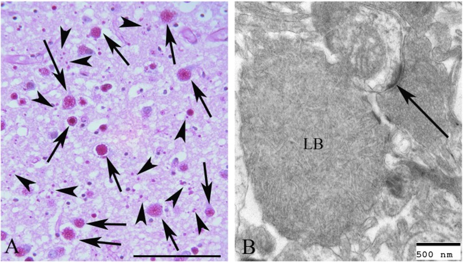
Figure 4. Lafora bodies in the frontal cortex of a dachshund dog seen with PAS diastase staining (A) and electron microscopy (B). Classical larger (arrows) and smaller (arrowheads) inclusions are evident. Bar = 50 µm in (A) and 500 nm in (B). The arrow in (B) identifies a synaptic density. The figure is reprinted with permission from[153]. Adhering to the CC BY 4.0 agreement.
Management of Lafora disease in humans and dogs is largely palliative, using anti-epileptic drugs and anti-ketogenic diets. Studies in dogs are especially insightful because, independent of Lafora disease, they develop generalized (inherited, idiopathic, and drug-resistant) epilepsy analogous to humans[157]. Therefore, pharmacokinetic properties of antiepileptic drugs are well characterized. In addition to medical management, more definitive therapies involving AAV transgenes and antisense oligonucleotides therapy to reduce glycogen accumulation in the Lafora bodies is being tested in mice and could be extended to dogs[158,159]. Humans and dogs should each benefit as newer treatment strategies are employed.
Speaking generally about conventional therapy of rare diseases, there has been considerable interest in either repurposing FDA-approved drugs or repositioning non-approved compounds that have been shelved because of problems such as toxicity[160,161]. Sometimes referred to as orphan drugs, these compounds have already been defined mechanistically and tested for safety, thus potentially reducing development costs and expediting approval[162,163]. This same approach has been utilized in veterinary medicine, particularly to treat cancer[164]. Outside of cancer treatment, a repurposed drug example for pets is the use of GS-441524 to treat FIP[52,165]. This compound was originally developed by Gilead Sciences to treat rare human viral diseases such as Ebola and later SARS-CoV-2 (COVID-19) (see under infectious diseases below and the Special Issue paper by Cerna et al., “From ebola to FIP: the cross-species journey of GS-441524 and the future of antiviral translation”)[166].
Radiation/Radiotherapy. The use of radiation to treat disease (radiotherapy) illustrates graphically the potential for translating findings from animals to humans (and back). Because of their relatively larger size and longer life spans, dogs were preferred over rodents to study radiation toxicity in the aftermath of World War II and have subsequently been used as sentinels to assess effects of the Chernobyl reactor accident[167]. These toxicity studies were critical in establishing basic principles of radiotherapy to better ensure that tumor cells are destroyed while minimizing damage to normal healthy tissue. Building on these experimental studies, radiation alone or as part of a multimodal regimen is now a mainstay in managing human cancer, including rare tumors such as mucosal melanoma, hepatobiliary and ovarian malignancies, and pancreatic and bronchopulmonary neuroendocrine neoplasms[168]. Using experimental data from dogs and subsequent human clinical studies as a base, similar radiotherapy strategies are now routinely used in dogs and cats with naturally occurring cancer[169].
Brain tumors illustrate the role radiation has played in managing cancer in companion animals. Going back to my own clinical practice in veterinary neurology, cobalt radiation therapy was shown to substantially improve survival in dogs with brain tumors, ranging from relatively common meningiomas to rare gliomas[170]. These benefits have been substantiated by more recent studies using linear accelerators as the radiation source and focused stereotactic delivery[171-173]. To increase the translational value of canine studies, the National Cancer Institute (NCI) established a Brain Tumor Consortium[174] to classify brain tumors of humans and dogs through their Comparative Oncology Program (see further discussion of comparative oncology below)[175,176].
Adding more support for the value of comparative studies, I have collaborated with radiation scientists and other clinical specialists from veterinary and human medicine to demonstrate benefits of whole-body hyperthermia as a radiation adjuvant and radiosensitizer in dogs with brain tumors[177]. Additional whole-body and localized hyperthermia studies have been done on other naturally occurring canine tumors[178,179] and the deadliest of human brain tumors, glioblastoma multiforme[180].
Radiopharmaceuticals bridge drug and radiation approaches[181] and can entail drug repurposing/repositioning[182,183]. Conventional repurposed drugs may either serve as radiosensitizers, thus increasing the therapeutic effects of radiation potentially at lower doses[182], or be bound to radionuclides that are already being used diagnostically in a field known as theranostics[183]. These compounds potentially qualify as orphan drugs[184], thus facilitating approval[185]. The emerging use of radiopharmaceuticals in companion animals further illustrates the value of comparative oncology[186].
Surgery. Extending beyond medical and radiation therapy, surgery offers a chance for a definitive cure in certain forms of cancer[187] (see the Special Issue paper by van Stee et al., “Rare pituitary diseases in dogs and cats and their human companions”)[188] and some developmental diseases[189,190] (see the Special Issue paper by Jones et al., “Computational modelling of cerebrospinal fluid (CSF) flow in a translational canine model”)[191]. Management of pituitary tumors is a classic example in which companion animals have ultimately benefitted from early research. The American neurosurgeon, Harvey Cushing, performed experiments in dogs to study the pituitary’s function and explore surgical approaches for its removal when diseased[192]. These techniques have subsequently been applied in treating dogs and cats with pituitary tumors[53,193]. Recognizing his role in defining clinical features of compensatory adrenal gland hyperplasia after pituitary removal or disease, hyperadrenocorticism of pituitary origin is often called Cushing’s syndrome (disease) in humans[194] and animals[195]. Further emphasizing comparative aspects, the incidence of different types of pituitary tumors varies among humans, dogs, and cats, offering insight into disease mechanisms[196-198].
Regarding developmental diseases and the role of animal models, surgery is often employed to correct primary anatomical anomalies or to lessen physical complications of rare diseases[199-201]. Models in large animals, including dogs and cats, have been used extensively because their size better models the human surgical experience, for instance, with medical device implantation[7,15,199]. Notably, joint replacement surgery was facilitated by experimental studies in dogs. These same methods are now being used on clinically affected animals[200,201]. While surgical procedures are often employed for common disorders such as degenerative joint disease[202,203], they are also used for rare conditions. An example is hip replacement to treat avascular necrosis of the femoral head (Legg-Calvé-Perthes disease) in humans and dogs[204,205].
Lessons learned from experimental canine studies have been especially impactful in enhancing our understanding of cardiovascular anomalies in companion animals. Dogs and cats with common disorders such as mitral valve insufficiency[206] and rare congenital anomalies[207] are now undergoing advanced surgical techniques that were refined in canine models[208].
CGTs
Considerable recent progress has been made in treating rare diseases by modifying the genome or using stem cells to promote tissue repair and regeneration. These CGT approaches, in principle, provide an opportunity to achieve substantial improvement and even cures[209,210]. The public is generally receptive to CGTs because recent successes have tempered concerns caused by early well-publicized failures[211]. With notable exceptions, genetic treatments have been used primarily for inherited diseases, whereas cell-based therapies are used mainly for acquired conditions. Studies of genetic treatments on canine models of rare inherited diseases, including hemophilia, congenital blindness, muscular dystrophy, and lysosomal storage diseases (LSDs), began in the 1990s on dogs raised in colonies[212].
Early attention to the benefits of gene therapy was provided by studies on a blind dog named “Lancelot” with an RPE65 gene mutation[213,214] [Figure 5]. He and other affected dogs recovered vision after intraocular injection of an AAV-RPE65 gene construct. Subsequently, genetic constructs to restore vision[126,215] and correct defects in other body systems[30,216-220] have been used experimentally in a wide array of inherited canine and feline diseases. Encouragingly, as noted in articles of this RDODJ Special Issue, companion animals with these conditions are now benefiting themselves (see the Special Issue paper by Doshi et al., “Adeno-associated viral vector gene therapy for canine hemophilia”)[129].
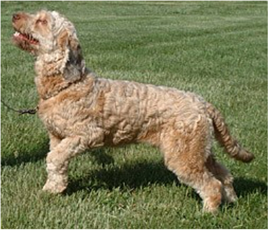
Figure 5. Photograph of “Lancelot,” a Briard-mix dog affected by congenital blindness, treated with gene therapy. From ref.[214], Reprinted with permission.
Undoubtedly, as these studies increasingly improve the lives of pets with rare diseases, additional genetic treatments will be explored (see the Special Issue paper by Vite et al., “Diagnosis and therapy of Niemann-Pick disease type C1- what have we learned that can help veterinary patients”?)[221].
Speaking to cellular therapy, the experimental use of canine stem cells dates to at least 1955 when Donnall Thomas defined immunity of bone marrow graft rejection[222]. His initial work in New York and later at Seattle’s Fred Hutchinson Cancer Center with Rainer Storb paved the way for using hematopoietic cell transplantation (HCT) to treat human leukemias[223] and lymphoma[224]. Pet dogs with analogous conditions are now being treated with HCT using this same approach[55].
Canine models have also demonstrated limitations to the efficacy of stem cell therapy for monogenic diseases. Promising results of muscle myoblast (satellite cells) transplantation and HCT in the mdx mouse model of Duchenne muscular dystrophy (DMD) could not be replicated in a canine model[225,226]. However, while neither approach has gained acceptance in DMD, stem cell studies continue in humans and dystrophic dogs, focusing on myogenic and progenitor cells to restore muscle fibers or MSCs to modify the immune/inflammatory response[227].
More broadly, MSCs have been used for a range of conditions in humans to take advantage of their anti-inflammatory and pro-regenerative effects[228,229]. There has been even greater use in veterinary medicine, especially for inflammatory musculoskeletal diseases and often as part of a multimodal treatment regimen[56,230,231] (see the discussion of using MSCs for Osteoarthritis [OA] and rheumatoid arthritis under degenerative diseases below and the Special Issue paper by Windsor, “Stem cell therapy for rare neurological diseases: translational research opportunities”. on their use in rare neurological diseases)[134].
The comparative aspects of brain cancer have been a particularly fertile ground for research in cell-based immunotherapy. CAR-T cells[232], tumor checkpoint inhibitors[232,233], T-regulator cell targeting[234], and vaccines[233,235] are being developed using canine glioma models. These treatments could ultimately benefit dogs and humans with aggressive brain tumors.
Disease specific treatments
This section will use the traditional division between inherited and acquired conditions, even though this distinction is now somewhat artificial. With the definition of the human and various animal genomes, it is clear that essentially all diseases have a genetic component[236]. For this discussion, the inherited category will largely focus on monogenic diseases that are passed through DNA from generation to generation. The acquired group will then be reviewed across four disease groups, first cancer followed by infectious, developmental, and degenerative conditions.
Importantly, there is overlap within and across these categories. Genetic influences vary among diseases classically considered to be acquired vs. inherited. Susceptibility to human breast cancer has perhaps been best characterized[237]. Here, human and veterinary oncology once more converge, as analogous breast cancer genes 1 and 2 (BRCA1 and BRCA2) that influence the incidence of mammary adenocarcinomas in humans have been identified in dogs. This provides an opportunity for comparative therapeutic targeting studies[238].
Developmental and degenerative diseases are often discussed together because of their frequent shared genetic origin[239] and pathogenetic mechanisms[240] despite having different patterns of onset (early/late) and progression (stable/progressive). The two groups are covered separately in this review, emphasizing those in which companion animals have played important experimental roles and are now benefiting from resultant treatments.
Notably, some progressive degenerative conditions, for instance the LSDs, are inherited as monogenic traits. However, because of their mechanistic association with human neurodegenerative diseases[241,242], they will be discussed primarily in the degenerative category of the acquired group.
Inherited diseases
Approximately 80% of rare human diseases are genetic and an estimated 95% of these conditions have no treatments[58]. Relevant to this RDODJ Special Issue and as mentioned under challenges for developing rare disease treatments above, the lack of animal models has limited studies of disease pathogenesis[30,96]. Nonetheless, rare inherited diseases in dogs and cats that share features with homologous human conditions have increasingly been identified[8,29,30]. This has been greatly facilitated by identification of the canine[243] and feline[244] genomes and greater availability of advanced genetic diagnostic testing, such as whole-genome sequencing[245,246]. An example is globoid cell leukodystrophy and the characteristic histopathologic changes seen in both affected humans and dogs[247] [Figure 6].
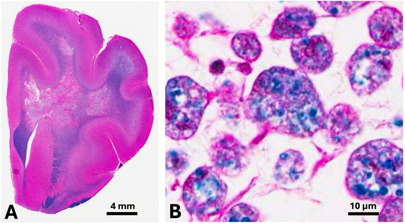
Figure 6. Histopathologic lesions of globoid cell leukodystrophy in a 6-month-old West Highland white terrier dog. Note the loss of blue-stained myelin within the white matter of the corona radiata (A) and characteristic large macrophages (globoid cells) containing phagocytized myelin lipids (B). Hematoxylin and eosin with a Luxol fast blue counterstain (H&E-LFB). Bar = 4 mm in (A) and 10 µm in (B).
Inherited diseases are more common in purebred dogs and cats that tend to be inbred, although mixed breeds may be equally affected, especially with ancient mutations[248-250]. Pedigree analysis in purebred animals allows the mode of inheritance to be defined[251-254]. Colonies of genetically homologous animals, often supported by the NIH and other funding agencies worldwide, have subsequently been established[30,217,226]. As an added advantage, sophisticated tests and diagnostic procedures like those used in humans with homologous conditions can be used to collect preclinical data[19] (see the Special Issue papers by Doshi et al., “Adeno-associated viral vector gene therapy for canine hemophilia”[129] and Vite et al., “Diagnosis and therapy of Niemann-Pick disease type C1- what have we learned that can help veterinary patients”?)[221].
Acquired diseases
Numerous rare acquired cancerous[255,256], infectious[18,257], developmental[258,259], and degenerative[260,261] diseases have been recognized in companion animals, offering valuable models for their human counterparts.
Cancer. The many parallels between canine and human cancer led the NCI to establish a Comparative Oncology Program through which naturally occurring tumors in dogs are studied as human models[262,263]. Notably, cancer incidence varies across species and among canine breeds[264,265]. Cancer susceptibility was initially presumed to track directly with cellularity (mass/size) but no association was found. Termed “Peto’s paradox,” this is one of the interesting riddles underlying the field of comparative oncology[266].
Osteosarcoma (OSA) is a notable example of a tumor with several common features between dogs and humans yet markedly variable incidence[267,268] [Figure 7]. Cases are rare in humans and relatively common in dogs. One epidemiological review projected an annual OSA incidence of 1/100,000 in humans vs.
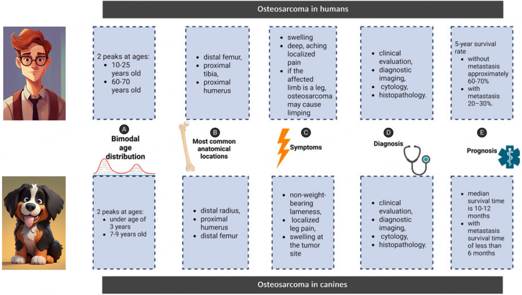
Figure 7. Clinical features of osteosarcoma in humans and dogs, including bimodal age distribution, common locations, symptoms/clinical signs, basis for diagnosis, and prognosis. Adapted with permission from[268]. Adhering to the CC BY 4.0 agreement. The young man and dog cartoons are Magnific open-source images (www.magnific.com).
Lymphoma/leukemia is another disease complex in which involvement varies among species. Recent data estimate the lymphoma incidence per 100,000 individuals at ~10 for humans (of which 90% are non-Hodgkin)[274]; ~20 for dogs[275]; and ~30 for cats[276]. At the same time, the occurrence of lymphoma subtypes varies among species. Some more common forms in dogs are rare in humans[275,277]. Similarly, gastrointestinal lymphoma is rare in humans[278], while occurring more frequently in cats[279].
Greater lymphoma involvement in cats likely occurs primarily due to oncogenic effects of the feline leukemia (FeLV) and immunodeficiency (FIV) retroviruses[280,281]. Both increase the risk of tumor development, with FeLV playing a direct role and FIV predisposing to tumor formation through immunosuppression. Indeed, the incidence of feline lymphoma was even higher (200+/100,000), before widespread FeLV testing and vaccination led to a marked decline in positive cats[282,283]. Offering further clues to disease pathogenesis, FeLV positivity varies with the site/organ affected. For instance, cats with mediastinal forms are far more likely to be FeLV positive than those with gastrointestinal involvement[284]. This could provide insight on the role of oncogenic viruses in human lymphoma, especially with certain rare forms[285].
Treatment of lymphoma in humans and companion animals has been complementary, with experiences in one field benefiting the other. Generally, the standard multiagent CHOP (cyclophosphamide, doxorubicin, vincristine, prednisone) chemotherapy protocol has been adapted from humans for affected dogs and cats and modified to enhance efficacy or reduce side effects[286,287]. The dosage for these drugs, including the risk of toxicity, was determined through a combination of preclinical studies in normal animals, including dogs[288,289], and veterinary clinical trials using escalating doses[290,291].
An additional translational example of the feline leukemia/lymphoma clinical complex is the study of antivirals in FIV-infected cats to assess potential efficacy and side effects when used against the human immunodeficiency virus (HIV)[292,293]. Interestingly, although antiretrovirals have shown efficacy in HIV-infected humans[293], they have minimal benefit in FeLV and/or FIV-positive cats and are only recommended in severe cases[292,294,295].
Infectious diseases. The potential value of companion animal models of rare and emerging infectious diseases is evident from the discussion of FIV and became even more obvious with the recent COVID-19 (SARS-CoV-2) pandemic. Both dogs and cats can be infected by coronaviruses[296], with FIP (see earlier discussion) being a rare especially virulent condition[297,298]. Cats with FIP have a spectrum of disease that has been characterized historically based on whether ascites (effusive/non-effusive; wet/dry) is present[298]
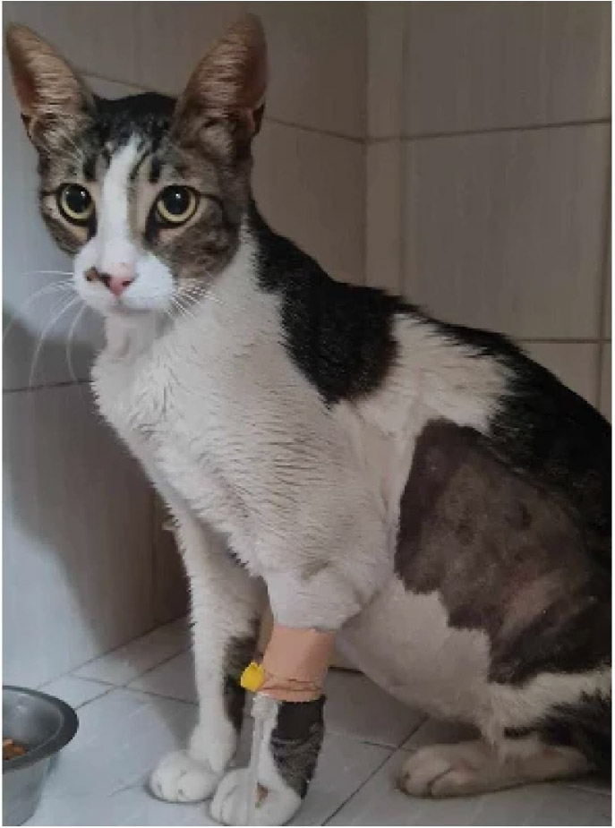
Figure 8. Cat with the effusive (“wet”) form of FIP with characteristic abdominal distention due to ascites. Reprinted with permission from[298]. Adhering to CC BY agreement.
Germane to this Special Issue, effective FIP antivirals were recently developed coincident with studies documenting their efficacy against COVID-19[52,173,300]. The case of GS-441524 is curious in that Gilead Sciences[301] patented the compound in 2009 but chose not to license it for FIP[302]. Despite this, it can be used “legally” with associated quality control in many countries[303,304]. In the US, the FDA has chosen not to enforce requirements for drugs compounded from GS-441524 when it is prescribed by a veterinarian for a specific affected cat[305] (see the Special Issue paper by Cerna et al., “From ebola to FIP: the cross-species journey of GS-441524 and the future of antiviral translation”)[166].
Prior to the advent of these new antivirals, veterinarians generally felt FIP was fatal[306]. This was certainly the experience of veterinary neurologists (including myself) in treating cats with the neurologic form[52,307]. But, amazingly, FIP cats treated with GS-441524 have achieved dramatic clinical improvement even when there is neurologic involvement[52]. Of further interest to dog owners and clinical veterinarians, these same antivirals may be effective against the canine distemper virus[308]. This virus has been studied extensively in dogs because of its potential linkage to multiple sclerosis and other rare forms of immune-mediated encephalitis[309] (see the Special Issue paper by Windsor, “Stem cell therapy for rare neurological diseases: translational research opportunities” relating to the use of MSCs in diseases affecting the nervous system)[134]. Antiviral pharmacokinetic data from normal dogs and cats have facilitated the use of these drugs in FIP and could potentially facilitate their use in canine distemper[127].
Central nervous system infections bring an added treatment variable into play as drugs must cross the blood-brain barrier or be delivered directly (intrathecally) into CSF[310]. Studies of CSF flow and drug delivery are a specialized area in which variables can be assessed better in large animals vs. rodents[311]. Dogs have been especially useful because they develop naturally occurring genetic conditions such as LSDs, allowing proof-of-concept treatment studies[312].
Commenting more broadly, approximately 75% of emerging infectious diseases are zoonotic[313], a term coined by Virchow, an early advocate of principles underlying One Health - One Medicine[77] (see prior discussion). Put another way, zoonotic diseases arise originally in animals and then “spill over” or “jump” to humans[313]. Not surprisingly, given their close association with people, dogs and cats can transmit zoonotic diseases[314] and together with other animals, serve as important sentinels in recognizing infections[18].
Reflecting on viral pathogenesis and FIP, cross-species transmission is facilitated by viral recombination and is particularly common with coronaviruses[315]. To this point, recombination between canine and feline coronaviruses recently led to a particularly virulent FIP outbreak in Cyprus[298].
Developmental diseases. Musculoskeletal and cardiovascular developmental diseases were discussed under surgery in the treatment approach section. Neurodevelopmental disorders likely take an even greater toll on humans through relatively common conditions such as childhood epilepsy[316], cerebral palsy[317], and Down’s syndrome[318]. Autism spectrum disorder and attention deficit and hyperactivity disorder have also been increasingly recognized and are no longer considered rare in children[319].
Disorders of cerebellar development and CSF flow, occurring separately or together, provide clearer examples of rare neurodevelopmental disorders in which companion animals have analogous diseases and have benefitted from experimental studies. Three conditions, feline cerebellar hypoplasia, a canine disease analogous to Dandy-Walker syndrome of humans in which the cerebellar vermis is selectively hypoplastic, and a disease complex in dogs like Chiari malformation, are noteworthy.
Cerebellar hypoplasia was described sporadically in kittens beginning in the latter part of the 19th century and originally thought to be inherited[320]. Studies conducted in the 1960s indicated that experimental and spontaneous in utero infection of pregnant cats with a virus[258], subsequently determined to be panleukopenia[321], caused cerebellar hypoplasia in the progeny. These and other in utero infection studies defined pathogenetic mechanisms of failed cerebellar development[322], helping to explain the origin of rare cerebellar developmental disorders that follow in utero infections in humans[323,324]. Otherwise, disease pathogenesis and cerebellar development in general have mainly been studied in murine models[325,326].
Distinctive hypoplasia of the caudal cerebellar vermis, analogous to Dandy-Walker syndrome in humans, has been seen in dogs worldwide[327,328] [Figure 9]. While its pathogenesis is poorly understood, passage of CSF from the fourth ventricle to the subarachnoid space is likely restricted due to anomalous brain development. Resultant ballooning of the fourth ventricle presumably impedes differentiation of the caudal vermis from the metencephalic alar plates[329]. Genetic factors[330,331] and in utero infections[332,333] have been implicated in both humans and dogs.
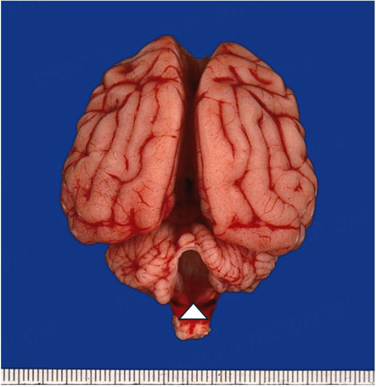
Figure 9. Brain at postmortem from a young dog with ataxia and tremor due to cerebellar hypoplasia. Note absence of caudal cerebellar vermis (arrowhead). This photograph was taken by the author who has the sole copyright[327].
A fascinating disease complex analogous to Chiari malformation of humans, with caudal skull fossa anomalies, craniosynostosis, and syringomyelia, has been well described in Cavalier King Charles Spaniel dogs[54,334]. Characteristic features include brain overcrowding due to brachycephaly, insufficiency of the caudal fossa, and craniofacial hypoplasia.
Ventricular dilatation in clinical syndromes of hydrocephalus/hydranencephaly occurs concomitant with cerebellar hypoplasia in people[335], dogs[327], and cats[336] as well as in dogs with Chiari-like malformation[54,334]. The cerebellar developmental defects of these three conditions are largely untreatable but mostly nonprogressive. In fact, cats with cerebellar hypoplasia due to in utero panleukopenia virus infections will generally adapt to their environments through other senses and lead relatively normal lives.
People, dogs, and cats with hydrocephalus, Chiari-like malformation, and syringomyelia can be managed surgically with various procedures, including ventricular shunting, decompression of the foramen magnum, and fenestration of fourth ventricular cysts[54,337]. Critically, it is not always clear which surgical procedure is appropriate. Modeling patient-specific surgical procedures using DICOM software may help to avoid multiple surgeries and poor outcomes[338,339] (see Special Issue paper by Jones et al., “Computational modelling of CSF flow in a translational canine model”)[191].
Degenerative diseases. OA, intervertebral disk disease (IVD), and other progressive degenerative diseases are relatively common worldwide. Dogs and, to a lesser degree, cats have analogous spontaneous OA[340,341] and IVD[342,343] syndromes that serve as valuable models for humans. Beyond these natural conditions, experimental studies have been done in dogs to guide treatment of humans with OA[344] and IVD[345]. As with other diseases, findings from research on common maladies such as these have helped guide management of rare conditions. For instance, lessons learned from using MSCs for canine OA[346] are being applied in rheumatoid arthritis[347], a rare disease seen in only 2/25,000 dogs[348].
A similar translational scenario for IVD is shown by an interesting condition in which disk material herniates within the dura (intradural) as opposed to its typical extradural location. This syndrome is seen rarely in humans[349] and cats[350] while occurring more commonly in dogs[351]. Similar interspecies differences are evident in the spinal infarction syndrome of fibrocartilaginous embolism (FCE), which is relatively common in dogs[352,353] but rare in cats[354] and humans[355]. Species predilections for intradural disk herniation and FCE likely reflect common underlying mechanisms and offer clues for disease management[356].
A wide array of spontaneous rare genetic neurodegenerative diseases has been described in dogs and cats, with many being promoted as translational models[30,357-360]. Degenerative myelopathy, a spinal cord disease that typically affects German Shepherds and several other canine breeds, is a prominent example[89,260]. On histopathology, there is degeneration of axons and their myelin sheaths, with lesions being most severe in the thoracolumbar spinal cord and extending to spinal nerve roots causing neurogenic muscle atrophy[361]. Toxic gain-of-function mutations in the SOD1 gene, with misfolding of the protein, have been associated with degenerative myelopathy in several breeds[89,362] and in humans with familial amyotrophic lateral sclerosis (ALS)[363]. This suggests degenerative myelopathy in dogs could serve as a model for certain forms of this devastating human disease[89].
The standard of care for dogs with degenerative myelopathy is physiotherapy (active and passive exercise, hydrotherapy, and massage) coordinated by veterinary neurologists and rehabilitation specialists[364,365]. Addition of MSCs to a regimented exercise program improved survival in one blinded study[366] (see the Special Issue paper by Windsor, “Stem cell therapy for rare neurological diseases: translational research opportunities”)[134]. In another recent therapeutic development for ALS patients, the FDA approved an antisense oligonucleotide that targets the SOD1 protein[367]. A similar treatment strategy in degenerative myelopathy dogs was safe and resulted in an ~50% reduction in SOD1 protein[368]. These results suggest that a multimodal approach, including MSCs and antisense oligonucleotides, could be beneficial in affected dogs.
Turning to LSDs, numerous conditions in which there is an inherited deficiency of enzymes necessary for hydrolysis of proteins, polysaccharides, and complex lipids have been described in humans[369] and domestic animals, including dogs and cats[30,261,370,371]. These enzymes are contained in cellular organelles termed lysosomes. When deficient, the substrate accumulates intracellularly, hence the collective term of LSDs. More broadly, lysosomes have been implicated in aging and neurodegenerative diseases[241,242]. Accordingly, although LSDs are inherited as monogenic traits, they are often discussed with degenerative conditions that are not strictly inherited. Neurons are preferentially affected by most of them, while others principally affect white matter and are classified as leukodystrophies.
Canine and feline LSDs have served as valuable models to guide treatment of homologous diseases in humans[30]. Bone marrow transplantation has been used in both species to treat mucopolysaccharidosis[372,373]. Gene therapy, usually employing AAV vectors to deliver the deficient transgene, has been studied in several LSDs of dogs and cats[30,216,312,374]. Clinical diagnostic tests have documented efficacy. Hopefully, over time, these experimental therapies will come full circle and be used in client-owned animas (see the Special Issue paper by Vite et al., “Diagnosis and therapy of Niemann-Pick disease type C1- what have we learned that can help veterinary patients”?)[221].
OPTIMIZING PRECLINICAL RESEARCH AND CLINICAL TRIALS
Providing care for people with rare diseases emerged as a worldwide priority with the identification of numerous inherited disorders in concert with the genomic age[375]. In this Special Issue, we focus on the role animal studies have played in the development of innovative treatments that are now being used in veterinary patients after a foundation for their effectiveness was established in these same species. An interrelated issue involves the current low success rate of human clinical trials and whether preclinical animal studies should be deemphasized and replaced with alternatives. The involved issues are complicated, prompting considerable debate. To understand underlying factors, we will first discuss the drug approval process used by regulatory bodies. This will be followed by a review of the current approach to, and potential problems with, preclinical research and clinical trials.
The drug discovery process
Drug approval in the US occurs through the FDA’s Center for Drug Evaluation and Research (CDER)[376] for humans and the Center for Veterinary Medicine (CVM)[377] for animals. Similar programs for human drug approval are conducted through the European Medicines Agency (EMA)[378], Japan’s Pharmaceuticals and Medical Devices Agency (PMDA)[379], and analogous entities in other countries. Efforts have been made to harmonize drug approval policies through the Council for Harmonization of Technical Requirements for Pharmaceuticals for Human Use[380]. Animal drug approval in Europe is conducted through the EMA’s Committee for Veterinary Medicinal Products[381]. A similar program exists in Japan’s Ministry of Agriculture, Forestry, and Fisheries[382].
Human patients
Standard approval. Drug development is deliberate and protracted, requiring as long as 10-15 years to complete. The FDA CDER defines five steps centered on developing an IND application: (1) Discovery and Development; (2) Preclinical Research; (3) Clinical Research; (4) FDA Review; and (5) FDA Post-Marketing Safety Monitoring (sometimes called Phase IV)[383,384]. These steps fall into three general stages of preclinical testing, clinical trials, and the review/approval process [Figure 10][385,386].
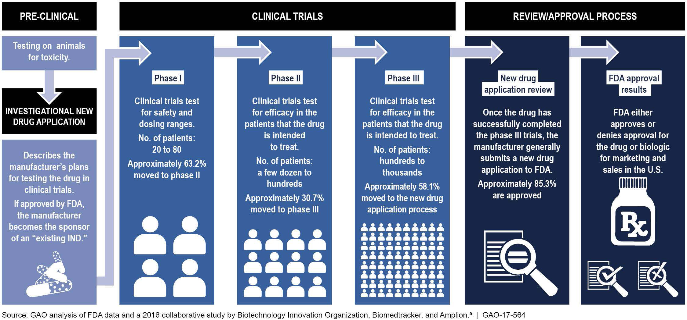
Figure 10. Schematic showing the three general stages (pre-clinical testing, clinical trials, and the review/approval process) of the FDA’s drug approval process. Drug approval percentages reflect FDA data from 2006-2015 analyzed by the US Government Accountability Office (GAO) through a collaborative study with the Biotechnology Innovation Organization, Biomedtracker, and Amplion (GAO 117-564)[385,386].
This approval process extends to CGT approaches through the FDA’s Center for Biologics Evaluation and Research (CBER)[387]. As noted in a January 2026 FDA news release, “The transformative potential of these therapies has captivated the imagination of the patient community and ignited product development”[388]. While CBER generally follows even more rigorous guidelines, the urgency associated with severe progressive rare diseases has prompted the FDA to maximize flexibility, as reflected in the accelerated approval process discussed below. Another FDA effort, directed at ultra-rare diseases, amounts to essentially “individualized therapies”. Termed the “Plausible Mechanism Framework”, this program would ideally allow development of effective (and safe) personalized biological products[389].
The FDA makes a special distinction for stem cells, which are classified as either a 351 or 361 product depending on the level of manipulation, as with expansion in culture[390]. Stem cells in the 351 (biological drug) category require greater manipulation and are subject to FDA approval[391].
The preclinical phase of drug development is intended to establish drug safety and efficacy in advance of human use[392]. Literally, thousands of compounds are screened during the discovery phase, progressing through target validation, compound screening, and lead optimization. Going back many years, (see discussion of alternatives below), this phase has included not just animal (in vivo) testing but also in vitro (cell culture, etc.) and in silico (computer modeling) methods[44,45,393,394]. More sophisticated in vitro techniques have regularly been implemented, such as the use of 3D culture systems to better replicate the in vivo environment[45].
Clinical research is divided across three phases (I, II, and III) in which the candidate drug is initially given to 20-100 healthy volunteers to establish safety (phase 1) followed by studies in a relatively small number (“up to several hundred”) of patients with the target disease in phase II and then a much larger (“300 to 3,000”) affected group in phase III[395]. This process is intended to be rigorous, as evidenced by the FDA’s approval of only around 50 new drugs each year[396,397]. Emphasis is placed on rare diseases for which few treatments exist; 26 of 50 (52%) novel drug approvals for 2024 were in this category[398]. For 2025, 46 novel drugs, including 34 “new molecular entities” and 12 biologics, were approved[397,399]. Exactly half (23) of these approvals were designated as orphan drugs.
While neither the FDA nor other groups involved in the drug discovery process have defined an optimal success level for drug approval, concerns have been raised about a persistently low (10%-15%) rate across the US, Europe, and Japan[400]. The two major reasons for failed clinical trials have historically been a lack of efficacy and safety concerns[401]. However, recent data suggest a potential shift during the 2013-2023 period, with strategic/business considerations exceeding efficacy as the most common reason for stopping trials[402] [Figure 11]. Moreover, enrollment and operational issues outranked safety as causes for trial termination. The implications of this apparent shift will be discussed below.
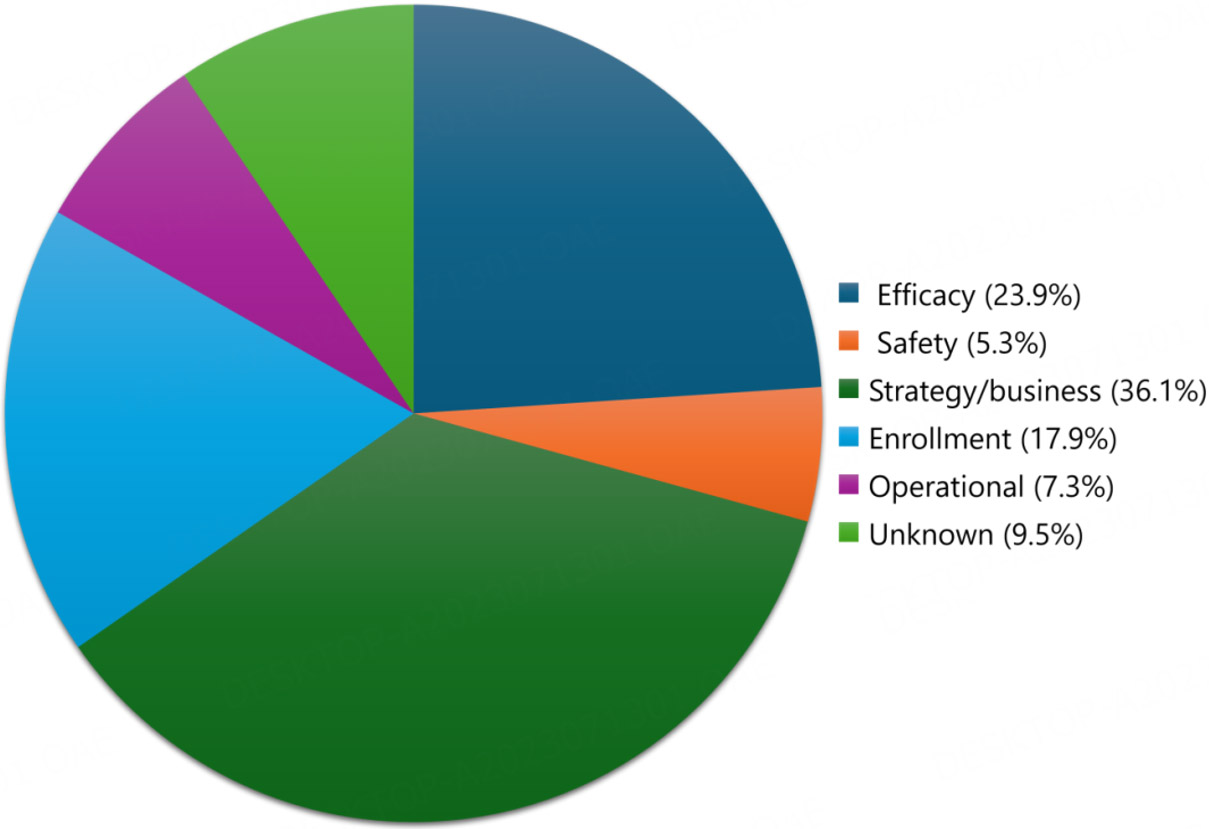
Figure 11. Reasons for phase II and III clinical trial terminations from 2013-2023. Data from Ref.[402]. Note the shift from prior studies generally showing that efficacy and safety concerns predominate to a higher frequency of issues tied to business strategies.
Orphan drugs. As discussed earlier under the “rare disease incidence criteria and drug development incentive programs” section, the US 1983 Orphan Drug Act[62] was intended to lessen barriers for developing rare disease treatments[47]. This law has been called, at least potentially, the best healthcare legislation of the 20th century, with over 6,000 new drugs and biologics being designated as orphan drugs since 1983[403]. While not the act’s specific intent, the streamlined process for drug approval has resulted in a slightly higher (~17%) approval rate compared to the standard FDA approach[403,404]. The top three therapeutic designations for 1983-2019 were oncology (37%), neurology (13%), and infectious diseases (9%)[404].
With the success of the Orphan Drug Act, companies have increasingly relied on this route for drug approval [Figure 12]. Consistent with the 2025 data mentioned above, one half of the 368 total drugs approved by the FDA between 2016 and 2023 were orphan drugs[405]. This emphasis on orphan drugs has caused concerns that rigor is being compromised in the approval process. Supporting this point, orphan drugs have been found to undergo fewer studies, with a reduction in randomized clinical trials and less supportive evidence, than their nonorphan counterparts[405]. Adding context to these concerns, pharmaceutical companies have promoted broad off-label usage of orphan drugs to reach a wider patient market[406], and over half of them have had changes to safety labels after approval[407]. This has led to questions on whether the Orphan Drug Act’s original intent has been lost[406].
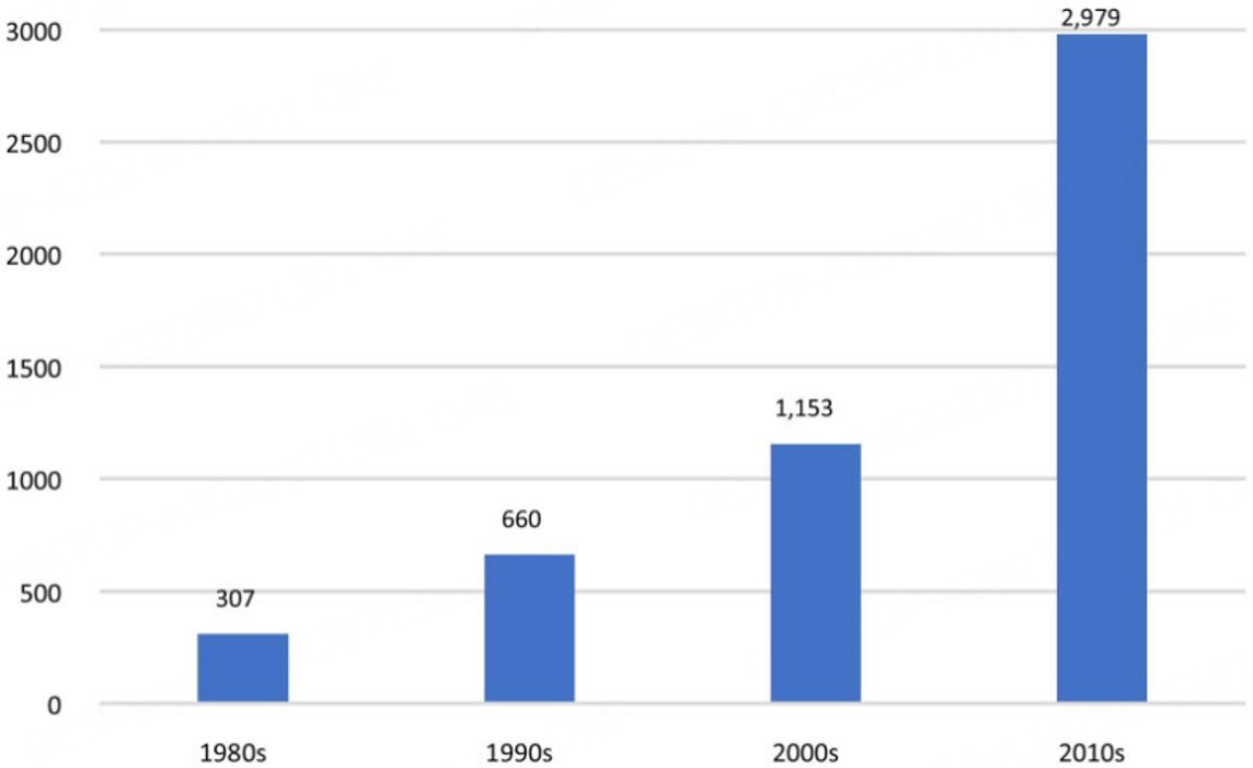
Figure 12. Histogram showing the progressive increase of FDA-approved orphan drugs by decade from the 1980s to 2010s. Reprinted with permission from[404]. Adhering to CC BY agreement.
Accelerated approval. Over and above the Orphan Drug Act, the FDA has initiated four other programs to expedite approval of drugs for serious diseases in which they “are the first available treatment or … have advantages over existing treatments[408]”. Termed fast track, breakthrough therapy, accelerated approval, and priority review, these programs streamline the approval process, for instance allowing surrogate biomarkers[409] and/or a modified phase 2 study to substitute for phase 3[46]. Accelerated approval has been used extensively for drugs to treat cancer and hematological malignancies[410,411] and has extended to CGT approaches[412]. Incentives like those of the orphan drug program are sometimes provided. In fact, the orphan drug and accelerated approval programs may be used simultaneously[407,413].
Drugs submitted through the accelerated process are typically approved faster and in some cases at a considerably higher rate. For instance, in the first year of the new breakthrough program initiated in 2012, 27 of 92 (29.3%) applications were approved with 24 still pending when these data were published[409]. As a safeguard, drugs approved through an accelerated program must ultimately undergo confirmatory studies[414] as part of the FDA’s Post-Marketing Safety Monitoring (Phase IV) process[383,384]. Notably, confirmation challenges can be more onerous for rare diseases because of fewer patients, especially for non-oncologic indications in which there is less experience[410].
Regarding the post-marketing safety program, over 80% of orphan drugs approved through expedited review between 1999-2018 were subsequently confirmed[407]. Although many had postmarketing safety concerns, severe events were rare. Another analysis of the program’s 30-year history was also generally supportive, showing that half of accelerated approvals were confirmed in a median time of 3.2 years[415]. Still, questions regarding reduced rigor within the accelerated approval program persist because of fewer randomized clinical trials[416], delays in confirmatory studies (“dangling approvals”)[417], higher drug costs[418], and the questionable validity of some surrogate endpoints[419].
Veterinary patients
As mentioned earlier, veterinary drug approval occurs through the FDA’s CVM[377] in the US and analogous programs in Europe, Japan, and other countries. Similar accelerated approval programs have also been implemented.
Just as with humans, the veterinary drug approval process is focused on ensuring efficacy and safety[420]. However, the process is complicated by different species indications and the need to ensure safety of products intended for human consumption[421]. The standard full-approval process entails an Investigational New Animal Drug (INAD) application analogous to the IND for humans. Beyond the INAD, there are four other applications intended to provide flexibility in meeting the public’s needs. These include abbreviated for generic drugs, conditional tied to the MUMS program, indexing for non-food producing animals such as those in zoos and ornamental fish, and rare instances of emergency use[422,423].
In considering the shift to orphan drug approval for humans cited above, relatively few new drugs[424], as cited in the FDA’s Green Book[425], have been approved through the MUMS program. While this suggests the program has remained true to its original intent, a new provision allows for “expanded conditional approval” of drugs to treat certain serious or life-threatening diseases (see discussion of MUMS in the “rare disease incidence criteria and drug development incentive programs” section)[72]. The FDA CVM also has an Innovation in Animal Drug Development program for accelerated drug approval[426]. This allows existing literature or alternative testing procedures to be substituted for laboratory animal studies.
Improving clinical translation
The low (~10%) clinical trial success rate may logically arise primarily because of the intentionally rigorous drug approval process. However, while an optimal rate has not been suggested, there is a consensus that potential problems with preclinical research and the clinical trials process should be addressed. These issues are inherently complex and impacted by other variables such as the shifting landscape of clinical trials discussed above and the ongoing strong push to replace in vivo animal studies with NAMs. Major factors involved in both areas are discussed here.
Preclinical research
Shortcomings in this area have generally been divided between issues of either internal or external validity[427]. Internal validity concerns include factors such as planning, conduct, and reporting of preclinical animal studies. Involved variables include sample size calculations, adequate statistical methods, concealed and randomized allocation of animals to treatment groups, blinded outcome assessment, and recording of animal flow through the experiment. The Animal Research: Reporting of In Vivo Experiments (ARRIVE) Guidelines, developed by the National Centre for the Replacement, Refinement, and Reduction of Animals in Research, provide a checklist of 10 recommendations for the full and transparent reporting of research involving animals[428,429] [Table 1]. Importantly, experimental variables such as these are not consistently mentioned in papers, raising questions on whether the studies have been conducted with sufficient rigor.
ARRIVE Essential 10 Guidelines
| Study design |
| Sample size |
| Inclusion and exclusion criteria |
| Randomization |
| Blinding/Masking |
| Outcome measures |
| Statistical methods |
| Experimental animals |
| Experimental procedures |
| Results |
In contrast, external validity refers to either surmountable (e.g., using inbred, young, healthy mice to study progressive diseases of older humans) or insurmountable issues (species, setting, and population differences)[430]. These factors have become even more important with the advent of CGTs that offer a chance for cure but also carry considerable risk[209-211]. With gene therapy, the choice of a disease-specific animal model is critical to ensure that safety, efficacy, dosage, and localization of transgene expression are properly assessed[431].
In this context, the FDA has emphasized species selection in developing CGTs for rare diseases[432,433]. Prior to performing preclinical studies, they recommend that in vitro (e.g., functional assays, immunophenotyping, morphologic evaluation) and pilot in vivo studies be done to establish the biological relevance of a specific animal species to the product(s) being studied. For the sake of the IND preclinical section, sponsors are encouraged to conduct a detailed assessment of the relevancy of the animal species used in support of each potential clinical trial.
Scientists have generally supported continued use of animal models to study recombinant proteins and CGT products for which clear translational value has been demonstrated[434]. While certainly not optimal, the 18.5% clinical trial success rate for durable CGTs approved by the FDA since 1987 was found in one study to be over twice that of the average drug[435].
As treatments are developed, it is critical to remember that preclinical studies and initial phase I human trials are principally intended to ensure safety[147,436], as exemplified by the Hippocratic Oath, “First, do no Harm”[437]. With CGT, an animal model’s ability to detect immunogenicity and associated severe adverse events varies among species. To this point, inbreeding among mice lessens their genetic diversity and immune response to foreign and/or auto-antigens[438,439]. In contrast, large animals are generally outbred and relatively long-lived, making them more suitable to detect acute and more chronic adverse effects after CGT[17,440].
With that said, large animal models have not consistently predicted adverse events in humans after AAV gene therapy[441]. As an example, genetically homologous canine models of hemophilia, DMD, and X-linked myotubular myopathy did not predict immune responses to AAV-transgene therapy in recent human trials[219,442,443]. Phenotypic differences, such as variable organ involvement between diseased humans and dogs, likely contribute to their relative immune response[444] (see phenotypic variation in genetic diseases above). Using NAMs, as with organoids and body-on-a-chip platforms, to guide AAV capsid selection could have value[445,446]. There are also limitations for large animal models in studying stem cells, including differences in pregnancy and fetal development, the lack of high-throughput approaches for screening, and cumbersome methods for genetic modification[447,448].
Human clinical trials
As discussed above, the FDA drug approval process is conducted in phases, progressing from phase 1 safety studies in normal volunteers through small phase 2 and then larger phase 3 studies of the target disease population. The general paradigm to calculate clinical trial success in Hay et al.’s 2014[396] paper provides a useful marker. They multiplied the success for each phase transition of drugs evaluated between 2003-11, i.e., Phase 1 to 2 (64.5%) X Phase 2 to 3 (32.4%) X Phase 3 to new drug application (NDA) (60.1%) X NDA to approval (83.2%), to show a 10.4% approval rate. Using a similar equation, another study found an approval value of 9.6% (63% X 31% X 58% X 85%) for 2006-2015[397,449] (see data depicted in Figure 10). In that study, hematology had the highest success rate (26%) and oncology the lowest (5%). Rare non-cancer disease drugs had a 25% success rate, outperforming other indications.
A decision to terminate a clinical trial is generally made by the sponsoring agency/company, often with input from its Data and Safety Monitoring Board[450]. There are three general reasons for early termination of a clinical trial: (1) one arm shows a clear benefit, such that it would be unethical to deprive the placebo group of the therapy, (2) safety concerns, and (3) a clear “no effect” based on interim data analysis[451]. As discussed in greater detail below, clinical trials are terminated for a combination of reasons, reflecting deficiencies in preclinical research, faulty trial design, and strategic business considerations.
While shortcomings of preclinical research clearly must be addressed to improve clinical translation, human clinical trials face similar challenges. These include biologic variability (phenotypic variation) of the patient population, underpowered studies due to inadequate patient accrual, and faulty trial design[452-454]. The trial design issues mirror those of preclinical studies, including lack of blinding or randomization of study participants, inappropriate inclusion/exclusion criteria, and invalid endpoints. Study design problems become especially problematic when advancing to phase 3[455]. Significant drug efficacy seen during the more focused phase 2 period is sometimes lost, potentially due to increased heterogeneity across patients, study sites, and outcome parameters[456]. Whether this appropriately weeds out ineffective compounds or leads to a loss of potentially valuable drugs has been debated[457].
Some of these issues have come into sharper focus with a recent analysis of reasons for clinical trial failures during 2013-2023[402,458]. In contrast to the 2013-15 data from the same group mentioned earlier[401], strategic/business reasons were cited most frequently (36%) followed by efficacy (24%), limited enrollment (low patient accrual) (18%), operational issues (7%), and safety (5%) [Figure 11]. The operational concerns included challenges to executing the trial, failed good manufacturing practice compliance, protocol deviations, supply chain issues, and poor data quality.
For the sake of strategic/business reasons to withdraw a candidate drug, companies considered factors such as its performance compared to competitor drugs or the possibility that it might perform better in other indications[402]. Other business-related reasons were suggested, for instance a downgrade in the drug’s commercial value, redundancy with other compounds under development, or a lack of funding. Based on comments in the paper, it appeared that some trial terminations in which efficacy was previously cited as the reason for withdrawal might now be placed in the strategic/business category. This points out the potential for overlap among reasons given for trial termination, as with a drug reaching its target efficacy but being terminated because it failed to outperform a competing compound.
The problem of patient enrollment/recruitment (accrual) is particularly problematic for the rare disease community. Other studies have emphasized the role that eligibility criteria complexity plays in this issue[452,459,460]. Carlisle et al.[461] found that 19% of trial terminations in 2011 were due to failed accrual or had less than 85% of their expected enrollment, compromising statistical power. Similar concerns were expressed in a literature review of factors causing clinical trials to fail over a 30-year period[453]. Accrual challenges must be considered in the context of broader changes in clinical trials, including the shift towards using orphan drug and expedited approval processes and the greater role that business concerns are playing in their termination.
When considering the apparent tendency for companies to terminate clinical trials for business reasons, the differing cultures in basic science and clinical medicine could be a factor[462]. As opposed to giving up on a compound, the preclinical mindset is to define the underlying cause and adjust. This is highlighted by
ALTERNATIVES TO PRECLINICAL ANIMAL RESEARCH
Concerns about animal experimentation were raised in the 17th century, first scientifically and later morally[1]. Objections to using dogs were especially noteworthy, as these concerns played a major role in seminal legislation such as the Cruelty to Animals Act (1876) in the United Kingdom[465,466] and the US Animal Welfare Act (1966)[467,468]. Further momentum and critical scientific focus were gained by publication of Russell and Burch’s The Principles of Humane Experimental Technique in 1959, defining the 3Rs (replace, reduce, and refine) that guide current day animal research[469,470]. Regarding the current debate on the use of animals in research, the public is generally not aware of these provisions and is more supportive when informed of existing guidelines to ensure animal welfare[471].
The development of alternatives to the use of animals in research, now collectively termed NAMs, is a longstanding mainstay for applying the 3Rs. Examples of NAMs include in vitro systems (cell/tissue culture, organoids), chemical/toxicologic assays, and computational (in silico) models[44,45,472-474] [Figure 13]. These alternative methods have been prioritized by governmental regulatory agencies such as the FDA[475], US Environmental Protection Agency[476], EMA[477], and Japan’s PMDA[478]. This has led to formal legislation, for example the FDA Modernization Act 2.0 passed by the US Congress in 2022[40-42] and further clarified by the 3.0 Act now under consideration[43], as well as policies such as the FDA’s Roadmap to Reducing Animal Testing in Preclinical Safety Studies[479].
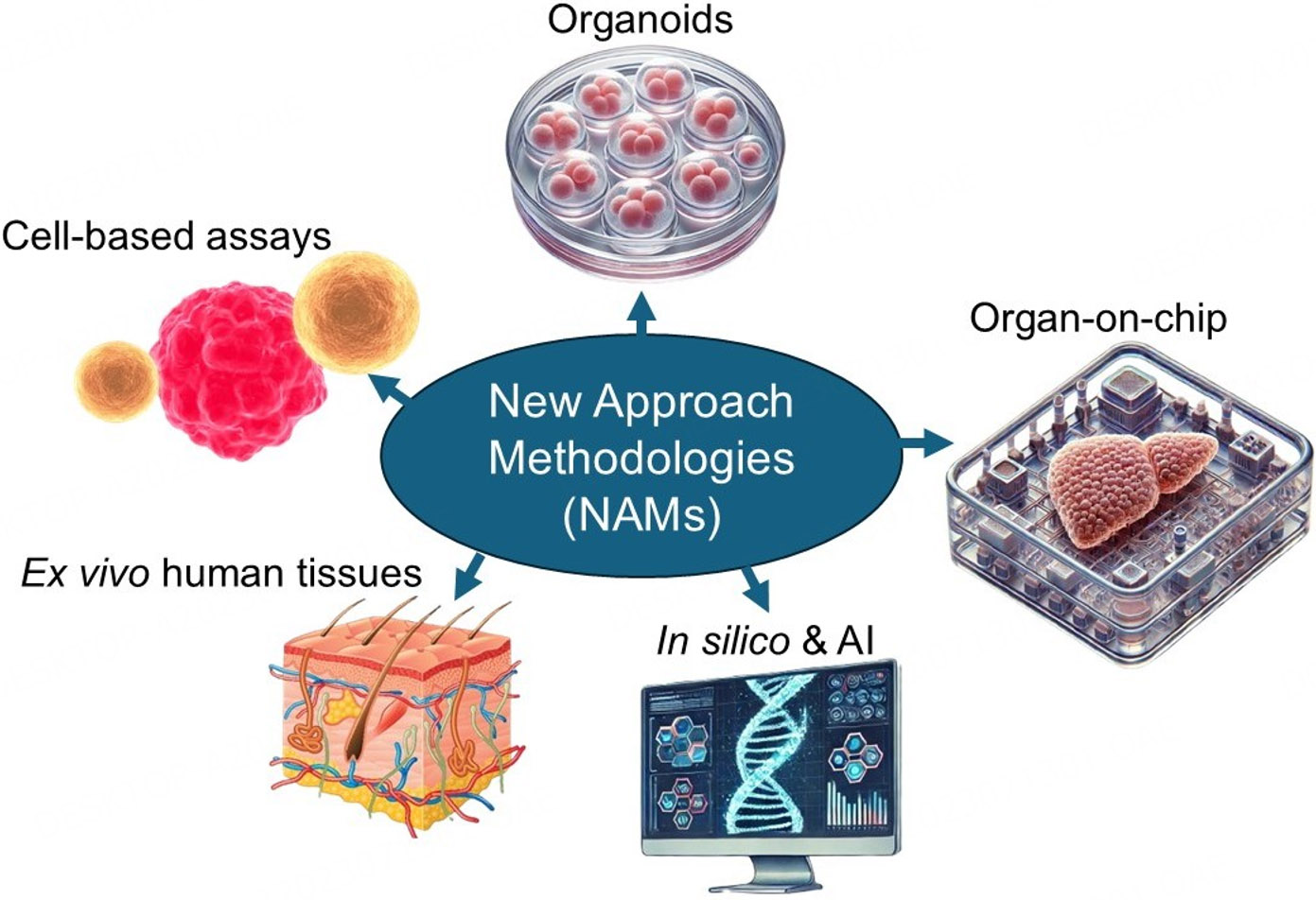
Figure 13. Schematic showing examples of new approach methodologies/methods (NAMs). Reprinted with permission from reference[474].
For the sake of the theme of this Special Issue, NAMs offer an opportunity to not only replace dogs and cats in research but also benefit them. However, there is a risk of overreach and implementation of ill-conceived policies. This concern was reflected by the Society of Toxicology’s statement on the use of animals in research and NAMS, which included the recommendation, “Studies that deliver consistent, reliable, and accurate data on the public health and environmental impacts of biological agents, chemicals, and compounds should be recognized, funded, and incorporated into decision-making—regardless of the model or technology used[480].
While NAMs are uniformly seen as extremely valuable tools in biomedical research, animal studies are for the most part still felt to be necessary to define body-wide disease mechanisms such as immunity[481,482]. Both the promise and shortcomings of NAMs, at least in the short term, were captured by a recent review published in Scientific American[483], in which one scientist opined, “Unless we hold NAMs to the same level of rigour and transparency that we expect of animal models, there will be harm done”.
ETHICAL ISSUES RAISED BY USE OF ANIMALS IN RESEARCH OR IN PROVIDING HUMANS AND ANIMALS ADVANCED MEDICAL CARE
Interrelated ethical issues face biomedical scientists in animal research and caregivers involved in human and veterinary medicine. In the case of biomedical research, the major justification for using animals has always been the potential benefit to humans[39]. This presupposes a moral hierarchy in which humans are superior to other species, an attitude that has been criticized as speciesism (like racism and sexism)[484]. In this spirit, those who wholly oppose animal research claim moral equivalency between humans and other species[485]. Another view recognizes the potential value of experiments on animals but points to their capacity to feel pain and suffer (so-called sentience) in arguing that proposed studies must be critically assessed[486]. This harkens back to the 3Rs and emphasizes the need for a harm-benefit analysis for any research involving animals[487,488]. Some have suggested that Russell and Burch’s original concept should be updated to reflect practical considerations, such as expense and availability of expertise to conduct the proposed research, including alternatives, to facilitate review and ultimate approval of studies by regulatory bodies[489].
The major responsibility in the US for ensuring animal research is done ethically resides with individual Institutional Animal Care and Use Committees (IACUCs)[490]. As mandated by the Animal Welfare Act[467,468], added governmental oversight is provided through the Department of Agriculture’s Animal and Plant Health Inspection Service[491] and the NIH’s Office of Laboratory Animal Welfare[492]. The use of laboratory animals in Europe is regulated by EU Directive 2010/63/EU17[493] and guided by the 3Rs. Relevant to this RDODJ Special Issue, the potential for animals themselves to benefit is considered in approving their experimental use[494].
Additional ethical concerns are faced by physicians and veterinarians in clinical settings, especially in managing rare diseases for which treatments are often seen as experimental and require enrollment of patients in highly regulated clinical trials. In the era of precision medicine, Institutional Review Boards (IRBs) have assumed an increasingly important role in human medicine to ensure patient safety and privacy[495]. Reflecting the severity of these conditions, the patients themselves or parents of affected children are often willing to accept considerable risk despite a low chance of substantial improvement provided by the treatment. Indeed, their tolerance for risk may exceed that of the caregivers themselves, creating an ethical risk-reward dilemma[496] and emphasizing the need for careful trial design[497].
Importantly, owners of companion animals with naturally occurring rare diseases often seek veterinary care, at times involving referral from general practice veterinarians to specialists. These dogs and cats can be entered into clinical trials akin to those used for humans with analogous diseases distinct from experimental studies done in colony-raised animals. Just as with humans, once efficacy has been shown in clinical trials, the treatment can be made more widely available to veterinary patients. This scenario constitutes the ideal continuum by which pets ultimately benefit from earlier experimental studies. And in the spirit of the bidirectional nature of human and animal studies that typifies the One Health - One Medicine concept[31], further valuable preclinical data can be translated to humans with the same conditions.
Veterinary clinical trials are generally overseen through IACUCs[498] or, if not available, by veterinary Clinical Studies Committees analogous to IRBs, as recommended by the American Veterinary Medical
CONCLUSIONS
• Companion animals, especially dogs and cats, have been used extensively as experimental models for rare human diseases. A long-held justification for their use has been the potential for them to benefit from the original experimental studies. These findings, in turn, have informed human treatment, in keeping with the bidirectional nature of One Health - One Medicine.
• Developing rare human disease treatments is challenging due to small patient populations, cost, protracted development, and lack of animal models. Patient and funding limitations are even more pronounced in affected companion animals. These challenges have prompted the FDA and analogous international agencies to enact laws, e.g., the 1983 Orphan Drug Act[62] (humans) and the MUMS Animal Health Act of 2004 (animals)[67] to expedite drug development. While the MUMS Act has helped facilitate treatment development, animal patient advocacy groups, funding agencies, and pioneering pet insurance companies will continue to play indispensable roles in making rare disease treatments more readily available to companion animals.
• Legitimate concerns have been raised about inconsistent translation of experimental findings to humans. Problems with internal and external validity have been identified. Alternatives to in vivo animal studies, generally grouped under so-called NAMs, have been increasingly sought. Development of NAMs should not only reduce the use of animals in research but also benefit them.
• Biomedical research funding directed at animal models, consistent with One Health - One Medicine, has been a major source of support for companion animal research that has enhanced our understanding of their rare diseases. This dynamic should be considered in the current debate on the role of animals in research and the concerted shift to NAMs.
• Clinical trials are subject to some of the same limitations as preclinical research, especially relating to trial design and the potential for underpowered studies. While trial failures have generally been attributed to a lack of efficacy or toxicity, not detected by animal studies, other variables such as limited enrollment and operational issues also play a major role. This points to the potential for overlap among reasons given for trial termination, emphasizing the complexity of drug approval and marketing.
• Beyond the introductory review presented here, this RDODJ Special Issue includes a series of papers on advanced therapies now being used in companion animals with rare diseases. These treatments have improved the pets’ lives and enriched the human-animal bond. Taken together, the Special Issue supports the long-held belief that animals themselves may benefit from biomedical research in keeping with the bidirectional nature of One Health - One Medicine.
DECLARATIONS
Acknowledgments
An initial draft of this paper was shared with authors of articles committed to the RDODJ Special Issue of the same name. Suggested edits were incorporated. The Graphical Abstract, “Translational Journey One Health - One Medicine,” was kindly provided by Lucinda van Stee and Bjorn Meij. Insightful discussions with Brian Berridge, Jeffrey Everitt, Wendy Hauser, Barbara Kaplan, and Dennis Rushovich are acknowledged.
Authors’ contributions
The author contributed solely to the article.
Availability of data and materials
Not applicable.
AI and AI-assisted tools statement
During the preparation of this manuscript, two stock Magnific cartoon images (www.magnific.com) of a young man and dog were used in Figure 7 to better distinguish the two sets of OSA clinical features for people and dogs. No AI tools were otherwise used. The author takes full responsibility for the final content of the manuscript.
Financial support and sponsorship
None.
Conflicts of interest
Kornegay JN is an Associate Editor and a Guest Editor of Special Issue: Clinical Translation of Findings from Rare Disease Animal Models to Veterinary Patients: Practical Reality or Elusive Dream of the journal Rare Disease and Orphan Drugs Journal, but was not involved in any steps of editorial processing, notably including reviewer selection, manuscript handling, and decision-making.
Ethical approval and consent to participate
Not applicable.
Consent for publication
Not applicable.
Copyright
@The Author 2026
REFERENCES
1. Franco N. Animal experiments in biomedical research: a historical perspective. Animals. 2013;3:238-73.
2. Kinter LB, Dehaven R, Johnson DK, Degeorge JJ. A brief history of use of animals in biomedical research and perspective on non-animal alternatives. ILAR J. 2021;62:7-16.
3. European Animal Research Association. Rare diseases - individually rare but collectively common. Available from: https://www.eara.eu/diseases/rare-diseases [Last accessed on 9 Aug 2026].
4. Murillo-Cuesta S, Artuch R, Asensio F, et al. The value of mouse models of rare diseases: a spanish experience. Front Genet. 2020;11:583932.
5. Vallender EJ, Hotchkiss CE, Lewis AD, et al. Nonhuman primate genetic models for the study of rare diseases. Orphanet J Rare Dis. 2023;18:20.
7. Pinnapureddy AR, Stayner C, Mcewan J, Baddeley O, Forman J, Eccles MR. Large animal models of rare genetic disorders: sheep as phenotypically relevant models of human genetic disease. Orphanet J Rare Dis. 2015;10:107.
9. Niesman IR. Stress and the domestic cat: have humans accidentally created an animal mimic of neurodegeneration? Front Neurol. 2024;15:1429184.
10. Cannarozzi G, Schneider A, Gonnet G. A phylogenomic study of human, dog, and mouse. PLoS Comput Biol. 2007;3:e2.
11. Lyons LA. Precision medicine in cats—The right biomedical model may not be the mouse! PLoS Genet. 2020;16:e1009177.
12. Jagadesan S, Mondal P, Carlson MA, Guda C. Evaluation of five mammalian models for human disease research using genomic and bioinformatic approaches. Biomedicines. 2023;11:2197.
13. Maynard LH, Humbert O, Peterson CW, Kiem H. Genome editing in large animal models. Mol Ther. 2021;29:3140-52.
15. Lelovas PP, Kostomitsopoulos NG, Xanthos TT. A comparative anatomic and physiologic overview of the porcine heart. J Am Assoc Lab Anim Sci. 2014;53:432-8.
16. Ziegler A, Gonzalez L, Blikslager A. Large animal models: the key to translational discovery in digestive disease research. Cell Mol Gastroenterol Hepatol. 2016;2:716-24.
18. Halliday JE, Meredith AL, Knobel DL, Shaw DJ, Bronsvoort BMDC, Cleaveland S. A framework for evaluating animals as sentinels for infectious disease surveillance. J Royal Soc Interface. 2007;4:973-84.
19. Partridge B, Rossmeisl JH. Companion animal models of neurological disease. J Neurosci Methods. 2020;331:108484.
20. The role of companion animals as sentinels for predicting environmental exposure effects on aging and cancer susceptibility in humans. Washington DC: National Academies of Sciences, Engineering, and Medicine, 2021. Available from: https://nap.nationalacademies.org/resource/26547/HLs_Companion_Animals.pdf [Last accessed on 9 Aug 2026].
21. Sanvictores T, Mahabadi N, Rehman CI. Classical conditioning (archived). Treasure Island (FL): StatPearls Publishing, 2024.
22. Creed RS, Denny-Brown D, Eccles JC, Liddell EGT, Sherrington CS. Reflex activity of the spinal cord. Oxford: The Clarendon Press, 1932. Available from: https://archive.org/details/reflexactivityof0000rscr_s0e5 [Last accessed on 9 Aug 2026].
24. Parrey MUR. From cats to the cortex: unravelling the hierarchical processing system of vision and brain plasticity. Cureus. 2024;16:e68424.
25. Klumpers F, Morgan B, Terburg D, Stein DJ, Van Honk J. Impaired acquisition of classically conditioned fear-potentiated startle reflexes in humans with focal bilateral basolateral amygdala damage. Soc Cogn Affect Neurosci. 2015;10:1161-8.
26. Matesanz S, Mcguire JL, Hopkins S. Acute flaccid myelitis: characteristics and outcomes of 2014 and 2016 cases at a single center. J Pediatr. 2019;215:272-276.e1.
27. Hailaiti A, Yu X, Yao K, Shentu X. Postoperative amblyopia in children with congenital cataracts: a systematic review and meta-analysis. Ital J Pediatr. 2025;51:141.
28. Heinonen T, Flegel T, Müller H, et al. A loss-of-function variant in canine GLRA1 associates with a neurological disorder resembling human hyperekplexia. Hum Genet. 2023;142:1221-30.
29. Shelton GD, Engvall E. Canine and feline models of human inherited muscle diseases. Neuromuscul Disord. 2005;15:127-38.
30. Gurda BL, Bradbury AM, Vite CH. Canine and feline models of human genetic diseases and their contributions to advancing clinical therapies. Yale J Biol Med. 2017;90:417-31.
31. King TA. The one medicine concept: its emergence from history as a systematic approach to re-integrate human and veterinary medicine. Emerg Top Life Sci. 2021;5:643-54.
32. Jacobs AC, Hatfield KP. History of chronic toxicity and animal carcinogenicity studies for pharmaceuticals. Vet Pathol. 2012;50:324-33.
33. Lehman AJ, Patterson WI, Davidow B, et al. Procedures for the appraisal of the toxicity of chemicals in foods, drugs and cosmetics. Food Drug Cosmet Law J. 1955;10:679-748.
34. Bower N, Achanzar WE, Boulifard V, Brinck PR, Kittel B, Vahle JL. The dog as a second species for toxicology testing provides value to drug development. Int J Toxicol. 2022;41:431-41.
35. Monticello TM, Jones TW, Dambach DM, et al. Current nonclinical testing paradigm enables safe entry to First-In-Human clinical trials: the IQ consortium nonclinical to clinical translational database. Toxicol Appl Pharmacol. 2017;334:100-9.
36. Leenaars CHC, Kouwenaar C, Stafleu FR, et al. Animal to human translation: a systematic scoping review of reported concordance rates. J Transl Med. 2019;17:223.
37. Ferreira GS, Veening-Griffioen DH, Boon WPC, Moors EHM, Van Meer PJK. Levelling the translational gap for animal to human efficacy data. Animals. 2020;10:1199.
38. Ineichen BV, Furrer E, Grüninger SL, Zürrer WE, Macleod MR. Analysis of animal-to-human translation shows that only 5% of animal-tested therapeutic interventions obtain regulatory approval for human applications. PLoS Biol. 2024;22:e3002667.
39. Kiani AK, Pheby D, Henehan G, et al. Ethical considerations regarding animal experimentation. J Prev Med Hyg. 2022;63:E255-66.
40. Congress.Gov. S.5002 - FDA modernization act 2.0. Available from: https://www.congress.gov/bill/117th-congress/senate-bill/5002 [Last accessed on 24 Aug 2026].
41. Han JJ. FDA modernization act 2.0 allows for alternatives to animal testing. Artif Organs. 2023;47:449-50.
43. Congress.Gov. S.355. FDA modernization act 3.0. Available from: https://www.congress.gov/bill/119th-congress/senate-bill/355 [Last accessed on 14 Aug 2026].
44. Imberechts D, Ny A, Copmans D. Established and emerging new approach methodologies in neuroscience. Front Neurosci. 2025;19:1696937.
45. Mirlohi MS, Yousefi T, Aref AR, Seyfoori A. Integrating new approach methodologies (NAMs) into preclinical regulatory evaluation of oncology drugs. Biomimetics. 2025;10:796.
46. Michaeli DT, Michaeli T, Albers S, Boch T, Michaeli JC. Special FDA designations for drug development: orphan, fast track, accelerated approval, priority review, and breakthrough therapy. Eur J Health Econ. 2023;25:979-97.
47. Saltonstall P, Ross H, Kim PT. The Orphan Drug Act at 40: legislative triumph and the challenges of success. Milbank Q. 2023;102:83-96.
48. Mao X, Alexander GC, Li G. Accelerated approvals: early-phase success or premature authorization? Cancer Cell. 2024;42:1799-802.
49. Soave OA. Animal experimentation leading to better care of laboratory and pet animals. Am J Public Health Nations Health. 1967;57:1621-6.
51. Loew FM. Animals as beneficiaries of biomedical research originally intended for humans. ILAR J. 2021;62:367-8.
52. Dickinson PJ, Bannasch M, Thomasy SM, et al. Antiviral treatment using the adenosine nucleoside analogue GS-441524 in cats with clinically diagnosed neurological feline infectious peritonitis. J Vet Intern Med. 2020;34:1587-93.
53. Meij BP, Van Stee LL. Transsphenoidal surgery for pituitary tumors. Vet Clin North Am Small Anim Pract. 2025;55:95-118.
54. Rusbridge C. Chiari-like malformation with syringomyelia in the cavalier king charles spaniel: long-term outcome after surgical management. Vet Surg. 2007;36:396-405.
55. Graves SS, Storb R. Evolution of haematopoietic cell transplantation for canine blood disorders and a platform for solid organ transplantation. Vet Med Sci. 2021;7:2156-71.
56. Windsor RC, Stewart S, Cassano JM, et al. Intravenous allogeneic mesenchymal stromal cell therapy in 13 Pugs with presumptive early necrotizing meningoencephalitis. J Am Vet Med Assoc. 2025;263:1-10.
57. Doshi BS, Samelson-Jones BJ, Nichols TC, et al. AAV gene therapy in companion dogs with severe hemophilia: real-world long-term data on immunogenicity, efficacy, and quality of life. Mol Ther Methods Clin Dev. 2024;32:101205.
58. Gürkan H, Satkın NB. The importance of genetic diagnosis in rare diseases. Balkan Med J. 2025;42:92-3.
59. National Organization for Rare Disorders (NORD). Available from: https://rarediseases.org/ [Last accessed on 16 Aug 2026].
60. European accessibility act vs. Americans with disabilities act (ADA): key differences. Available from: https://usattorneys.com/european-accessibility-act-vs-americans-with-disabilities-act-ada-key-differences/ [Last accessed on 16 Aug 2026].
61. Auvin S, Irwin J, Abi-Aad P, Battersby A. The problem of rarity: estimation of prevalence in rare disease. Value Health. 2018;21:501-7.
62. H.R.5238 - Orphan Drug Act. Available from: https://www.congress.gov/bill/97th-congress/house-bill/5238 [Last accessed on 16 Aug 2026].
63. Act on Medical Care for Patients with Intractable Diseases (Act No. 50 of 2014). Available from: https://www.japaneselawtranslation.go.jp/en/laws/view/3363/en [Last accessed on 16 Aug 2026].
64. Regulation (EC) No 141/2000 of the European parliament and of the council of 16 December 1999 on orphan medicinal products. Available from: https://eur-lex.europa.eu/eli/reg/2000/141/oj/eng [Last accessed on 16 Aug 2026].
65. Song P, Gao J, Inagaki Y, Kokudo N, Tang W. Rare diseases, orphan drugs, and their regulation in Asia: current status and future perspectives. Intractable Rare Dis Res. 2012;1:3-9.
66. Rosenberg N, Van Den Berg S, Stolwijk NN, et al. Access to medicines for rare diseases: a European regulatory roadmap for academia. Front Pharmacol. 2023;14:1142351.
67. Federal Registry. Defining small number of animals for minor use determination; Periodic reassessment. Available from: https://www.federalregister.gov/documents/2022/09/15/2022-19954/defining-small-number-of-animals-for-minor-use-determination-periodic-reassessment [Last accessed on 16 Aug 2026].
68. FDA provides for more treatments for dogs and cats to qualify for minor use status. Available from: https://www.fda.gov/animal-veterinary/cvm-updates/fda-provides-more-treatments-dogs-and-cats-qualify-minor-use-status#:~:text=The%20FDA%20has%20increased%20the%20threshold%20of,Conditional%20Approval%20*%20Waivers%20from%20user%20fees [Last accessed on 16 Aug 2026].
69. US pet ownership statistics. Available from: https://www.avma.org/resources-tools/reports-statistics/us-pet-ownership-statistics [Last accessed on 16 Aug 2026].
70. Karyopharm announces: FDA considers the effectiveness and safety technical sections complete to support conditional approval for the new animal drug application for verdinexor (KPT-335) to treat lymphoma in client owned dogs. Available from: https://investors.karyopharm.com/2014-05-16-Karyopharm-Announces-FDA-Considers-the-Effectiveness-and-Safety-Technical-Sections-Complete-to-Support-Conditional-Approval-for-the-New-Animal-Drug-Application-for-Verdinexor-KPT-335-to-Treat-Lymphoma-in-Client-Owned-Dogs [Last accessed on 16 Aug 2026].
71. Loss CM. Veterinarians should understand what it means to use conditionally approved drugs, with a focus on conditional approval of companion animal oncology drugs. J Am Vet Med Assoc. 2022;260:1562-5.
72. CVM GFI #261 - Eligibility criteria for expanded conditional approval of new animal drugs. Available from: https://www.fda.gov/regulatory-information/search-fda-guidance-documents/cvm-gfi-261-eligibility-criteria-expanded-conditional-approval-new-animal-drugs [Last accessed on 18 Aug 2026].
73. FDA conditionally approves drug for management of ventricular hypertrophy in cats. Available from: https://www.fda.gov/animal-veterinary/cvm-updates/fda-conditionally-approves-drug-management-ventricular-hypertrophy-cats [Last accessed on 16 Aug 2026].
74. FDA completes safety for gallant’s first ready-to-use stem cell therapy for refractory FCGS. Available from: https://www.gallant.com/news/fda-completes-safety-for-gallants-first-ready-to-use-stem-cell-therapy-for-refractory-fcgs/ [Last accessed on 16 Aug 2026].
75. Soltero-Rivera M, Hart S, Blandino A, Vapniarsky N, Arzi B. Mesenchymal stromal cell therapy for feline chronic gingivostomatitis: Long term experience. Front Vet Sci. 2023;10:1171922.
76. Veterinary limited markets. Available from: https://www.ema.europa.eu/en/veterinary-regulatory-overview/research-development-veterinary-medicines/veterinary-limited-markets#:~:text=The%20Regulation%20aims%20to%20further,requirements%20and%20enhanced%20regulatory%20assistance [Last accessed on 16 Aug 2026].
77. Weiss MG, Agashe M, Gupte MD. The concept of one health: cultural context, background & prospects in India. Indian J Med Res. 2021;153:333-7.
78. One Health History. In one health, centers for disease control. Available from: https://www.cdc.gov/one-health/about/one-health-history.html [Last accessed on 14 Aug 2026].
79. Cardiff RD, Ward JM, Barthold SW. ‘One medicine—one pathology': are veterinary and human pathology prepared? Lab Investig. 2008;88:18-26.
80. Pettan-Brewer C, Penn G, Biondo AW, Jaenisch T, Grützmacher K, Kahn LH. Who coined the term “One Health”? Cooperation amid the siloization. One Health. 2024;18:100678.
81. Stroud C, Dmitriev I, Kashentseva E, et al. A one health overview, facilitating advances in comparative medicine and translational research. Clin Transl Med. 2016;5:e26.
82. Rath A, Salamon V, Peixoto S, et al. A systematic literature review of evidence-based clinical practice for rare diseases: what are the perceived and real barriers for improving the evidence and how can they be overcome? Trials. 2017;18:556.
83. Macy J, Horvath TL. Comparative medicine: an inclusive crossover discipline. Yale J Biol Med. 2017;90:493-8.
84. Mcentire KD, Gage M, Gawne R, et al. Understanding drivers of variation and predicting variability across levels of biological organization. Integr Comp Biol. 2021;61:2119-31.
85. Frey HH, Löscher W. Clinical pharmacokinetics of phenytoin in the dog: a reevaluation. Am J Vet Res. 1980;41:1635-8.
86. Chow L, Wheat W, Ramirez D, Impastato R, Dow S. Direct comparison of canine and human immune responses using transcriptomic and functional analyses. Sci Rep. 2024;14:2207.
87. Reinholdt L, Chesler E, Pera M, Rosenthal N. The rare-to-common disease journey: a winding road to new therapies. Trends Genet. 2025;41:762-73.
88. Davidson BA, Hassan S, Garcia EJ, Tayebi N, Sidransky E. Exploring genetic modifiers of Gaucher disease: the next horizon. Hum Mutat. 2018;39:1739-51.
89. Crisp MJ, Beckett J, Coates JR, Miller TM. Canine degenerative myelopathy: biochemical characterization of superoxide dismutase 1 in the first naturally occurring non-human amyotrophic lateral sclerosis model. Exp Neurol. 2013;248:1-9.
90. Kingdom R, Wright CF. Incomplete penetrance and variable expressivity: from clinical studies to population cohorts. Front Genet. 2022;13:920390.
91. Rahit KMTH, Tarailo-Graovac M. Genetic modifiers and rare mendelian disease. Genes. 2020;11:239.
92. Fehr A, Prütz F. Rare diseases: a challenge for medicine and public health. J Health Monit. 2023;8:3-6.
93. Dhengale M, Bathe H, Hage P, Challenges and innovations in conducting clinical trials in rare and orphan diseases, et al. Int J Sci Res Technol 2025;2:427-41.
94. Vavassori S, Russell S, Scotti C, Benvenuti S. Unlocking the full potential of rare disease drug development: exploring the not-for-profit sector’s contributions to drug development and access. Front Pharmacol. 2024;15:1441807.
95. Sutherland S. Why are there so few drugs approved specifically for veterinary use. Available from: https://www.anivive.com/learn/article/why-are-there-so-few-drug-approved-specifically-for-veterinary-use [Last accessed on 15 Aug 2026].
96. Vashi N, Justice MJ. Treating rett syndrome: from mouse models to human therapies. Mamm Genome. 2019;30:90-110.
97. Gunes D, Karaca M, Durmus A, et al. Challenges in the clinical management of rare diseases and center-based multidisciplinary approach to creating solutions. Eur J Pediatr. 2025;184:281.
98. Yokoshiki S, Arato T. Implementation status of pharmacological studies in the development of orphan drugs. Orphanet J Rare Dis. 2024;19:5.
99. Yates N, Hinkel J. The economics of moonshots: value in rare disease drug development. Clin Transl Sci. 2022;15:809-12.
100. Phares S, Trusheim M, Emond SK, Pearson SD. Managing the challenges of paying for gene therapy: strategies for market action and policy reform in the United States. J Comp Eff Res. 2024;13:e240118.
101. Patterson AM, O’boyle M, Vannoy GE, Dies KA. Emerging roles and opportunities for rare disease patient advocacy groups. Ther Adv Rare Dis. 2023;4:26330040231164425.
102. Chehade M, Meyers S, Mccright-Gill T, et al. Collaboration as a catalyst for advancing rare disease research: the experience of the rare diseases clinical research network. Clin Transl Sci. 2026;19:e70585.
103. The Comparative Oncology Trials Consortium. Available from: https://ccr.cancer.gov/comparative-oncology-program/trials-consortium [Last accessed on 15 Aug 2026].
104. Lenz JA, Atherton MJ. Maximizing the dual benefit of pet dogs in cancer trials. Nat Rev Cancer. 2025;25:147-8.
105. Moore SA, O’kell A, Borghese H, et al. A CTSA one health alliance guidance on institutional review of veterinary clinical studies. BMC Vet Res. 2021;17:83.
106. Tan MH, Thomas M, Maceachern MP. Using registries to recruit subjects for clinical trials. Contemp Clin Trials. 2015;41:31-8.
107. Augustine EF, Adams HR, Mink JW. Clinical trials in rare disease: challenges and opportunities. J Child Neurol. 2013;28:1142-50.
108. American Kennel Club Canine Health Foundation. Participate in research. Available from: https://www.akcchf.org/research/participate-in-research/ [Last accessed on 15 Aug 2026].
109. Quigley M, Mckenna C, Webb TL. Best practices for recruitment in veterinary clinical trials. Front Vet Sci. 2024;11:1418747.
110. AKC Canine Health Foundation awards first research grant through the comparative brain tumor consort. Available from: https://www.akc.org/expert-advice/news/chf-awards-first-research-grant/ [Last accessed on 15 Aug 2026].
111. Zhu Q, Nguyễn Ð, Sheils T, et al. Scientific evidence based rare disease research discovery with research funding data in knowledge graph. Orphanet J Rare Dis. 2021;16:483.
112. NIH under fire for funding dog tests despite vow to cut animal research. The Guardian. Available from: https://www.theguardian.com/us-news/2025/jul/16/nih-animal-testing-dogs-cats [Last accessed on 15 Aug 2026].
113. The NHGRI Dog Genome Project. Available from: https://research.nhgri.nih.gov/dog_genome/ [Last accessed on 15 Aug 2026].
114. Acland GM, Ray K, Mellersh CS, et al. A novel retinal degeneration locus identified by linkage and comparative mapping of canine early retinal degeneration. Genomics. 1999;59:134-42.
115. Karyadi DM, Karlins E, Decker B, et al. A copy number variant at the KITLG locus likely confers risk for canine squamous cell carcinoma of the digit. PLoS Genet. 2013;9:e1003409.
116. NIH to prioritize human-based research technologies. Available from: https://www.nih.gov/news-events/news-releases/nih-prioritize-human-based-research-technologies [Last accessed on 15 Aug 2026].
117. Rare disease advocacy organizations. Available from: https://www.gaucherdisease.org/about-gaucher-disease/resources/rare-disease-advocacy-organizations/ [Last accessed on 15 Aug 2026].
118. National organization for rare disorders (NORD) announces annual seed grant RFPs totaling $245,000 to accelerate research for five rare diseases. Available from: https://rarediseases.org/national-organization-for-rare-disorders-nord-announces-annual-seed-grant-rfps-totaling-245000-to-accelerate-research-for-five-rare-diseases/ [Last accessed on 15Aug 2026].
119. American Kennel Club Canine Health Foundation. Advancing scientific discovery in canine health. Available from: https://www.akcchf.org/ [Last accessed on 15 Aug 2026].
120. Every Cat Health Foundation. Funding groundbreaking research to advance feline medicine. Available from: https://everycat.org/the-latest/grants/ [Last accessed on 15 Aug 2026].
121. Yahoo!finance. AKC Canine Health Foundation earns top ratings from two leading independent charity evaluations. Available from: https://finance.yahoo.com/healthcare/articles/akc-canine-health-foundation-earns-150000801.html?guccounter=1&guce_referrer=aHR0cHM6Ly93d3cuZ29vZ2xlLmNvbS8&guce_referrer_sig=AQAAAAmc-M3N8IOKCeyeysj6U0QFG0rXPsBIrsD6iW7yNk2ieSCPHaezm1G08R-Npk44lZikn_VTKr90AntltwNG-vues6b1n-7GP1TUMpuS1jppKczf_aVSyRxWATeXtluoeJQC3RzVLyi8gHpwt3fgfFKD4Gq79iJnB1Tc3ThgzG-6 [Last accessed on 15 Aug 2026].
122. AKC Canine Health Foundation. 01185-A: Hemophilia A in golden retriever search for the causative mutation. Available from: https://www.akcchf.org/grant-directory/01185-a/ [Last accessed on 15 Aug 2026].
123. AKC Canine Health Foundation. AKC Canine Health Foundation-fund the cutting edge: canine osteosarcoma hope in viral therapy treatment. Available from: https://www.akcchf.org/press-release/osteosarcoma-canine-health/ [Last accessed on 15 Aug 2026].
124. CABI Digital Library. Stem cell study to aid dogs suffering from degenerative myelopathy. Available from: https://www.cabidigitallibrary.org/do/10.5555/collection-news-17015/full/ [Last accessed on 15 Aug 2026].
125. Glikin GC, Finocchiaro LME. Clinical trials of cancer immunogene therapies in companion animals: an update (2017-2024). Vet Sci. 2025;12:329.
126. Petersen-Jones SM, Beckwith-Cohen B. Gene therapy advances using canine and feline animal models of inherited retinal degeneration. Eye. 2025;39:2143-50.
127. Rasmussen HB, Thomsen R, Hansen PR. Nucleoside analog GS-441524: pharmacokinetics in different species, safety, and potential effectiveness against Covid-19. Pharmacol Res Perspec. 2022;10:e00945.
128. Skipper AM, Packer RMA, O’neill DG. Researcher, research thyself? Mapping the landscape of canine health and welfare research funding provided by UK not-for-profit organisations from 2012-2022. PLoS ONE. 2024;19:e0303498.
129. Doshi BS, Crudele JM, Callan MB. Adeno-associated viral vector gene therapy for canine hemophilia. Rare Dis Orphan Drugs J. 2026;5:7.
130. Ceva Sante Animalé acquires Scout Bio, paving the way for groundbreaking innovations in pet therapeutics. Available from: https://www.ceva.com/press-release/ceva-sante-animale-acquires-scout-bio-paving-the-way-for-groundbreaking-innovations-in-pet-therapeutics/ [Last accessed on 15 Aug 2026].
131. Rejuvenate Bio. A gene therapy approach to treating chronic, age-related disease. Available from: https://rejuvenatebio.com/ [Last accessed on 15 Aug 2026].
132. Landfeldt E. Gene therapy for neuromuscular diseases: health economic challenges and future perspectives. J Neuromuscul Dis. 2022;9:675-88.
133. Williams C. At-cost AAV: from production cost to patient dose. 2024. Available from: https://desciappliedresearch.com/2024/01/at-cost-aav/ [Last accessed on 15 Aug 2026].
134. Windsor R. Stem cell therapy for rare neurological diseases: translational research opportunities. Rare Dis Orphan Drugs J. 2026;5:11.
135. Edmunds GL, Mason NJ. Immunotherapeutic advances for the treatment of osteosarcoma, driven by studies in pet dogs with spontaneous disease. Rare Dis Orphan Drugs J. 2026:5.
136. Ng QX, Ong C, Chan KE, et al. Comparative policy analysis of national rare disease funding policies in Australia, Singapore, South Korea, the United Kingdom and the United States: a scoping review. Health Econ Rev. 2024;14:42.
137. Metz J. Pet insurance statistics. 2025. Available from: https://www.forbes.com/advisor/pet-insurance/pet-insurance-statistics/#sources_section [Last accessed on 15 Aug 2026].
138. Pet insurance penetration rates by country: where are the biggest MGA opportunities? 2026. Available from: https://insurnest.com/blog/pet-insurance-mga-penetration-rates-by-country/ [Last accessed on 15 Aug 2026].
139. Pet Benefit Solutions. Pet insurance versus human health insurance: what’s the difference? Available from: https://www.petbenefits.com/blog/pet-insurance-versus-human-health-insurance-whats-the-difference/ [Last accessed on 15 Aug 2026].
140. Steere K. Pet insurance vs. human health insurance: key differences to know. Available from: https://www.pawlicy.com/blog/pet-insurance-vs-human-health-insurance/ [Last accessed on 15 Aug 2026].
141. Debes C, Wowra J, Manzoor S, Ruple A. Predicting health outcomes in dogs using insurance claims data. Sci Rep. 2023;13:9122.
142. Williams A, Williams B, Hansen CR, Coble KH. The impact of pet health insurance on dog owners’ spending for veterinary services. Animals. 2020;10:1162.
143. Applied Market Research. Pet insurance market (2023-2033). Available from: https://www.alliedmarketresearch.com/pet-insurance-market [Last accessed on 15 Aug 2026].
144. Stephens JL. How angel’s story could save your dog’s life. Pets Best, 2011. Available from: https://www.petsbest.com/blog/how-angels-story-could-save-dog [Last accessed on 15 Aug 2026].
145. Veterinary Practice News. Trupanion adds rare procedures to standard pet insurance policy. 2014. Available from: https://www.veterinarypracticenews.com/trupanion-adds-rare-procedures-to-standard-pet-insurance-policy/ [Last accessed on 15 Aug 2026].
146. CNBC Select. Best pet insurance for pre-existing conditions. 2026. Available from: https://www.cnbc.com/select/best-pet-insurance-pre-existing-conditions/ [Last accessed on 15 Aug 2026].
147. Braga LH, Farrokhyar F, Dönmez Mİ, et al. Randomized controlled trials - the what, when, how and why. J Pediatr Urol. 2025;21:397-404.
148. Cyske Z, Radzanowska-Alenowicz E, Rintz E, Gaffke L, Pierzynowska K. The rare disease burden: a multidimensional challenge. Acta Biochim Pol. 2025;72:14777.
149. Megalizzi D, Trastulli G, Colantoni L, et al. Deciphering the complexity of FSHD: a multimodal approach as a model for rare disorders. Int J Mol Sci. 2024;25:10949.
150. Adams LS, Miller JL, Grady PA. The spectrum of caregiving in palliative care for serious, advanced, rare diseases: key issues and research directions. J Palliat Med. 2016;19:698-705.
151. Verhalen B, Arnold S, Minassian B. Lafora disease: a review of molecular mechanisms and pathology. Neuropediatrics. 2018;49:357-62.
152. Hajek I, Kettner F, Simerdova V, et al. NHLRC1 repeat expansion in two beagles with Lafora disease. J Small Anim Pract. 2016;57:650-2.
153. Swain L, Key G, Tauro A, et al. Lafora disease in miniature Wirehaired Dachshunds. PLoS ONE. 2017;12:e0182024.
154. von Klopmann T, Ahonen S, Espadas-Santiuste I, et al. Canine lafora disease: an unstable repeat expansion disorder. Life. 2021;11:689.
155. Flegel T, Dirauf C, Kehl A, et al. Clinical signs in 166 beagles with different genotypes of lafora. Genes. 2024;15:122.
156. ZAFRA-Puerta L, Iglesias-Cabeza N, Burgos DF, et al. Gene therapy for Lafora disease in the Epm2a mouse model. Mol Ther. 2024;32:2130-49.
157. Charalambous M, Fischer A, Potschka H, et al. Translational veterinary epilepsy: a win-win situation for human and veterinary neurology. Vet J. 2023;293:105956.
158. Donohue KJ, Fitzsimmons B, Bruntz RC, et al. Gys1 antisense therapy prevents disease-driving aggregates and epileptiform discharges in a lafora disease mouse model. Neurotherapeutics. 2023;20:1808-19.
159. Williams MI, Donohue KJ, Sanz P, et al. The 9th annual Lafora science symposium: a rare epilepsy community makes progress towards clinical readiness. Epilepsy Behav. 2025;171:110654.
160. Jonker AH, O’connor D, Cavaller-Bellaubi M, et al. Drug repurposing for rare: progress and opportunities for the rare disease community. Front Med. 2024;11:1352803.
161. Jourdan J, Bureau R, Rochais C, Dallemagne P. Drug repositioning: a brief overview. J Pharm Pharmacol. 2020;72:1145-51.
162. Southall NT, Natarajan M, Lau LPL, et al. The use or generation of biomedical data and existing medicines to discover and establish new treatments for patients with rare diseases - recommendations of the IRDiRC Data Mining and Repurposing Task Force. Orphanet J Rare Dis. 2019;14:225.
164. Mag P, Nemes-Terényi M, Jerzsele Á, Mátyus P. Some aspects and convergence of human and veterinary drug repositioning. Molecules. 2024;29:4475.
165. Gokalsing E, Ferrolho J, Gibson MS, Vilhena H, Anastácio S. Efficacy of GS-441524 for feline infectious peritonitis: a systematic review (2018-2024). Pathogens. 2025;14:717.
166. Cerna P, Coggins S, Blyth M, Reagan KL. From ebola to feline infectious peritonitis: the cross-species journey of GS-441524 and the future of antiviral translation. Rare Dis Orphan Drugs J. 2026;5:20.
167. Spatola GJ, Ostrander EA, Mousseau TA. The effects of ionizing radiation on domestic dogs: a review of the atomic bomb testing era. Biol Rev. 2021;96:1799-815.
168. Fiorentino A, Gregucci F, Desideri I, et al. Radiation treatment for adult rare cancers: oldest and newest indication. Crit Rev Oncol Hematol. 2021;159:103228.
169. Vanhaezebrouck IF, Scarpelli ML. Companion animals as a key to success for translating radiation therapy research into the clinic. Cancers. 2023;15:3377.
170. Heidner GL, Kornegay JN, Page RL, Dodge RK, Thrall DE. Analysis of survival in a retrospective study of 86 dogs with brain tumors. J Vet Intern Med. 1991;5:219-26.
171. MagalhÃes TR, BenoÎt J, NÉČovÁ S, North S, Queiroga FL. Outcome after radiation therapy in canine intracranial meningiomas or gliomas. In Vivo. 2021;35:1117-23.
172. Kundapur V, Mayer M, Auer RN, et al. Is mini beam ready for human trials? Results of randomized study of treating de-novo brain tumors in canines using linear accelerator generated mini beams. Radiat Res. 2022;198:162-71.
173. Gieger TL. Radiation therapy for brain tumors in dogs and cats. Vet Clin North Am Small Anim Pract. 2025;55:67-80.
174. Comparative Brain Tumor Consortium. Available from: https://ccr.cancer.gov/comparative-oncology-program/comparative-brain-tumor-consortium [Last accessed on 16 Aug 2026].
175. Beck JA, Mazcko C, Belluco S, et al. Comparative pathology boards facilitate the translation of knowledge between canine and human cancer patients. Brain Pathol. 2025;35:e70013.
176. Leblanc AK, Mazcko C, Brown DE, et al. Creation of an NCI comparative brain tumor consortium: informing the translation of new knowledge from canine to human brain tumor patients. Neuro Oncol. 2016;18:1209-18.
177. Thrall DE, Larue SM, Po BE. Use of whole body hyperthermia as a method to heat inaccessible tumours uniformly: a phase III trial in canine brain masses. Int J Hyperthermia. 2009;15:383-98.
178. Schuh EM, Portela R, Gardner HL, Schoen C, London CA. Safety and efficacy of targeted hyperthermia treatment utilizing gold nanorod therapy in spontaneous canine neoplasia. BMC Vet Res. 2017;13:294.
179. Wylleman B, Brancato L, Gorbaslieva I, et al. Tolerability of long-term temperature controlled whole-body thermal treatment in advanced cancer-bearing dogs. Int J Hyperthermia. 2021;39:48-56.
180. Obrador E, Moreno-Murciano P, Oriol-Caballo M, et al. Glioblastoma therapy: past, present and future. Int J Mol Sci. 2024;25:2529.
181. Zhang S, Wang X, Gao X, et al. Radiopharmaceuticals and their applications in medicine. Sig Transduct Target Ther. 2025;10:1.
182. Khan MK, Nasti TH, Buchwald ZS, Weichselbaum RR, Kron SJ. Repurposing drugs for cancer radiotherapy: early successes and emerging opportunities. Cancer J. 2019;25:106-15.
183. Salgueiro MJ, Zubillaga M. Strategic objectives of nanotechnology-driven repurposing in radiopharmacy—implications for radiopharmaceutical repurposing (beyond oncology). Pharmaceutics. 2025;17:1159.
184. Designating an orphan product: drugs and biological products. Available from: https://www.fda.gov/industry/medical-products-rare-diseases-and-conditions/designating-orphan-product-drugs-and-biological-products [Last accessed on 16 Aug 2026].
186. Maitz CA, Bryan JN. The role of companion animal models in radiopharmaceutical development and translation. Vet Comp Oncol. 2024;22:165-73.
187. Dare AJ, Anderson BO, Sullivan R, et al. Surgical services for cancer care. In: Gelband H, Jha P, Sankaranarayanan R, Horton S, editors. Disease control priorities, third edition; 2015. pp. 223-38.
188. Van Stee L, Van Der Steen F, Galac S, Kooistra H, Meij B. Rare pituitary diseases in dogs and cats and their human companions. Rare Dis Orphan Drugs J. 2026;5:19.
189. Ricca RL, Penn E. Surgical support of the developmentally delayed or neurologically impaired child. Surg Clin North Am. 2022;102:847-60.
190. Ghasemi Shayan R, Fatollahzadeh Dizaji M, Sajjadian F. Surgical and postoperative management of congenital heart disease: a systematic review of observational studies. Langenbeck's Arch Surg. 2025;410:113.
191. Jones R, Rusbridge C, Cirovic S. Computational modelling of cerebrospinal fluid flow in a translational canine model. Rare Dis Orphan Drugs J. 2026;5:24.
192. Cucu AI, Costea CF, Perciaccante A, Donell ST, Bianucci R. The evolution of pituitary gland surgery from the ancients to the millennials. World Neurosurg. 2023;180:52-65.
193. Van Stee LL, Van Rijn SJ, Galac S, Meij BP. Challenges of transsphenoidal pituitary surgery in severe brachycephalic dogs. Front Vet Sci. 2023;10:1154617.
195. Massardi E, Gaudenzi G, Carra S, et al. Cushing’s disease in the animal Kingdom: translational insights for human medicine. Int J Mol Sci. 2025;26:8626.
196. Polledo L, Grinwis GCM, Graham P, Dunning M, Baiker K. Pathological findings in the pituitary glands of dogs and cats. Vet Pathol. 2018;55:880-8.
198. Castellanos LE, Gutierrez C, Smith T, Laws ER, Iorgulescu JB. Epidemiology of common and uncommon adult pituitary tumors in the U.S. according to the 2017 World Health Organization classification. Pituitary. 2021;25:201-9.
199. Mukherjee P, Roy S, Ghosh D, Nandi SK. Role of animal models in biomedical research: a review. Lab Anim Res. 2022;38:18.
200. Kwananocha I, Magré J, Verseijden F, et al. Pilot study on the use of an off-the-shelf 3D-printed titanium acetabular rim extension implant for treating canine hip dysplasia. Res Vet Sci. 2025;191:105668.
201. Woody LR, Guiot LP, Garcia TC, Hudson CC, Sadowitz M, Marcellin-Little DJ. A lateral approach allows accurate and stable total elbow replacement in dogs. Am J Vet Res. 2024;85:ajvr.24.04.0100.
202. Ferguson RJ, Palmer AJ, Taylor A, Porter ML, Malchau H, Glyn-Jones S. Hip replacement. Lancet. 2018;392:1662-71.
203. Willemsen K, Möring MM, Harlianto NI, et al. Comparing hip dysplasia in dogs and humans: a review. Front Vet Sci. 2021;8:791434.
204. Tong Y, Ihejirika-Lomedico R, Rathod P, Deshmukh A. Approaching total hip arthroplasty after Legg-Calvé-Perthes disease: a case series and literature review. J Clin Orthop Trauma. 2024;53:102478.
205. Roh Y, Heo S, Yoon J, Park J, Jeong S, Lee H. Micro total hip replacement in two dogs with Legg-Calvé-Perthes disease. J Vet Clin. 2017;34:454-8.
206. Matsuura K, Yoshida T, Yamada S, et al. The outcome of surgical mitral valve repair with loop-in-loop technique in dogs with different stage myxomatous mitral valve disease. J Vet Cardiol. 2022;42:74-82.
207. Brockman DJ, Holt DE, Gaynor JW, Theman TE. Long-term palliation of tetralogy of Fallot in dogs by use of a modified Blalock-Taussig shunt. J Am Vet Med Assoc. 2007;231:721-6.
208. Bradshaw A, Bradshaw JC, Quinn R, Briscoe JB, Lawton JS. 80 years after the first blalock-thomas-taussig shunt: cardiac research in canines that saved lives. J Surg Res. 2025;312:252-62.
209. Elverum K, Whitman M. Delivering cellular and gene therapies to patients: solutions for realizing the potential of the next generation of medicine. Gene Ther. 2019;27:537-44.
210. Baek HJ, Nam Y, Rim YA, Ju JH. Advances in cell and gene therapy for rare disease treatment. Int J Stem Cells. 2025;18:324-37.
211. Locquet P, Reckelbus M, Van Steijvoort E, et al. Public perception and willingness to accept somatic gene therapy: a Belgian survey study. Open Res Eur. 2026;5:319.
212. Switonski M. Impact of gene therapy for canine monogenic diseases on the progress of preclinical studies. J Appl Genet. 2020;61:179-86.
213. Acland GM, Aguirre GD, Ray J, et al. Gene therapy restores vision in a canine model of childhood blindness. Nat Genet. 2001;28:92-5.
214. Segelken HR. Gene therapy restores vision to dogs blinded by inherited disease, bringing new hope to childhood sufferers of similar condition. Cornell Chronicle, 2001. Available from: https://news.cornell.edu/stories/2001/04/gene-therapy-restores-vision-dogs-blinded-inherited-disease-bringing-new-hope [Last accessed on 16 Aug 2026].
215. Gilger BC. How study of naturally occurring ocular disease in animals improves ocular health globally. J Am Vet Med Assoc. 2022;260:1887-93.
216. Bradbury AM, Gurda BL, Casal ML, Ponder KP, Vite CH, Haskins ME. A review of gene therapy in canine and feline models of lysosomal storage disorders. Hum Gene Ther Clin Dev. 2015;26:27-37.
217. Nichols T, Hough C, Agersø H, Ezban M, Lillicrap D. Canine models of inherited bleeding disorders in the development of coagulation assays, novel protein replacement and gene therapies. J Thromb Haemost. 2016;14:894-905.
218. Soufizadeh P, Mansouri V, Ahmadbeigi N. A review of animal models utilized in preclinical studies of approved gene therapy products: trends and insights. Lab Anim Res. 2024;40:17.
219. Birch SM, Lawlor MW, Conlon TJ, et al. Assessment of systemic AAV-microdystrophin gene therapy in the GRMD model of Duchenne muscular dystrophy. Sci Transl Med. 2023;15:eabo1815.
220. Teixeira J, Burke MJ, Herzog RW, Duan D. Canine model of duchenne muscular dystrophy. In: Flanigan K, Nicolau S, editors. Duchenne muscular dystrophy. New York, NY: Springer; 2025. pp. 203-38.
221. Vite CH, Gurda BL, Porter FD, Casal ML. Diagnosis and therapy of niemann-pick disease type C1- what have we learned that can help veterinary patients? Rare Dis Orphan Drugs J. 2026;5:23.
222. Thomas ED. Bone marrow transplantation - past, present, and future, 1990. Available from: www.nobelprize.org/prizes/medicine/1990/thomas/lecture/ [Last accessed on 16 Aug 2026].
223. Bair SM, Brandstadter JD, Ayers EC, Stadtmauer EA. Hematopoietic stem cell transplantation for blood cancers in the era of precision medicine and immunotherapy. Cancer. 2020;126:1837-55.
224. Cortez AJP, Dulley FL, Saboya R, et al. Autologous hematopoietic stem cell transplantation in classical Hodgkin's lymphoma. Rev Bras Hematol Hemoter. 2010;33:10-4.
225. Dell'agnola C, Wang Z, Storb R, et al. Hematopoietic stem cell transplantation does not restore dystrophin expression in Duchenne muscular dystrophy dogs. Blood. 2004;104:4311-8.
226. Kornegay JN, Bogan JR, Bogan DJ, et al. Canine models of Duchenne muscular dystrophy and their use in therapeutic strategies. Mamm Genome. 2012;23:85-108.
227. Tominari T, Sathyaprakash C, Aoki Y. Stem/progenitor cell-based therapy for Duchenne muscular dystrophy. Front Cell Dev Biol. 2025;13:1640275.
228. Newman R, Yoo D, Leroux M, Danilkovitch-Miagkova A. Treatment of inflammatory diseases with mesenchymal stem cells. Inflamm Allergy Drug Targets. 2009;8:110-23. 2009;8:110.
229. Han X, Liao R, Li X, et al. Mesenchymal stem cells in treating human diseases: molecular mechanisms and clinical studies. Sig Transduct Target Ther. 2025;10:262.
230. Guest DJ, Dudhia J, Smith RKW, et al. Position statement: minimal criteria for reporting veterinary and animal medicine research for mesenchymal stromal/stem cells in orthopedic applications. Front Vet Sci. 2022;9:817041.
231. Kim H, Shin T. Canine and feline mesenchymal stem cells: an integrative review of direct comparative studies by species and tissue source. J Vet Sci. 2025;26:e78.
232. Yin Y, Boesteanu AC, Binder ZA, et al. Checkpoint blockade reverses anergy in IL-13Rα2 humanized scFv-based CAR T cells to treat murine and canine gliomas. Mol Ther Oncolytics. 2018;11:20-38.
233. Olin MR, Ampudia-Mesias E, Pennell CA, et al. Treatment combining CD200 immune checkpoint inhibitor and tumor-lysate vaccination after surgery for pet dogs with high-grade glioma. Cancers. 2019;11:137.
234. Panek WK, Toedebusch RG, Mclaughlin BE, et al. The CCL2-CCR4 axis promotes regulatory T cell trafficking to canine glioma tissues. J Neurooncol. 2024;169:647-58.
235. Ammons DT, Guth A, Rozental AJ, et al. Reprogramming the canine glioma microenvironment with tumor vaccination plus oral losartan and propranolol induces objective responses. Cancer Res Commun. 2022;2:1657-67.
236. Jackson M, Marks L, May GH, Wilson JB. The genetic basis of disease. Essays Biochem. 2018;62:643-723.
237. Caterina Anton S, Lazan A, Grigore M, et al. Current trends in breast cancer genetics, risk factors, and screening strategies. J Biol Methods. 2025;12:e99010054.
238. Pasaol JC, Śmieszek A, Pawlak A. Exploring the therapeutic potential of BRCA1 and BRCA2 as targets in canine oncology: a comprehensive review of their role in cancer development and treatment. Int J Mol Sci. 2025;26:1768.
239. Ouellette J, Lacoste B. From neurodevelopmental to neurodegenerative disorders: the vascular continuum. Front Aging Neurosci. 2021;13:749026.
240. Shabani K, Hassan BA. The brain on time: links between development and neurodegeneration. Development. 2023;150:dev200397.
241. Lloyd-Evans E, Haslett LJ. The lysosomal storage disease continuum with ageing-related neurodegenerative disease. Ageing Res Rev. 2016;32:104-21.
242. Onyenwoke RU, Brenman JE. Lysosomal storage diseases-regulating neurodegeneration. J Exp Neurosci. 2016;9s2:JEN.S25475.
243. Lindblad-Toh K, Wade CM, Mikkelsen TS, et al. Genome sequence, comparative analysis and haplotype structure of the domestic dog. Nature. 2005;438:803-19.
244. Pontius JU, Mullikin JC, Smith DR, et al. Initial sequence and comparative analysis of the cat genome. Genome Res. 2007;17:1675-89.
245. Li RHL, Ontiveros E, Nguyen N, et al. Precision medicine identifies a pathogenic variant of the ITGA2B gene responsible for Glanzmann's thrombasthenia in a cat. J Vet Intern Med. 2020;34:2438-46.
246. Woerde DJ, Palm CA, Cosaro EC, et al. A novel missense variant in the calcium-sensing receptor gene in a dog with hypercalcemia. J Vet Intern Med. 2026;40:aalaf046.
247. Wenger DA. Globoid cell leukodystrophy in Cairn and West Highland white terriers. J Hered. 1999;90:138-42.
248. Bellumori TP, Famula TR, Bannasch DL, Belanger JM, Oberbauer AM. Prevalence of inherited disorders among mixed-breed and purebred dogs: 27,254 cases (1995-2010). J Am Vet Med Assoc. 2013;242:1549-55.
249. Donner J, Anderson H, Davison S, et al. Frequency and distribution of 152 genetic disease variants in over 100,000 mixed breed and purebred dogs. PLoS Genet. 2018;14:e1007361.
250. Hadar BN, Bonnett BN, Poljak Z, Bernardo TM. Morbidity of insured Swedish cats between 2011 and 2016: comparing disease risk in domestic crosses and purebreds. Vet Rec. 2023;192:e2778.
251. Kier AB, Bresnahan JF, White FJ, Wagner JE. The inheritance pattern of factor XII (Hageman) deficiency in domestic cats. Can J Comp Med. 1980;44:309-14.
253. Crawford KC, Dreger DL, Shelton GD, Ekenstedt KJ, Lewis MJ. Juvenile-onset motor polyneuropathy in Siberian cats. J Vet Intern Med. 2020;34:2595-604.
254. Melniczek JR, Dambach D, Prociuk U, et al. Sry-negative XX sex reversal in a family of norwegian elkhounds. J Vet Intern Med. 1999;13:564.
255. Oh JH, Cho J. Comparative oncology: overcoming human cancer through companion animal studies. Exp Mol Med. 2023;55:725-34.
256. Giuliano A. Companion animal model in translational oncology; feline oral squamous cell carcinoma and canine oral melanoma. Biology. 2021;11:54.
257. Stull JW, Anderson ME, Weese JS. The dynamic nature of canine and feline infectious disease risks in the twenty-first century. Vet Clin North Am Small Anim Pract. 2019;49:587-98.
258. Kilham L, Margolis G, Colby ED. Congenital infections of cats and ferrets by feline panleukopenia virus manifested by cerebellar hypoplasia. Lab Invest. 1967;17:465-80.
259. Crumlish PT, Sweeney T, Jones B, Angles JM. Hypertrophic osteodystrophy in the Weimaraner dog: lack of association between DQA1 alleles of the canine MHC and hypertrophic osteodystrophy. Vet J. 2006;171:308-13.
260. Coates JR, Wininger FA. Canine degenerative myelopathy. Vet Clin North Am Small Anim Pract. 2010;40:929-50.
262. Comparative Oncology Program. Available from: https://ccr.cancer.gov/comparative-oncology-program [Last accessed on 17 Aug 2026].
263. Leblanc AK, Breen M, Choyke P, et al. Perspectives from man’s best friend: national academy of medicine’s workshop on comparative oncology. Sci Transl Med. 2016;8:324ps5.
264. Compton ZT, Mellon W, Harris VK, et al. Cancer prevalence across vertebrates. Cancer Discov. 2025;15:227-44.
266. Nunney L, Maley CC, Breen M, Hochberg ME, Schiffman JD. Peto's paradox and the promise of comparative oncology. Phil Trans R Soc B. 2015;370:20140177.
267. Makielski KM, Mills LJ, Sarver AL, et al. Risk factors for development of canine and human osteosarcoma: a comparative review. Vet Sci. 2019;6:48.
268. Dolnicka A, Fosse V, Raciborska A, Śmieszek A. Building a therapeutic bridge between dogs and humans: a review of potential cross-species osteosarcoma biomarkers. Int J Mol Sci. 2025;26:5152.
269. Simpson S, Dunning MD, De Brot S, Grau-Roma L, Mongan NP, Rutland CS. Comparative review of human and canine osteosarcoma: morphology, epidemiology, prognosis, treatment and genetics. Acta Vet Scand. 2017;59:71.
271. Borges RSDM, Da Silva PHS, Pimentel PAB, et al. Surgical approaches to canine appendicular osteosarcoma part 2 - limb-sparing techniques. Front Vet Sci. 2025;12:1655874.
272. Chow L, Ammons D, Dow SW. Role of companion dogs in cancer immunotherapy development. J Immunol. 2025;214:2515-22.
273. NIH-funded canine immunotherapy data center charts a path toward transformative therapies. Available from: https://penntoday.upenn.edu/news/nih-funded-canine-immunotherapy-data-center-charts-path-toward-transformative-therapies [Last accessed on 17 Aug 2026].
274. Berndt SI, Cavalli F, Smedby KE. Non-hodgkin lymphoma: complex etiology, including the role of immune function. In: Wild CP, Weiderpass E, Stewart BW, editors. World Cancer report: cancer research for cancer prevention. Lyon (FR): International Agency for Research on Cancer, 2020. Available from: https://www.ncbi.nlm.nih.gov/books/NBK606529/ [Last accessed on 17 Aug 2026].
275. Ito D, Frantz AM, Modiano JF. Canine lymphoma as a comparative model for human non-Hodgkin lymphoma: recent progress and applications. Vet Immunol Immunopathol. 2014;159:192-201.
276. Economu L, Stell A, O'neill DG, Schofield I, Stevens K, Brodbelt D. Incidence and risk factors for feline lymphoma in UK primary‐care practice. J Small Anim Pract. 2020;62:97-106.
277. Seelig D, Avery A, Ehrhart E, Linden M. The comparative diagnostic features of canine and human lymphoma. Vet Sci. 2016;3:11.
279. Paulin MV, Couronné L, Beguin J, et al. Feline low-grade alimentary lymphoma: an emerging entity and a potential animal model for human disease. BMC Vet Res. 2018;14:306.
280. Parisi F, Fonti N, Millanta F, Freer G, Pistello M, Poli A. Exploring the link between viruses and cancer in companion animals: a comprehensive and comparative analysis. Infect Agent Cancer. 2023;18:40.
282. Schneider R. Comparison of age- and sex-specific incidence rate patterns of the leukemia complex in the cat and the dog. J Natl Cancer Inst. 1983;70:971-7.
283. Louwerens M, London CA, Pedersen NC, Lyons LA. Feline lymphoma in the post-feline leukemia virus era. J Vet Intern Med. 2005;19:329-35. [PMID:15954547 DOI:10.1892/0891.
284. Pimentel PAB, Giuliano A, Da Costa FVA, et al. Feline lymphoma associated with feline leukemia virus (FeLV) and feline immunodeficiency virus (FIV) infections in Brazil- Systematic review. Vet Anim Sci. 2025;30:100514.
285. Zhang Y, Guo W, Zhan Z, Bai O. Carcinogenic mechanisms of virus-associated lymphoma. Front Immunol. 2024;15:1361009.
286. Prevedel NE, Mee MW, Wood GA, Coomber BL. Effect of proteasome inhibitors on canine lymphoma cell response to CHOP chemotherapy in vitro. Vet Comp Oncol. 2024;22:96-105.
287. Pui Yung Anna L, Horta R, Sze CN, Giuliano A. Use of cyclophosphamide, vincristine, prednisolone and vinblastine for the treatment of large cell lymphoma in cats. J Vet Intern Med. 2025;39:jvim70066.
288. Wheeler A, Dansby D, Hawkins H, Payne H, Weikel J. A toxicologic and hematologic evaluation of cyclophosphamide (Cytoxan®) in experimental animals. Toxicol Appl Pharmacol. 1962;4:324-43.
289. Harrison SD. An investigation of the mouse as a model for vincristine toxicity. Cancer Chemother Pharmacol. 1983;11:62-5.
290. Ogilvie GK, Richardson RC, Curtis CR, et al. Acute and short-term toxicoses associated with the administration of doxorubicin to dogs with malignant tumors. J Am Vet Med Assoc. 1989;195:1584-7.
291. Siewert JM, Gustafson DL, Weishaar KM, Galloway AM, Thamm DH. Individualized chemotherapy drug dose escalation in dogs with multicentric lymphoma. J Vet Intern Med. 2023;37:2402-9.
292. Hartmann K, Wooding A, Bergmann M. Efficacy of antiviral drugs against feline immunodeficiency virus. Vet Sci. 2015;2:456-76.
293. Policicchio BB, Pandrea I, Apetrei C. Animal models for HIV cure research. Front Immunol. 2016;7:12.
294. Westman M, Hall E, Norris J, Meili T, Hofmann‐Lehmann R, Malik R. Antiviral therapy in cats progressively infected with feline leukaemia virus: lessons from a series of 18 consecutive cases from Australia. Aust Vet J. 2024;102:453-65.
295. Akhtar N, Mishra R, Tripathi S, et al. Feline immunodeficiency virus: current insights into pathogenesis, clinical impact, and advances in treatment and vaccine development. Front Vet Sci. 2025;12:1665999.
296. Le Poder S. Feline and canine coronaviruses: common genetic and pathobiological features. Adv Virol. 2011;2011:1-11.
297. Paltrinieri S, Giordano A, Stranieri A, Lauzi S. Feline infectious peritonitis (FIP) and coronavirus disease 19 (COVID-19): are they similar? Transbound Emerg Dis. 2020;68:1786-99.
298. Attipa C, Warr AS, Epaminondas D, et al. Feline infectious peritonitis epizootic caused by a recombinant coronavirus. Nature. 2025;645:228-34.
299. Sykes JE. Feline coronavirus infection. Canine and feline infectious diseases. Elsevier; 2014. pp. 195-208.
300. Cook SE, Vogel H, Castillo D, Olsen M, Pedersen N, Murphy BG. Investigation of monotherapy and combined anticoronaviral therapies against feline coronavirus serotype II in vitro. J Feline Med Surg. 2021;24:943-53.
301. Gilead Sciences. Available from: https://www.gilead.com/ [Last accessed on 17 Aug 2026].
302. Zuzzi-Krebitz A, Buchta K, Bergmann M, et al. Short treatment of 42 days with oral GS-441524 results in equal efficacy as the recommended 84-day treatment in cats suffering from feline infectious peritonitis with effusion—a prospective randomized controlled study. Viruses. 2024;16:1144.
303. Accessing antiviral treatment for FIP. Available from: https://www.fipvetguide.com/access-to-treatment#:~:text=Compounded%20GS%2D441524%20is%20available,preparation%20from%20specials%20compounder%20BOVA [Last accessed on 17 Aug 2026].
304. Taylor SS, Coggins S, Barker EN, et al. Retrospective study and outcome of 307 cats with feline infectious peritonitis treated with legally sourced veterinary compounded preparations of remdesivir and GS-441524 (2020-2022). J Feline Med Surg. 2023;25:1098612X231194460.
305. FDA announces position on use of compounded GS-441524 to treat FIP. Available from: https://www.fda.gov/animal-veterinary/cvm-updates/fda-announces-position-use-compounded-gs-441524-treat-fip#:~:text=Because%20no%20FDA%20approved%20drug,a%20nomination%20for%20GS%2D441524 [Last accessed on 17 Aug 2026].
306. Dowdy SM. Feline Infectious peritonitis: from fatal to treatable. DVM360. 2020. Available from: https://www.dvm360.com/view/feline-infectious-peritonitis-fatal-treatable [Last accessed on 19 Aug 2026].
307. Kornegay JN. Feline infectious peritonitis: the central nervous system form. J Am Anim Hosp Assoc. 1978;14:580-4. Available from: https://www.researchgate.net/publication/279597488_Feline_infectious_peritonitis_The_central_nervous_system_form [Last accessed on 19 Aug 2026].
308. Oliver-Guimera A, Murphy BG, Keel MK. The nucleoside analog GS-441524 effectively attenuates the in vitro replication of multiple lineages of circulating canine distemper viruses isolated from wild North American carnivores. Viruses. 2025;17:150.
309. Krakowka S, Mccullough B, Koestner A, Olsen R. Myelin-specific autoantibodies associated with central nervous system demyelination in canine distemper virus infection. Infect Immun. 1973;8:819-27.
310. Achar A, Myers R, Ghosh C. Drug delivery challenges in brain disorders across the blood-brain barrier: novel methods and future considerations for improved therapy. Biomedicines. 2021;9:1834.
311. Kadamus C, Sibi S, Bharadwaj R, Subramony JA. A multimodal perspective to overcome translational barriers in cerebrospinal drug delivery. Expert Opin Drug Deliv. 2025;23:209-20.
312. Gurda BL, De Guilhem De Lataillade A, Bell P, et al. Evaluation of AAV-mediated gene therapy for central nervous system disease in canine mucopolysaccharidosis VII. Mol Ther. 2016;24:206-16.
313. Nichol AA. Emerging infectious diseases at the intersections of human, animal, and environmental health. AMA J Ethics. 2024;26:E99-102.
315. Wells HL, Bonavita CM, Navarrete-Macias I, Vilchez B, Rasmussen AL, Anthony SJ. The coronavirus recombination pathway. Cell Host Microbe. 2023;31:874-89.
316. Junges L, Galvis D, Winsor A, et al. The impact of paediatric epilepsy and co-occurring neurodevelopmental disorders on functional brain networks in wake and sleep. PLoS ONE. 2024;19:e0309243.
317. Olusanya BO, Gladstone M, Wright SM, et al. Cerebral palsy and developmental intellectual disability in children younger than 5 years: findings from the GBD-WHO rehabilitation database 2019. Front Public Health. 2022;10:894546.
318. Presson AP, Partyka G, Jensen KM, et al. Current estimate of down syndrome population prevalence in the United States. J Pediatr. 2013;163:1163-8.
319. Scandurra V, Emberti Gialloreti L, Barbanera F, Scordo MR, Pierini A, Canitano R. Neurodevelopmental disorders and adaptive functions: a study of children with autism spectrum disorders (ASD) and/or attention deficit and hyperactivity disorder (ADHD). Front Psychiatry. 2019;10:673.
320. Résibois A, Coppens A, Poncelet L. Naturally occurring parvovirus-associated feline hypogranular cerebellar hypoplasia—a comparison to experimentally-induced lesions using immunohistology. Vet Pathol. 2007;44:831-41.
321. Johnson RH, Margolis G, Kilham L. Identity of feline ataxia virus with feline panleucopenia virus. Nature. 1967;214:175-7.
322. Csiza CK, De Lahunta A, Scott FW, Gillespie JH. Pathogenesis of feline panleukopenia virus in susceptible newborn kittens II. Pathology and immunofluorescence. Infect Immun. 1971;3:838-46.
323. Howley MM, Keppler-Noreuil KM, Cunniff CM, Browne ML; for the National Birth Defects Prevention Study. Descriptive epidemiology of cerebellar hypoplasia in the national Birth defects prevention study. Birth Defects Res. 2018;110:1419-32.
324. Patel S, Barkovich AJ. Analysis and classification of cerebellar malformations. AJNR Am J Neuroradiol. 2002;23:1074-87.
325. Jash S, Sharma S. Pathogenic infections during pregnancy and the consequences for fetal brain development. Pathogens. 2022;11:193.
326. Haldipur P, Millen KJ. What cerebellar malformations tell us about cerebellar development. Neurosci Lett. 2019;688:14-25.
328. Bernardino F, Rentmeister K, Schmidt MJ, et al. Inferior cerebellar hypoplasia resembling a dandy-walker-like malformation in purebred eurasier dogs with familial non-progressive ataxia: a retrospective and prospective clinical cohort study. PLoS ONE. 2015;10:e0117670.
329. Ten Donkelaar HJ, Lammens M, Wesseling P, Thijssen HO, Renier WO. Development and developmental disorders of the human cerebellum. J Neurol. 2003;250:1025-36.
330. Grinberg I, Northrup H, Ardinger H, Prasad C, Dobyns WB, Millen KJ. Heterozygous deletion of the linked genes ZIC1 and ZIC4 is involved in Dandy-Walker malformation. Nat Genet. 2004;36:1053-5.
331. Gerber M, Fischer A, Jagannathan V, et al. A deletion in the VLDLR gene in eurasier dogs with cerebellar hypoplasia resembling a dandy-walker-like malformation (DWLM). PLoS ONE. 2015;10:e0108917.
332. Cotes C, Bonfante E, Lazor J, et al. Congenital basis of posterior fossa anomalies. Neuroradiol J. 2015;28:238-53.
333. Schatzberg SJ, Haley NJ, Barr SC, et al. Polymerase chain reaction (PCR) amplification of parvoviral DNA from the brains of dogs and cats with cerebellar hypoplasia. J Vet Intern Med. 2003;17:538-44.
334. Jones R, Cirovic S, Rusbridge C. A review of cerebrospinal fluid circulation with respect to Chiari-like malformation and syringomyelia in brachycephalic dogs. Fluids Barriers CNS. 2025;22:25.
335. Ocampo-Navia MI, Perez-Mendez W, Rodriguez-Alvarez MP, Chadid-Contreras J, Vergara MF. Dandy-Walker syndrome: an updated literature review. Child's Nerv Syst. 2025;41:194.
336. Sharp N, Davis B, Guy J, Cullen J, Steingold S, Kornegay J. Hydranencephaly and cerebellar hypoplasia in two kittens attributed to intrauterine parvovirus infection. J Comp Pathol. 1999;121:39-53.
337. Schmidt MJ, Farke D. Surgical management of primary and idiopathic internal hydrocephalus in dogs and cats. Front Vet Sci. 2024;11:1435982.
338. Knowler SP, Mcfadyen AK, Freeman C, et al. Quantitative analysis of chiari-like malformation and syringomyelia in the griffon bruxellois dog. PLoS ONE. 2014;9:e88120.
339. Taylor DG, Mastorakos P, Jane JA, Oldfield EH. Two distinct populations of Chiari I malformation based on presence or absence of posterior fossa crowdedness on magnetic resonance imaging. J Neurosurg. 2016;126:1934-40.
340. Tellegen AR, Dessing AJ, Houben K, et al. Dog as a Model for osteoarthritis: the FGF4 retrogene insertion may matter. J Orthop Res. 2019;37:2550-60.
341. Lefort-Holguin M, Delsart A, Frézier M, et al. Osteoarthritis in cats: what we know, and mostly, what we don’t know. . . yet. J Feline Med Surg. 2025;27:1098612X251347999.
342. Bergknut N, Rutges JP, Kranenburg HJ, et al. The dog as an animal model for intervertebral disc degeneration? Spine. 2012;37:351-8.
343. Ebeling R, Lorenz N, Zablotski Y, Meyer-Lindenberg A, Kornmayer M. Feline intervertebral disc disease: a systematic review and meta-analysis. J Feline Med Surg. 2025;27:1098612X251385878.
344. van der Weiden GS, Meij BP, van de Belt A, et al. The modified canine groove model of osteoarthritis. Biomedicines. 2025;13:913.
345. Hong JY, Kim H, Jeon W, et al. Animal models of intervertebral disc diseases: advantages, limitations, and future directions. Neurol Int. 2024;16:1788-818.
346. Morawska-Kozłowska M, Pitas M, Zhalniarovich Y. Mesenchymal stem cells in veterinary medicine-still untapped potential. Animals. 2025;15:1175.
347. Seo M, Park S, Han S, et al. Long-term treatment of allogeneic adipose-derived stem cells in a dog with rheumatoid arthritis. J Vet Sci. 2022;23:e61.
348. Pedersen NC. A review of immunologic diseases of the dog. Vet Immunol Immunopathol. 1999;69:251-342.
349. Aprígio RM, Caramanti RL, Santos FOR, et al. Intradural disc herniation at the L1-L2 level: a case report and literature review. Surg Neurol Int. 2019;10:196.
350. Cooper L, Kent M. Lumbosacral intradural-extramedullary intervertebral disc extrusion in a cat. JFMS Open Rep. 2025;11:20551169251352386.
351. Kim J, Kim H, Hwang J, Eom K. Preliminary study of presumptive intradural-intramedullary intervertebral disc extrusion in 20 dogs. J Vet Sci. 2020;21:e52.
352. Cauzinille L, Kornegay JN. Fibrocartilaginous embolism of the spinal cord in dogs: review of 36 histologically confirmed cases and retrospective study of 26 suspected cases. J Vet Intern Med. 1996;10:241-5.
353. Bartholomew KA, Stover KE, Olby NJ, Moore SA. Clinical characteristics of canine fibrocartilaginous embolic myelopathy (FCE): a systematic review of 393 cases (1973-2013). Vet Rec. 2016;179:650-650.
354. Okada K, Kagawa Y. Fibrocartilaginous embolism of the cervical spinal cord and cerebellum in a cat. J Vet Med Sci. 2019;81:747-9.
355. Abdelrazek MA, Mowla A, Farooq S, Silvestri N, Sawyer R, Wolfe G. Fibrocartilaginous embolism: a comprehensive review of an under-studied cause of spinal cord infarction and proposed diagnostic criteria. J Spinal Cord Med. 2015;39:146-54.
356. De Risio L. A review of fibrocartilaginous embolic myelopathy and different types of peracute non-compressive intervertebral disk extrusions in dogs and cats. Front Vet Sci. 2015;2:24.
357. Katz ML, Rustad E, Robinson GO, et al. Canine neuronal ceroid lipofuscinoses: promising models for preclinical testing of therapeutic interventions. Neurobiol Dis. 2017;108:277-87.
358. Story BD, Miller ME, Bradbury AM, et al. Canine models of inherited musculoskeletal and neurodegenerative diseases. Front Vet Sci. 2020;7:80.
359. Bonifacino T, Zerbo RA, Balbi M, et al. Nearly 30 years of animal models to study amyotrophic lateral sclerosis: a historical overview and future perspectives. Int J Mol Sci. 2021;22:12236.
360. Mcgeachan RI, Ewbank L, Watt M, et al. Amyloid-beta pathology increases synaptic engulfment by glia in feline cognitive dysfunction syndrome: a naturally occurring model of Alzheimer's disease. Eur J Neurosci. 2025;62:e70180.
361. Morgan BR, Coates JR, Johnson GC, Shelton GD, Katz ML. Characterization of thoracic motor and sensory neurons and spinal nerve roots in canine degenerative myelopathy, a potential disease model of amyotrophic lateral sclerosis. J Neurosci Res. 2013;92:531-41.
362. Awano T, Johnson GS, Wade CM, et al. Genome-wide association analysis reveals a SOD1 mutation in canine degenerative myelopathy that resembles amyotrophic lateral sclerosis. Proc Natl Acad Sci USA. 2009;106:2794-9.
363. Kaur SJ, Mckeown SR, Rashid S. Mutant SOD1 mediated pathogenesis of amyotrophic lateral sclerosis. Gene. 2016;577:109-18.
364. Kathmann I, Cizinauskas S, Doherr MG, Steffen F, Jaggy A. Daily controlled physiotherapy increases survival time in dogs with suspected degenerative myelopathy. J Vet Intern Med. 2006;20:927-32. [PMID:16955818 DOI:10.1892/0891.
365. Bouché TV, Coates JR, Moore SA, Faissler D, Rishniw M, Olby NJ. Diagnosis and management of dogs with degenerative myelopathy: a survey of neurologists and rehabilitation professionals. J Vet Intern Med. 2023;37:1815-20.
366. Gouveia D, Correia J, Cardoso A, et al. Intensive neurorehabilitation and allogeneic stem cells transplantation in canine degenerative myelopathy. Front Vet Sci. 2023;10:1192744.
367. Van Daele SH, Masrori P, Van Damme P, Van Den Bosch L. The sense of antisense therapies in ALS. Trends Mol Med. 2024;30:252-62.
368. Olby NJ, Panek W, Galifi C, et al. Development and pilot testing of U1 adaptor therapy targeting SOD1 expression for dogs with degenerative myelopathy. J Vet Intern Med. 2026:aalaf072.
369. Platt FM, Boland B, van der Spoel AC. The cell biology of disease: lysosomal storage disorders: the cellular impact of lysosomal dysfunction. J Cell Biol. 2012;199:723-34.
370. Jolly RD, Walkley SU. Lysosomal storage diseases of animals: an essay in comparative pathology. Vet Pathol. 1997;34:527-48.
371. Skelly BJ, Franklin RJ. Recognition and diagnosis of lysosomal storage diseases in the cat and dog. J Vet Intern Med. 2002;16:133.
372. Ellinwood NM, Colle M, Weil MA, et al. Bone marrow transplantation for feline mucopolysaccharidosis I. Mol Genet Metab. 2007;91:239-50.
373. Haskins ME. Animal models for mucopolysaccharidosis disorders and their clinical relevance. Acta Paediatr. 2007;96:56-62.
374. Hordeaux J, Jeffrey BA, Jian J, et al. Efficacy and safety of a krabbe disease gene therapy. Hum Gene Ther. 2022;33:499-517.
375. Chung CCY, Chu ATW, Chung BHY; Hong Kong Genome Project. Rare disease emerging as a global public health priority. Front Public Health. 2022;10:1028545.
376. Center for Drug Evaluation and Research (CDER). Available from: https://www.fda.gov/about-fda/fda-organization/center-drug-evaluation-and-research-cder [Last accessed on 18 Aug 2026].
377. Center for Veterinary Medicine. Available from: https://www.fda.gov/about-fda/fda-organization/center-veterinary-medicine [Last accessed on 18 Aug 2026].
378. European Medicines Agency. Available from: https://www.ema.europa.eu/en/homepage [Last accessed on 18 Aug 2026].
379. Pharmaceuticals and Medical Devices Agency (PMDA). Available from: https://www.pmda.go.jp/english/ [Last accessed on 18 Aug 2026].
380. International Council for Harmonization of Technical Requirements for Pharmaceuticals for Human Use (ICH). Available from: https://www.ich.org/ [Last accessed on 18 Aug 2026].
381. Committee for Veterinary Medical Products (CVMP). Available from: https://www.ema.europa.eu/en/committees/committee-veterinary-medicinal-products-cvmp [Last accessed on 18 Aug 2026].
382. Outline of Regulation System of Veterinary Medicinal Products (VMPs) in Japan. Available from: https://www.maff.go.jp/nval/english/pdf/outline130325.pdf [Last accessed on 18 Aug 2026].
383. The Drug Development Process. Available from: https://www.fda.gov/patients/learn-about-drug-and-device-approvals/drug-development-process [Last accessed on 18 Aug 2026].
384. ALS Therapy Development Institute. The clinical trials process|The four phases explained. Available from: https://www.als.net/news/the-clinical-trial-process-the-four-phases-explained/ [Last accessed on 18 Aug 2026].
385. Clinical Development Success Rates. Available from: https://ptacts.uspto.gov/ptacts/public-informations/petitions/1498535/download-documents?artifactId=5v_9LXjCDowMBkWYk_yPRH4PU_rjV20njZu2PAaXZXkFMuAjrDmidjg [Last accessed on 21 Aug 2026].
386. Figure
387. Cellular & Gene Therapy Products. Available from: https://www.fda.gov/vaccines-blood-biologics/cellular-gene-therapy-products [Last accessed on 18 Aug 2026].
388. FDA increases flexibility on requirements for cell and gene therapies to advance innovation. Available from: https://www.fda.gov/news-events/press-announcements/fda-increases-flexibility-requirements-cell-and-gene-therapies-advance-innovation [Last accessed on 18 Aug 2026].
389. Considerations for the use of the plausible mechanism framework to develop individualized therapies that target specific genetic conditions with known biological cause. Available from: https://www.fda.gov/regulatory-information/search-fda-guidance-documents/considerations-use-plausible-mechanism-framework-develop-individualized-therapies-target-specific [Last accessed on 18 Aug 2026].
390. When a Product Becomes a 351 Biologic. Available from: https://www.novavitalabs.com/post/what-is-a-361-vs-351-product-understanding-fda-regulation-of-human-cells-and-tissues#:~:text=Culture%20expansion%20is%20the%20most,BLA%20before%20marketing%20the%20product [Last accessed on 18 Aug 2026].
391. Knoepfler PS. From bench to FDA to bedside: US regulatory trends for new stem cell therapies. Adv Drug Delivery Rev. 2015;82-3:192-6.
392. Preclinical Studies in Drug Development. Available from: https://www.ppd.com/what-is-a-cro/preclinical-studies-in-drug-development/ [Last accessed on 18 Aug 2026].
393. Ekwall B. Screening of toxic compounds in mammalian cell cultures. Ann N Y Acad Sci. 1983;407:64-77.
394. Goh JY, Weaver RJ, Dixon L, Platt NJ, Roberts RA. Development and use of in vitro alternatives to animal testing by the pharmaceutical industry 1980-2013. Toxicol Res. 2015;4:1297-307.
395. Clinical Research. Step 3. Available from: https://www.fda.gov/patients/drug-development-process/step-3-clinical-research [Last accessed on 18 Aug 2026].
396. Hay M, Thomas DW, Craighead JL, Economides C, Rosenthal J. Clinical development success rates for investigational drugs. Nat Biotechnol. 2014;32:40-51.
397. Mullard A. 2025’s FDA approvals by regulatory designations. Nat Rev Drug Discov. 2026;25:163-163.
398. New Drug Therapy Approvals 2024. Available from: https://www.fda.gov/files/drugs/published/new-drug-therapy-2025-annual-report.pdf#:~:text=Drugs%20for%20Rare%20Diseases.%20In%202024%2C%2026,no%20drugs%20available%20to%20treat%20their%20conditions [Last accessed on 18 Aug 2026].
399. FDA CDER releases 15th annual new drug therapy approvals report for 2025. Available from: https://oncodaily.com/public-health/fda-15#:~:text=Overview%20of%202025%20Drug%20Approvals,diverse%20and%20evolving%20development%20pipeline [Last accessed on 18 Aug 2026].
400. Yamaguchi S, Kaneko M, Narukawa M. Approval success rates of drug candidates based on target, action, modality, application, and their combinations. Clin Transl Sci. 2021;14:1113-22.
402. Bowling H, Cocucci A, Koo DCE, Harrison RK. Analysis of phase II and phase III clinical trial terminations from 2013 to 2023. Nat Rev Drug Discov. 2025;25:345-6.
403. Michaeli T, Jürges H, Michaeli DT. FDA approval, clinical trial evidence, efficacy, epidemiology, and price for non-orphan and ultra-rare, rare, and common orphan cancer drug indications: cross sectional analysis. BMJ. 2023;381:e073242.
404. Miller KL, Fermaglich LJ, Maynard J. Using four decades of FDA orphan drug designations to describe trends in rare disease drug development: substantial growth seen in development of drugs for rare oncologic, neurologic, and pediatric-onset diseases. Orphanet J Rare Dis. 2021;16:265.
405. Koong AJ, Irvin VL, Narayan A, Song S, Kaplan RM. Evidence available and used by the Food and Drug Administration for the approval of orphan and nonorphan drugs. Health Aff Sch. 2025;3:qxaf057.
406. Daniel MG, Pawlik TM, Fader AN, Esnaola NF, Makary MA. The Orphan Drug Act: restoring the mission to rare diseases. Am J Clin Oncol. 2016;39:210-3.
407. Fan M, Chan AYL, Yan VKC, et al. Postmarketing safety of orphan drugs: a longitudinal analysis of the US Food and Drug Administration database between 1999 and 2018. Orphanet J Rare Dis. 2022;17:3.
408. Fast track, breakthrough therapy, accelerated approval, priority review. Available from: https://www.fda.gov/patients/learn-about-drug-and-device-approvals/fast-track-breakthrough-therapy-accelerated-approval-priority-review [Last accessed on 18 Aug 2026].
409. Darrow JJ, Avorn J, Kesselheim AS. New FDA Breakthrough-drug category—implications for patients. N Engl J Med. 2014;370:1252-8.
410. Bhavsar K, Collins G. Data-driven insights|30+ years of accelerated approval: trends, timelines, and impact. Available from: https://friendsofcancerresearch.org/blog/30-years-of-accelerated-approval-trends-timelines-and-impact/ [Last accessed on 18 Aug 2026].
411. Domike R, Raju GK, Sullivan J, Kennedy A. Expediting treatments in the 21st century: orphan drugs and accelerated approvals. Orphanet J Rare Dis. 2024;19:418.
412. Seoane-Vazquez E, Rodriguez-Monguio R, Powers JH 3rd. Analysis of US food and drug administration new drug and biologic approvals, regulatory pathways, and review times, 1980-2022. Sci Rep. 2024;14:3325.
413. Monge AN, Sigelman DW, Temple RJ, Chahal HS. Use of US food and drug administration expedited drug development and review programs by orphan and nonorphan novel drugs approved from 2008 to 2021. JAMA Netw Open. 2022;5:e2239336.
414. Confirmatory trials: timing is everything. Available from: https://premier-research.com/perspectives/accelerated-approval-and-confirmatory-trials-timing-is-everything/ [Last accessed on 18 Aug 2026].
415. Beakes-Read G, Neisser M, Frey P, Guarducci M. Analysis of FDA's accelerated approval program performance December 1992-December 2021. Ther Innov Regul Sci. 2022;56:698-703.
416. Omae K, Onishi A, Sahker E, Furukawa TA. US food and drug administration accelerated approval program for nononcology drug indications between 1992 and 2018. JAMA Netw Open. 2022;5:e2230973.
418. Naci H, Shahzad M, Murphy P, et al. Costs and benefits of early access to new cancer drugs through the US Food and Drug Administration’s accelerated approval pathway: retrospective observational study and economic evaluation. BMJ Med. 2025;4:e001934.
419. Zhang AD, Puthumana J, Downing NS, Shah ND, Krumholz HM, Ross JS. Assessment of clinical trials supporting US food and drug administration approval of novel therapeutic agents, 1995-2017. JAMA Netw Open. 2020;3:e203284.
420. From an idea to the marketplace: the Journey of an animal drug through the approval process. Available from: https://www.fda.gov/animal-veterinary/animal-health-literacy/idea-marketplace-journey-animal-drug-through-approval-process#:~:text=CVM's%20approval%20of%20the%20NADA,impact%20on%20the%20environment;%20and [Last accessed on 18 Aug 2026].
421. FDA animal drug regulation: how it compares to human drugs. Available from: https://intuitionlabs.ai/articles/fda-animal-drug-regulation [Last accessed on 18 Aug 2026].
422. Understanding FDA pathways for animal drugs. Available from: https://www.avma.org/resources-tools/animal-health-and-welfare/animal-health/understanding-fda-pathways-animal-drugs#:~:text=Understanding%20the%20various%20pathways%20by,rare%20circumstances%E2%80%94emergency%20use%20authorization [Last accessed on 18 Aug 2026].
423. Minor Use/Minor Species. Available from: https://www.fda.gov/animal-veterinary/development-approval-process/minor-useminor-species [Last accessed on 18 Aug 2026].
424. Lau E. Tending to animals on 'the whole ark'. VIN News. 2022. Available from: https://news.vin.com/default.aspx?pid=210&catId=620&Id=11289647#:~:text=*%20The%20nearly%2020%2Dyear%2D,%2C%20honeybees%2C%20ferrets%20and%20cattle [Last accessed on 18 Aug 2026].
425. Approved Animal Drug Products (Green Book). Available from: https://www.fda.gov/animal-veterinary/products/approved-animal-drug-products-green-book [Last accessed on 18 Aug 2026].
426. Innovation in Animal Drug Development. Available from: https://www.fda.gov/animal-veterinary/animal-health-literacy/innovation-animal-drug-development#:~:text=In%20December%202021%2C%20FDA%20conditionally,associated%20with%20chronic%20kidney%20disease [Last accessed on 18 Aug 2026].
427. Vollert J, Schenker E, Macleod M, et al. Systematic review of guidelines for internal validity in the design, conduct and analysis of preclinical biomedical experiments involving laboratory animals. BMJ Open Sci. 2020;4:e100046.
428. ARRIVE Guidelines. Available from: https://arriveguidelines.org/ [Last accessed on 18 Aug 2026].
429. Percie Du Sert N, Hurst V, Ahluwalia A, et al. The ARRIVE guidelines 2.0: updated guidelines for reporting animal researchThe ARRIVE guidelines 2.0: updated guidelines for reporting animalresearch. BMJ Open Sci. 2020;4:e100115.
430. Pound P, Ritskes-Hoitinga M. Is it possible to overcome issues of external validity in preclinical animal research? Why most animal models are bound to fail. J Transl Med. 2018;16:304.
431. Gopinath C, Nathar T, Ghosh A, Hickstein D, Remington Nelson E. Contemporary animal models for human gene therapy applications. Curr Gene Ther. 2015;15:531-40.
432. Bailey AM, Mendicino M, Au P. An FDA perspective on preclinical development of cell-based regenerative medicine products. Nat Biotechnol. 2014;32:721-3.
433. Bailey AM, Arcidiacono J, Benton KA, Taraporewala Z, Winitsky S. United States food and drug administration regulation of gene and cell therapies. In: Galli MC, Serabian M, editors. Regulatory aspects of gene therapy and cell therapy products. Cham: Springer International Publishing; 2015. pp. 1-29.
434. Meer PJ, Graham ML, Schuurman H. The safety, efficacy and regulatory triangle in drug development: Impact for animal models and the use of animals. Eur J Pharmacol. 2015;759:3-13.
435. Phares SE, Young CM, Phillip KI, Trusheim MR. The next decade in cell and gene therapy. Drug Discov Today. 2026;31:104551.
436. Johnson RA, Rid A, Emanuel E, Wendler D. Risks of phase I research with healthy participants: a systematic review. Clin Trials. 2015;13:149-60.
437. Askitopoulou H. The Hippocratic principle “to help or at least to do no harm”. Transfus Clin Biol. 2024;31:174-80.
438. Brinks V, Jiskoot W, Schellekens H. Immunogenicity of therapeutic proteins: the use of animal models. Pharm Res. 2011;28:2379-85.
439. Enriquez J, Mims BMD, Trasti S, Furr KL, Grisham MB. Genomic, microbial and environmental standardization in animal experimentation limiting immunological discovery. BMC Immunol. 2020;21:50.
440. Lehr EJ, Rayat GR, Desai LS, Coe JY, Korbutt GS, Ross DB. Inbred or outbred? An evaluation of the functional allogenicity of farm sheep used in cardiac valve studies. J Thorac Cardiovasc Surg. 2006;132:1156-61.
441. Martino AT, Markusic DM. Immune response mechanisms against AAV vectors in animal models. Mol Ther Methods Clin Dev. 2020;17:198-208.
442. Shirley JL, De Jong YP, Terhorst C, Herzog RW. Immune responses to viral gene therapy vectors. Mol Ther. 2020;28:709-22.
443. Shieh PB, Kuntz NL, Dowling JJ, et al. Safety and efficacy of gene replacement therapy for X-linked myotubular myopathy (ASPIRO): a multinational, open-label, dose-escalation trial. Lancet Neurol. 2023;22:1125-39.
444. Kornegay JN, Stedman HH, Lawlor MW, Byrne BJ, Childers MKC. First, do no harm: the role of preclinical animal models in predicting adverse events in gene therapy clinical trials for Duchenne muscular dystrophy and X-Linked myotubular myopathy. Rare Dis Orphan Drugs J. 2024;3:31.
445. Depla JA, Sogorb-Gonzalez M, Mulder LA, et al. Cerebral organoids: a human model for AAV capsid selection and therapeutic transgene efficacy in the brain. Mol Ther Methods Clin Dev. 2020;18:167-75.
446. Ramamurthy RM, Atala A, Porada CD, Almeida-Porada G. Organoids and microphysiological systems: promising models for accelerating AAV gene therapy studies. Front Immunol. 2022;13:1011143.
447. Harding J, Roberts RM, Mirochnitchenko O. Large animal models for stem cell therapy. Stem Cell Res Ther. 2013;4:23.
448. Rogers CS. Engineering large animal species to model human diseases. Curr Protoc Hum Genet. 2016;90:15.9.1-14.
450. Data Safety and Monitoring Board. Available from: https://toolkit.ncats.nih.gov/module/clinical-trials-and-fda-review/serving-on-boards-to-review-and-monitor-clinical-trials/data-safety-and-monitoring-board/ [Last accessed on 19 Aug 2026].
451. Largent EA, Walter S, Childs N, et al. Putting participants and study partners FIRST when clinical trials end early. Alzheimers Dement. 2022;18:2736-46.
452. Ferrill MJ, Fakhriravari A, Hong L, Wedret JJ. A focus on evaluating major study limitations in order to apply clinical trials to patient care: implications for the healthcare team. Hosp Pharm. 2020;56:597-603.
453. Fogel DB. Factors associated with clinical trials that fail and opportunities for improving the likelihood of success: a review. Contemp Clin Trials Commun. 2018;11:156-64.
454. Byrne EM, Pascoe M, Cooper D, Armstrong TS, Gilbert MR. Challenges and limitations of clinical trials in the adolescent and young adult CNS cancer population: a systematic review. Neurooncol Adv. 2024;6:vdad159.
455. Marder SR, Laughren T, Romano SJ. Why are innovative drugs failing in phase III? Am J Psychiatry. 2017;174:829-31.
456. Van Norman GA. Phase II trials in drug development and adaptive trial design. JACC Basic Transl Sci. 2019;4:428-37.
457. Khan A, Fahl Mar K, Brown WA. The impact of underpowered studies on clinical trial results. Am J Psychiatry. 2018;175:188.
458. LinkedIn.com discussion. Peter Janiszewski, Ph.D. Available from: https://www.linkedin.com/posts/peterjaniszewski_analysis-of-phase-ii-and-phase-iii-clinical-activity-7418973763951554560-TD5f/ [Last accessed on 21 Aug 2026].
459. Feijoo F, Palopoli M, Bernstein J, Siddiqui S, Albright TE. Key indicators of phase transition for clinical trials through machine learning. Drug Discov Today. 2020;25:414-21.
460. Markey N, Howitt B, El-Mansouri I, Schwartzenberg C, Kotova O, Meier C. Clinical trials are becoming more complex: a machine learning analysis of data from over 16,000 trials. Sci Rep. 2024;14:3514.
461. Carlisle B, Kimmelman J, Ramsay T, Mackinnon N. Unsuccessful trial accrual and human subjects protections: an empirical analysis of recently closed trials. Clin Trials. 2014;12:77-83.
462. Fernandez-Moure JS. Lost in translation: the gap in scientific advancements and clinical application. Front Bioeng Biotechnol. 2016;4:43.
463. Sun D, Gao W, Hu H, Zhou S. Why 90% of clinical drug development fails and how to improve it? Acta Pharm Sin B. 2022;12:3049-62.
464. Why do drugs fail. Available from: https://www.weatherden.com/why-do-drugs-fail-in-clinical-trials [Last accessed on 19 Aug 2026].
465. Cruelty to Animals. Available from: https://www.legislation.gov.uk/ukpga/1876/77/pdfs/ukpga_18760077_en.pdf [Last accessed on 19 Aug 2026].
466. Clutton RE. An anglocentric history of anaesthetics and analgesics in the refinement of animal experiments. Animals. 2020;10:1933.
467. Animal Welfare Act. Available from: https://www.nal.usda.gov/animal-health-and-welfare/animal-welfare-act [Last accessed on 21 Aug 2026].
468. Kirk RGW, Ramsden E. “Havens of mercy”: health, medical research, and the governance of the movement of dogs in twentieth-century America. Hist Philos Life Sci. 2021;43:126.
469. Russell WMS, Burch RL. The principles of humane experimental technique. London: Methuen, 1959. Available from: https://norecopa.no/textbase/the-principles-of-humane-experimental-technique [Last accessed on 21 Aug 2026].
470. Balls M. Replacement of animal procedures: alternatives in research, education and testing. Lab Anim. 1994;28:193-211.
471. Metzger MM. Knowledge of the animal welfare act and animal welfare regulations influences attitudes toward animal research. J Am Assoc Lab Anim Sci. 2015;54:70-5.
472. Kaplan BL, Hoberman AM, Slikker W, et al. Protecting human and animal health: the road from animal models to new approach methods. Pharmacol Rev. 2024;76:251-66.
473. Sewell F, Alexander-White C, Brescia S, et al. New approach methodologies (NAMs): identifying and overcoming hurdles to accelerated adoption. Toxicol Res. 2024;13:tfae044.
474. Marin Biologic Laboratories. Transforming preclinical safety testing: The FDA’s roadmap to replacing animal studies with new approach methodologies (NAMs). MAvailable from: https://www.marinbio.com/transforming-preclinical-safety-testing-the-fdas-roadmap-to-replacing-animal-studies-with-human-relevant-models-and-technologies/ [Last accessed on 28 Sep 2026].
475. FDA announces plan to phase out animal testing requirement for monoclonal antibodies and other drugs. Available from: https://www.fda.gov/news-events/press-announcements/fda-announces-plan-phase-out-animal-testing-requirement-monoclonal-antibodies-and-other-drugs [Last accessed on 19 Aug 2026].
476. EPA new approach methods work plan: reducing use of vertebrate animals in chemical testing. Available from: https://www.epa.gov/chemical-research/epa-new-approach-methods-work-plan-reducing-use-vertebrate-animals-chemical [Last accessed on 19 Aug 2026].
477. Regulatory acceptance of new approach methodologies (NAMs) to reduce animal testing. Available from: https://www.ema.europa.eu/en/human-regulatory-overview/research-development/ethical-use-animals-medicine-testing/regulatory-acceptance-new-approach-methodologies-nams-reduce-animal-use-testing#:~:text=New%20approach%20methodologies%20(NAMs)%20refer,used%20in%20regulatory%20decision%2Dmaking [Last accessed on 19 Aug 2026].
478. PMDA’s Initiatives. Available from: https://www.pmda.go.jp/english/review-services/0022.html#:~:text=PMDA's%20Initiatives,international%20stakeholders%20including%20regulatory%20authorities [Last accessed on 19 Aug 2026].
479. Roadmap to Reducing Animal Testing in Preclinical Safety Studies. Available from: https://www.fda.gov/files/newsroom/published/roadmap_to_reducing_animal_testing_in_preclinical_safety_studies.pdf#:~:text=1%20Over%2090%25%20of%20drugs%20that%20appear,due%20to%20safety%20and/or%20efficacy%20issues%20(1) [Last accessed on 19 Aug 2026].
480. Rider C. SOT statement on the use of animals in research and new approach methodologies, 2025. Available from: https://toxchange.toxicology.org/blogs/cynthia-rider1/2025/05/01/sot-statement-on-the-use-of-animals-in-research-an [Last accessed on 19 Aug 2026].
481. Stein M, Elefteriou F, Busse B, et al. Why animal experiments are still indispensable in bone research: a statement by the european calcified tissue society. J Bone Miner Res. 2023;38:1045-61.
482. Laman JD, Kooistra SM, Clausen BE. Reproducibility issues: avoiding pitfalls in animal inflammation models. In: Clausen BE, Laman JD, editors. Inflammation. New York, NY: Springer; 2017. pp. 1-17.
483. The age of animal experiments may be waning. Available from: https://www.scientificamerican.com/article/the-age-of-animal-experiments-may-be-waning/ [Last accessed on 19 Aug 2026].
484. Hopster J. The speciesism debate: intuition, method, and empirical advances. Animals. 2019;9:1054.
485. Singer P. All animals are equal. In: Regan T, Singer P, editors. Animal rights and human obligations. New Jersey: Prentice-Hall, 1989, pp. 148-62. Available from: https://spot.colorado.edu/~heathwoo/phil1200,Spr07/singer.pdf [Last accessed on 19 Aug 2026].
487. Brønstad A, Newcomer CE, Decelle T, Everitt JI, Guillen J, Laber K. Current concepts of harm-benefit analysis of animal experiments - report from the AALAS-FELASA working group on harm-benefit analysis - part 1. Lab Anim. 2016;50:1-20.
488. Landi M, Everitt J, Berridge B. Bioethical, reproducibility, and translational challenges of animal models. ILAR J. 2022;62:60-5.
489. Grimm H, Dusseldorp M. The (re)turn of the 3Rs: an inquiry into the normative nature of Russell and Burch’s principles of humane experimental technique - their misunderstanding, reform and implementation through an ethics tool. Lab Anim. 2025;59:556-69.
490. Silk SB, Hampton LL, Brown PA. What investigators need to know about the use of animals. ILAR J. 2014;54:324-8.
491. Animal and Plant Health Inspection Service. Available from: https://www.aphis.usda.gov/ [Last accessed on 19 Aug 2026].
492. Animal Welfare (OLAW). Available from: https://grants.nih.gov/policy-and-compliance/policy-topics/animal-welfare [Last accessed on 19 Aug 2026].
493. Directives. Available from: https://eur-lex.europa.eu/LexUriServ/LexUriServ.do?uri=OJ:L:2010:276:0033:0079:en:PDF [Last accessed on 19 Aug 2026].
494. Petetta F, Ciccocioppo R. Public perception of laboratory animal testing: historical, philosophical, and ethical view. Addict Biol. 2020;26:e12991.
495. Matrana MR, Campbell B. Precision medicine and the institutional review board: ethics and the genome. Ochsner J. 2020;20:98-103.
496. The ethics of rare disease clinical trials: balancing innovation, urgency, and responsibility. Available from: https://uncommoncures.com/the-ethics-of-rare-disease-clinical-trials-balancing-innovation-urgency-and-responsibility/ [Last accessed on 19 Aug 2026].
497. Coors M, Bauer L, Edwards K, et al. Ethical issues related to clinical research and rare diseases: 15th Gordon L. Snider critical issues workshop, April 1, 2016, Bethesda, Maryland. Transl Sci Rare Dis. 2017;2:175-94.
498. Bertout JA, Baneux PJR, Robertson-Plouch CK. Recommendations for ethical review of veterinary clinical trials. Front Vet Sci. 2021;8:715926.
499. Baneux PJR, Bertout JA, Middleton JR, et al. Perspectives on implementing veterinary clinical studies committees. J Am Vet Med Assoc. 2023;261:1-6.
500. Quain A, Ward MP, Mullan S. Ethical challenges posed by advanced veterinary care in companion animal veterinary practice. Animals. 2021;11:3010.
501. Principles of veterinary medical ethics of the AVMA. Available from: https://www.avma.org/resources-tools/avma-policies/principles-veterinary-medical-ethics-avma [Last accessed on 19 Aug 2026].
Cite This Article
 Open Access
How to Cite
Kornegay JN. Clinical translation of findings from rare disease animal models to veterinary patients: practical reality or elusive dream. Rare Dis Orphan Drugs J. 2026;5:33. https://dx.doi.org/10.20517/rdodj.2026.17
Download Citation
If you have the appropriate software installed, you can download article citation data to the citation manager of your choice. Simply select your manager software from the list below and click on download.
Export Citation File
Type of Import
Tips on Downloading Citation
Citation Manager File Format
Type of Import
Direct Import: When the Direct Import option is selected (the default state), a dialogue box will give you the option to Save or Open the downloaded citation data. Choosing Open will either launch your citation manager or give you a choice of applications with which to use the metadata. The Save option saves the file locally for later use.
Indirect Import: When the Indirect Import option is selected, the metadata is displayed and may be copied and pasted as needed.
About This Article
Special Topic
Copyright

Data & Comments
Data
0










Comments
Comments must be written in English. Spam, offensive content, impersonation, and private information will not be permitted. If any comment is reported and identified as inappropriate content by OAE staff, the comment will be removed without notice. If you have any queries or need any help, please contact us at [email protected].