
Glucose transporters in health and disease: from molecular mechanisms to therapeutic applications
 0
Graphical Abstract
Abstract
The purpose of this review is to provide a comprehensive analysis of glucose transporters, integrating molecular mechanisms, physiological roles, and therapeutic applications across health and disease states. Glucose transport across cellular membranes, mediated by facilitative glucose transporters (GLUTs) and sodium-glucose cotransporters (SGLTs), represents a fundamental control point in glucose homeostasis. Since the initial discovery of specific membrane components for glucose transfer in 1948, our understanding has evolved substantially, revealing complex tissue-specific expression patterns and regulatory mechanisms. Recent structural biology advances, genetic studies, and clinical developments, particularly the success of SGLT2 inhibitors in treating type 2 diabetes and their unexpected benefits in heart failure and chronic kidney disease, have transformed our understanding of these essential proteins. This review examines the structural and functional properties of glucose transporters, their tissue-specific distribution and regulation, their roles in metabolic organs, and their dysfunction in various diseases. We specifically address unresolved questions regarding tissue-specific regulation, expanding roles in disease pathogenesis beyond diabetes, and emerging therapeutic strategies. By synthesizing recent technological advances, clinical implications, and areas of continuing controversy, this review provides a framework for understanding both the fundamental biology of glucose transporters and their growing therapeutic significance in metabolic diseases.
Keywords


 Download PDF
INTRODUCTION
Glucose transport across cellular membranes constitutes a fundamental process in human metabolism and acts as a critical control point for energy homeostasis. Since LeFevre’s pioneering hypothesis in 1948 that specific membrane components mediate glucose transfer across lipid bilayers[1], research has since uncovered complex, tissue-specific transporter families with distinct regulatory mechanisms and pathophysiological roles. Studies of erythrocyte plasma membranes in the 1970s confirmed protein-mediated transport[2], launching decades of investigation into these essential membrane transport proteins.
Two principal families facilitate cellular glucose transport: facilitative glucose transporters (GLUTs, SLC2 family) and sodium-glucose cotransporters (SGLTs, SLC5 family). GLUTs facilitate passive transport down concentration gradients through ATP-independent mechanisms, while SGLTs actively transport glucose against gradients using sodium-coupling and energy-dependent processes[3]. Both families regulate glucose homeostasis across multiple organ systems, including the brain, liver, kidney, intestine, pancreas, and muscles. Each transporter demonstrates distinct kinetic properties and tissue-specific expression patterns suited to their physiological roles[4]. Later discovery of the SWEET class of glucose uniporters (SLC50) added another dimension, with SLC50A1 being the sole member in the human genome[5,6]. SWEET homologs are better characterized in plants, where they facilitate glucose efflux particularly in interactions with symbionts and pathogens[7].
Recent therapeutic developments underscore the clinical significance of glucose transporters. SGLT2 inhibitors were initially designed to treat type 2 diabetes (T2D) through enhanced renal glucose excretion[8]. Unexpectedly, large-scale clinical trials revealed substantial cardiovascular and renal protective effects[9,10]. These agents decrease heart failure hospitalizations and decelerate chronic kidney disease progression, transforming treatment paradigms far beyond glycemic control[11,12]. The therapeutic success has sparked a renewed investigation into the molecular mechanisms, tissue-specific regulation, and broader disease applications of glucose transporters.
This review addresses glucose transporter structure, function, and regulation while examining their tissue-specific roles and control mechanisms. We explore how these transporters contribute to disease pathogenesis in metabolic, cardiovascular, renal, and oncological conditions, and assess both established and emerging therapeutic strategies. Our goal is to bridge basic science with clinical applications, offering insights into both fundamental biology of glucose transporters and their expanding therapeutic significance.
FACILITATIVE GLUCOSE TRANSPORTER FAMILY (GLUTS)
Structure and general properties
The GLUT family represents the most extensively characterized glucose transporter system. Encoded by SLC2A genes, GLUTs belong to the major facilitator superfamily (MFS) and transport monosaccharides, polyols, and other small carbon compounds. These integral membrane proteins span approximately 500 amino acids and share a conserved architecture: 12 hydrophobic transmembrane alpha-helices (TMH), both NH2 and COOH termini facing the cytoplasmic, and a characteristic N-linked oligosaccharide positioned between TMH 1 and 2[13,14]. Fourteen GLUT family members exist in the human genome, classified into three groups by sequence homology: Class 1 (GLUTs 1 - 4), Class 2 (GLUTs 5, 7, 9, and 11), and Class 3 (GLUTs 6, 8, 10, 12, and HMIT)[15,16] [Table 1] adapted from Wright and Thorens[5,17]. Most members operate through facilitative diffusion, though HMIT (H+/myo-inositol transporter, GLUT13) functions as an H+/myo-inositol symporter[18,19]. This structural diversity enables both unidirectional and bidirectional transport with symmetric or asymmetric kinetics[15]. Specific inhibitors, particularly phloretin and cytochalasin B, have proven instrumental in characterizing this family[20-22].
SLC2: facilitative GLUT transporter family
| GLUT classes | GLUT isoforms | Gene | Main tissue expression | Substrate specificity | Physiological function | Disorder involved |
| I | GLUT1 | SLC2A1 | Ubiquitous, erythrocytes, brain-barrier[13] | Glucose/Galactose/Mannose/Glucosamine[13] | Basal glucose uptake[306] | GLUT1 deficiency syndrome (epilepsy)[26], cognitive decline[36], gastric cancer[24,28], osteonecrosis[307] |
| I | GLUT2 | SLC2A2 | Liver, islets, small intestine, kidney[40] | Glucose/ Fructose/ Galactose/ Mannose/ Glucosamine[13,39] | Glucoses absorption in intestine and kidney Glucose sensor[40] | Fanconi-Bickel syndrome[58,59], T2D[43,55] |
| I | GLUT3 | SLC2A3 | Brain (neuronal), testis[27] | Glucose/Galactose/ Mannose/Xylose[13] | Basal Glucose uptake[27] | Breast cancer[28] |
| I | GLUT4 | SLC2A4 | Muscle, adipose tissue and heart[4] | Glucose/ Glucosamine[13] | Mediates insulin-stimulated glucose uptake in skeletal, cardiac muscle, and fat cells[60] | Gastric cancer[28], T2D[61] |
| I | GLUT14 | SLC2A14 | Testis[17] | Glucose/Galactose[3] | Duplicon of GLUT3[13] | - |
| II | GLUT5 | SLC2A5 | Small intestine, kidney, muscle and adipose tissue[73] | Fructose[73] | Fructose transporter[79,88] | Colon cancer[293], Crohn’s disease[73,76] |
| II | GLUT7 | SLC2A7 | Colon, testis, prostate[35] | Fructose/Glucose[87] | Unknown | - |
| II | GLUT9 | SLC2A9 | Kidney, liver[90,91] | Urate/Fructose/ Glucose[13,89] | Urate homeostasis[89] | Renal hypouricemia[92] |
| II | GLUT11 | SLC2A11 | Heart, skeletal muscle, pancreas[94,95] | Glucose/Fructose[65] | Unknown | - |
| III | GLUT6 | SLC2A6 | Spleen, peripheral leucocytes and brain[6] | Glucose[6] | Lysosomal transporter; modulates glycolysis in macrophages[97] | Endometrial cancer[99] |
| III | GLUT8 | SLC2A8 | Testes, mammary gland, kidney, intestine epithelia, skeletal muscle, blastocyst, liver, brain and adipose tissue[6,99] | Glucose[99] | Insulin-responsive transport in blastocyst[99] | Hepatic steatosis[101,102] |
| III | GLUT10 | SLC2A10 | Heart, lung, brain, liver, skeletal muscle, pancreas, placenta and kidney[103] | 2-Deoxy-glucose,dehydroascorbic acid[6,99] | Not determined | Arterial tortuosity syndrome[105,106], Leukemia[99] |
| III | GLUT12 | SLC2A12 | Spleen, skeletal muscle, kidney, testes, mammary gland, liver, lung and intestine[107-110] | Glucose/Fructose[96] | Insulin-stimulated glucose uptake[108,109] | Breast cancer[112] |
| III | GLUT13 - HMIT | SLC2A13 | Brain[18] | Myoinositol[13] | H+/myo-inositol transporter | - |
Class I GLUT transporters
Class I transporters comprise the most well-characterized GLUT family members.
GLUT1 and GLUT3: central glucose transporters
GLUT1 (SLC2A1), the first membrane transporter purified and cloned[23,24], serves a vital function in glucose homeostasis. This transporter exhibits moderate glucose affinity (Km 1-5 mM) and accepts multiple substrates, including mannose, galactose, glucosamine, and ascorbic acid[13]. Expression levels are particularly high in erythrocytes and at the blood-brain barrier (BBB)[25]. The critical importance of GLUT1 for cerebral energy metabolism becomes evident in GLUT1 deficiency syndrome, a rare genetic disorder marked by seizures, developmental delay, and movement abnormalities[26]. GLUT3 offers higher glucose affinity (Km ~ 1.5 mM) and localizes predominantly to neurons[27]. These two transporters work in concert to ensure adequate glucose delivery to metabolically demanding tissues, especially the brain.
GLUT1 and GLUT3 in cancer metabolism: Cancer cells commonly upregulate both GLUT1 and GLUT3, driving the enhanced glucose uptake seen in the Warburg effect[28]. Within hypoxic tumor microenvironments, hypoxia-inducible factor-1α (HIF-1α) becomes activated[29] and promotes transcription of both transporters, facilitating metabolic adaptation under oxygen and nutrient stress[30].
Several oncogenic signaling pathways selectively enhance GLUT3 expression. mTORC1 and NF-κB signaling[31,32], loss of p53 function[33], and oncogenic EGFR signaling all enhance GLUT3 levels, sustaining glycolytic metabolism and tumor cell proliferation. GLUT3 thus emerges as a key driver of metabolic plasticity in cancer[34]. Both transporters represent attractive, though technically difficult, therapeutic targets in oncology.
Beyond cancer, disrupted GLUT1 and GLUT3 function contributes to neurodegenerative and neurological disorders[35]. Decreased GLUT1 expression or impaired glucose transport at the BBB correlates with cerebral hypometabolism characteristic of Alzheimer’s disease and related conditions[36]. Neuronal GLUT3 expression changes may likewise restrict glucose availability to vulnerable neuronal populations, worsening energy failure, synaptic dysfunction, and neurodegeneration[37]. These metabolic defects appear early in disease progression, pointing to GLUT1 and GLUT3 as promising targets for interventions designed to maintain neuronal energy homeostasis.
GLUT2: the low-affinity metabolic sensor
GLUT2 (SLC2A2) displays distinctive kinetic properties, with unusually low glucose affinity (Km ~ 17 mM) that renders its activity largely insensitive to normal physiological blood glucose fluctuations[38]. Beyond glucose, this transporter accepts diverse substrates, glucosamine with notably high affinity (Km ~ 0.8 mM), alongside fructose (Km ~ 76 mM), galactose (Km ~ 92 mM), and mannose (Km ~ 125 mM)[39]. These kinetic characteristics match GLUT2's tissue distribution in insulin-secreting beta cells, liver, intestine, kidney, and select central nervous system regions, including neurons, astrocytes, and tanycytes[40].
Tissue distribution and regulation across metabolic organs: In pancreatic beta cells, GLUT2 works together with glucokinase to create a glucose-sensing apparatus that responds rapidly to blood glucose changes. GLUT2-deficient mice demonstrate the vital importance of this system; loss of Slc2a2 in beta cells impairs glucose-stimulated insulin secretion (GSIS) and causes neonatal death[40,41]. At the molecular level, GLUT2 surface expression requires interaction with galectin 9, a lectin that binds specific N-linked glycan structures[42]. Glucolipotoxicity destabilizes these N-glycan modifications, triggering transporter internalization and potentially contributing to impaired insulin secretion and T2D progression[43].
Hepatic GLUT2 follows distinct regulatory pathways, controlled primarily by the transcription factor hepatic nuclear factor-1alpha (HNF1A)[44,45]. In hepatocytes, GLUT2 acts as the primary glucose transporter in both rodents and humans, proving essential for glucose uptake though not for glucose output[23,41,45].
In kidney and intestine, GLUT2 acts with SGLTs to establish an efficient transport system where SGLTs mediate apical uptake while GLUT2 handles basolateral export[46]. Renal expression shows segment-specific patterns, with higher levels in the proximal convoluted tubule (S1/S2) alongside SGLT2, but lower expression in the proximal straight tubule (S3) where SGLT1 predominates[47].
Intestinal GLUT2 localizes predominantly to the duodenum and proximal jejunum, facilitating glucose and fructose absorption from dietary sources into the bloodstream. While GLUT2 absence leads to significant glucosuria, this transporter is not essential for intestinal glucose absorption under normal conditions[35]. GLUT2 displays dynamic trafficking behavior, particularly SGLT1-dependent translocation to the brush-border membrane (BBM) in murine models. Increased SGLT1-mediated glucose uptake depolarizes the membrane, raising cytoplasmic calcium through Cav1.3 channels[48,49]. Elevated calcium activates protein kinase C (PKC) βII in intestine and PKC βI in kidney, phosphorylating myosin II and driving GLUT2 translocation to the apical surface[50,51], allowing rapid adaptation to dietary glucose and fructose loads[52,53]. Chronic dietary patterns modulate this system mainly through SGLT1 regulation[54]. Because apical GLUT2 translocation depends on SGLT1-mediated glucose transport, long-term dietary habits substantially affect glucose absorption capacity[48,49]. In T2D, increased GLUT2 recruitment to the BBM occurs in both intestine and kidney[55]. Insulin normally promotes apical GLUT2 internalization, but insulin resistance disrupts this control, resulting in increased glucose absorption[56].
Loss-of-function mutations in SLC2A2 cause Fanconi-Bickel syndrome (FBS, OMIM 227810), a rare autosomal recessive disorder[57,58]. Affected individuals develop hepatomegaly from glycogen storage, glucose and galactose intolerance, fasting hypoglycemia, and renal dysfunction manifesting as glucosuria and aminoaciduria, all while maintaining normal insulin secretion[59].
GLUT4: the insulin-responsive transporter
GLUT4 (SLC2A4) represents the primary insulin-responsive glucose transporter and controls postprandial glucose homeostasis. This transporter effectively removes glucose (Km ~ 5 mM) from the bloodstream in muscle, adipose tissue, and heart[4], functioning as the rate-limiting step in glucose disposal. Unlike other GLUTs, GLUT4 remains sequestered in intracellular storage vesicles under basal conditions[60], translocating to the plasma membrane only following insulin stimulation to enhance glucose uptake and metabolism[61]. Exercise and muscle contraction can also drive GLUT4 translocation through insulin-independent pathways[62].
Mouse model insights: GLUT4-null mice develop cardiac hypertrophy, reduced adipose tissue, postprandial hyperinsulinemia, and blunted insulin responsiveness[63]. Compensatory regulation of GLUT2 in the liver or GLUT1 in heart allows survival, though skeletal muscle function suffers[4]. Heterozygous GLUT4+/- mice show peripheral insulin resistance with progression toward hypertension and diabetes, whereas GLUT4 overexpression improves insulin sensitivity and glucose tolerance[64,65].
Tissue-specific knockouts revealed critical inter-organ crosstalk: muscle-specific Slc2a4 depletion (MG4KO) impairs insulin responsiveness in adipose tissue and liver, while adipose tissue-specific deletion (AG4KO) produces insulin resistance in muscle and liver[66,67]. These cross-tissue effects highlight the metabolic interdependence of insulin target organs. During obesity and T2D, GLUT4 expression declines in adipose tissue but remains stable in skeletal muscle[68].
Class II GLUT transporters
Class II GLUTs preferentially transport fructose over glucose, distinguishing them functionally from Class I members.
GLUT5: the fructose specialist
GLUT5 (SLC2A5), the first characterized Class 2 member, exhibits the highest fructose transport capacity (Km ~ 6-15 mM) among glucose transporters[69]. This transporter primarily facilitates dietary fructose absorption across the apical membrane of small intestine enterocytes[70] and mediates fructose reabsorption in kidney proximal straight tubule (S3) cells[71].
Tissue distribution and regulation: GLUT5 expression predominates in proximal intestinal segments (duodenum and proximal jejunum) compared to distal regions (distal jejunum and ileum) in both humans and rodents[35]. Like GLUT2, GLUT5 localizes to the basolateral membrane of human jejunum and shuttles between membranes with assistance from the cytosolic Ras-related protein-in-brain 11[72].
Intestinal GLUT5 displays remarkable substrate-specific regulation[73]: high-fructose diets elevate mRNA and protein levels, whereas high-glucose diets produce no such increase[74]. Conversely, lipid intake may suppress intestinal GLUT5 expression[75]. Aberrant GLUT5 expression correlates with fructose malabsorption, inflammatory bowel disease, and metabolic disorders including obesity, diabetes, and cancer[76].
Fructose malabsorption (FM, OMIM 229600) represents a common digestive disorder marked by impaired fructose absorption in the small intestine[77]. Paradoxically, adult FM patients show normal GLUT5 levels compared to healthy individuals[78] though Slc2a5 deletions produce intestinal FM in mice[79]. Slc2a5 knockout mice absorb 75% less dietary fructose and maintain 90% lower blood fructose concentrations. Under high-fructose feeding, these mice develop hypertension, malabsorption syndrome, and cecal distention[77,79].
The fructose-GLUT5 pathway contributes to cancer progression. Elevated fructose intake promotes colorectal cancer metastasis and enhances nucleotide synthesis in hepatic and pancreatic cancer cells[80]. Since GLUT5 is absent from pancreas[81], other transporters must mediate these effects. Diabetic patients express elevated intestinal GLUT5 levels[75], while Zucker rats (a T2D model) do not[82,83]. In diabetic rats, increased intestinal Glut5 expression does not correlate with enhanced activity, as fructose uptake normalizes[72].
Beyond the intestine, GLUT5 appears in insulin-sensitive tissues including skeletal muscle and adipose tissue[70], particularly in adipocyte membranes and skeletal sarcolemma. Insulin upregulates GLUT5 expression in these tissues, with T2D patients showing substantial increases in skeletal muscle SLC2A5 mRNA and protein[70]. Obesity-induced adipose tissue hypoxia also drives GLUT5 expression. In streptozotocin-induced diabetic rats, glycemia regulates SLC2A5 mRNA levels in adipocytes[69,84]. While GLUT5 represents an interesting therapeutic target across multiple pathologies, its complete physiological and pathological roles require further investigation.
Additional Class II GLUT Transporters
GLUT7 (SLC2A7) displays approximately 50% sequence identity with GLUT5 and localizes predominantly to the small intestine and colon[35]. Early reports proposed high-affinity fructose transport[85,86] but mammalian expression systems reveal minimal fructose activity relative to GLUT5[87]. Chimeric protein studies suggest GLUT7 may recognize distinct substrates or perform alternative functions[88], underscoring the need for deeper structural and functional characterization.
GLUT9 (SLC2A9) acts primarily as a urate transporter while retaining glucose transport capacity[89]. Two splice variants, GLUT9a and GLUT9b, show tissue-specific membrane targeting[90]: GLUT9a localizes predominantly to basolateral membranes in liver, kidney, and placenta, whereas GLUT9b targets the apical membrane in kidney proximal tubules[91]. Loss-of-function mutations in SLC2A9 cause renal hypouricemia type 2 (RHUC2) and modulate serum uric acid concentrations[92], connecting GLUT9 dysfunction to gout and cardiometabolic disorders[93]. Although urate transport is well-characterized, the physiological significance of GLUT9-mediated glucose transport remains unclear, particularly in diabetic tissues. This dual substrate specificity may mechanistically link fructose consumption, hyperuricemia, and metabolic syndrome[4]. GLUT11 (SLC2A11) exhibits approximately 42% sequence identity with GLUT5 and generates three splice variants that localize predominantly to skeletal muscle, heart, and pancreas[94,95]. This transporter accepts both glucose and fructose as substrates, with fructose competitively inhibiting glucose transport[65]. Localization to insulin-sensitive tissues implicates GLUT11 in glucose homeostasis, with potential involvement in cancer metabolism[96].
Class III GLUT transporters
Class III GLUTs display a distinctive structural feature: a large extracellular loop positioned between transmembrane helices 9 and 10, contrasting with the Class I and II arrangement between helices 1 and 2. These transporters also possess unique trafficking motifs that govern intracellular localization and regulation[65].
GLUT6 (SLC2A6) exhibits low-glucose affinity and is expressed in brain, spleen, and leukocytes, where an N-terminal dileucine motif drives intracellular retention[6]. Expression increases in pancreatic islets of obese and insulin-resistant mice. Rather than mediating direct glucose uptake, GLUT6 localizes to lysosomes and modulates glycolytic flux in inflammatory macrophages[97]. NF-κB induces its expression, though knockout mice display only modest phenotypic changes during inflammatory challenge[97,98].
GLUT8 (SLC2A8) is highly expressed in testis, albeit less in the brain, liver, and adipose tissue[99]. A dileucine motif controls subcellular targeting, and GLUT8 participates in blastocyst development, neuronal function[35] and sperm glucose supply[100]. This transporter proves essential for hepatic fructose metabolism, where it influences insulin[101]. Liver-specific GLUT8 deletion protects female mice from fructose-induced macrosteatosis and inflammation[4,102], linking this transporter to NAFLD-NASH pathogenesis[101].
GLUT10 (SLC2A10) targets mitochondria in liver, pancreas, and vascular smooth muscle cells[103] and may transport dehydroascorbic acid[104]. Loss-of-function mutations produce arterial tortuosity syndrome (ATS), characterized by arterial anomalies, ventricular hypertrophy, skin hyperextensibility, and connective tissue abnormalities[105]. Defective monosaccharide transport likely disrupts glycosylation pathways critical for extracellular matrix integrity[106].
GLUT12 (SLC2A12) transports D-glucose and 2-deoxy-D-glucose[96] and localizes to insulin-sensitive tissues including heart, skeletal muscle, intestine, and white adipose tissue[107-110]. Insulin stimulates GLUT12 translocation to the plasma membrane in these metabolic organs[108]. Overexpression enhances whole-body insulin sensitivity and glucose clearance[111]. In cancer, GLUT12 sustains glucose uptake under hormonal and hypoxic conditions[96] fueling the Warburg effect and tumor progression[112-114].
SODIUM GLUCOSE-CO-TRANSPORTER FAMILY (SGLTS)
Structure and general properties
The Na+/glucose co-transporter family (SGLTs, SLC5A) comprises 12 members that transport diverse substances including sugars, anions, vitamins, and short-chain fatty acids[14]. Beyond substrate transport, SGLTs serve additional roles as water and urea channels, glucose sensors, and mediators of coupled water and urea flux[115]. The SLC5A1-3 genes encode proteins spanning approximately 665 amino acids with 14 transmembrane helices (TMHs). Both NH2 and COOH termini face the extracellular space, and N-linked glycosylation occurs between TMH 6 and 7[116,117]. The most thoroughly characterized members are summarized in Table 2 adapted from Wright and Hediger[5,118].
SLC5: sodium-glucose co-transporter family
| SGLT isoforms | Gene | Main tissue expression | Substrate specificity | Physiological function | Disorder involved |
| SGLT1 | SLC5A1 | Small intestine, kidney (proximal convoluted tubule S3)[128,167] | Glucose/Galactose/[35,246] Proposed Urea[199] | Absorption of dietary glucose[144] /Reabsorption in kidney[167] | Glucose-galactose malabsorption (GGM)[163,164] |
| SGLT2 | SLC5A2 | Kidney (proximal convoluted tubule S1-S2)[187] | Glucose[192] | Reabsorption in proximal tubule of kidney[187] | Familial Renal glucosuria[189] and diabetic nephropathy[170] |
| SGLT3 | SLC5A4 | Cholinergic neurons of enteric nervous system[219] | - | Glucose sensor only[219] | Attention deficit hyperactivity disorder[14,15] |
| SGLT4 | SLC5A9 | Small intestine, pancreas[198,227] | Mannose, glucose, fructose, galactose[227] | Intestinal absorption of mannose, fructose and glucose[227] | Diabetic proliferative retinopathy[115] |
| SGLT5 | SLC5A10 | Kidney cortex[235] | Mannose, fructose, galactose, glucose and 1,5-anhydroglucitol[190,235] | Fructose transporter[190,236] | Neutropenia G6PC3 deficiency-related[190,232] |
| SGLT6 | SLC5A11 | Proximal convoluted tubule[14] | Myo-inositol and glucose[14] | Not available | Rheumatological disease, Ischemic stroke, and Schizophrenia/bipolar disorder[14] |
Building on Crane’s 1960 sodium-glucose cotransport hypothesis[119], the sodium gradient across cell membranes powers active glucose transport. Na+/K+-ATPase in the basolateral membrane maintains this gradient by expelling three sodium ions while importing two potassium ions. The transport mechanism follows a six-state model with mirror symmetry. Under saturating external Na+, two cations bind first, creating an outward-facing open conformation. Glucose binding triggers external gate closure and rapid isomerization, releasing both sugar and ions into cytoplasm through an internal vestibule. The protein then isomerizes back to its ligand-free outward-facing state, completing the cycle. Voltage influences the distribution between conformational states[120].
SGLT1: high-affinity intestinal and renal transporter
Semeza and Wright first cloned the phlorizin-sensitive SGLT1 in the 1980s[121]. SGLT1 (SLC5A1) operates as a high-affinity, low-capacity glucose transporter (Km ~ 0.5 mM) with 2 Na+:1 sugar coupling stoichiometry. This transporter responds to natural sugars, sweet proteins, and certain artificial sweeteners (sucralose, acesulfame K, though not aspartame in vitro)[122,123]. Whether artificial sweeteners induce SGLT1 activity in vivo remains debated[124-126].
Intestinal expression and function: SGLT1 localizes primarily to the BBM of mature enterocytes in intestinal villi, where it mediates glucose and galactose uptake (Km ~ 1 mM) from the lumen[35,46][Figure 1]. Expression predominates in the proximal jejunum compared to distal segments[56,127,128], though SGLT1 also appears in crypt cells[127,129]. Immunoreactivity occurs in enteroendocrine K and L cells, which secrete gastric inhibitory peptide (GIP) and glucagon-like peptide-1 (GLP1), respectively[130,131]. These hormones stimulate pancreatic beta cells for insulin release[132,133] while suppressing glucagon secretion and appetite[134,135].
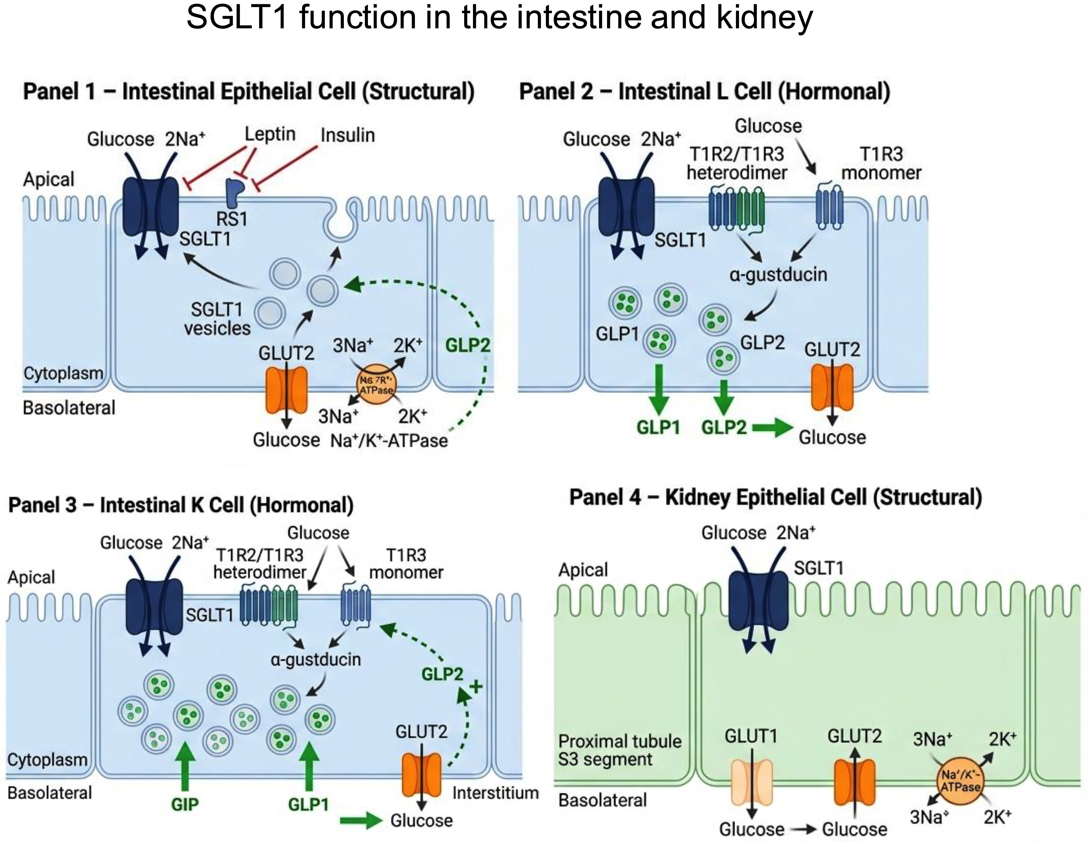
Figure 1. SGLT1 function in the intestine and kidney. Four cell types are depicted, separated into structural (glucose transport) and hormonal (incretin secretion) roles. Panel 1 - Intestinal epithelial cell (structural): SGLT1 mediates apical co-transport of glucose with 2Na+ from the intestinal lumen. RS1 protein inhibits SGLT1 activity at the apical membrane. Leptin and insulin exert inhibitory regulation of SGLT1 expression and trafficking. Intracellular SGLT1-containing vesicles are recruited to the apical membrane in response to luminal glucose, a process stimulated by GLP2 via a neuro-paracrine feedback loop (dashed arrow). GLUT2 on the basolateral membrane exports glucose to the interstitium. The Na+/K+-ATPase maintains the electrochemical sodium gradient driving apical SGLT1 activity. Panel 2 - Intestinal L cell (hormonal): SGLT1 on the apical membrane senses and transports luminal glucose. The T1R2/T1R3 heterodimer and T1R3 monomer sweet taste receptors activate α-gustducin, stimulating intracellular synthesis and basolateral secretion of GLP1 and GLP2. GLUT2 mediates basolateral glucose export. Panel 3 - Intestinal K cell (hormonal): Identical apical glucose-sensing machinery (SGLT1, T1R2/T1R3, T1R3, α-gustducin) drives secretion of GIP and GLP1 from the basolateral membrane. GLP2 arriving from L cells provides a positive feedback signal (+) stimulating SGLT1 activity. GLUT2 exports glucose basolaterally. Panel 4 - Kidney epithelial cell (structural): SGLT1 in the proximal tubule S3 segment recovers residual filtered glucose from the tubular lumen with 2Na+. The Na+/K+-ATPase (3Na+/2K+) maintains the driving gradient. GLUT1 and GLUT2 on the basolateral membrane return glucose to the interstitium. Figure created with FigureLabs. SGLT: sodium-glucose cotransporter; GLUT: facilitative glucose transporter; GIP: gastric inhibitory peptide; GLP: glucagon-like peptide.
Regulation of intestinal SGLT1: Dietary and hormonal factors modulate intestinal SGLT1 expression. Luminal nutrients prove essential for maintaining expression levels[136], with changes occurring independently of how sugars are metabolized[137]. Chronic high-carbohydrate diets increase intestinal glucose transport rates in parallel with Sglt1 expression[56]. Short-term glucose exposure enhances protein levels at the BBM, while sustained exposure drives SLC5A1 gene translation. Post-transcriptionally, SGLT1 accumulates in subapical vesicles[35], with glucose accelerating RS1 protein-mediated exocytosis[138]. The sweet taste receptor T1R2+T1R3 complex senses luminal sugar concentrations and activates G protein alpha-gustducin[139], upregulating SLC5A1 mRNA expression. This sweet receptor also triggers GLP2 expression in L-cells[140], which then regulates SGLT1 activity through neuro-paracrine pathways[140,141]. Deletion of alpha-gustducin, T1R3, or GLP2 receptors prevents dietary monosaccharide-induced Sglt1 upregulation[141,142]. Long-term regulation occurs at the crypt-villus junction, where SLC5A1 mRNA levels peak[143]. In mice, this heterodimer also appears in endocrine L and K cells. Sglt1-/- mice show reduced glucose-triggered GIP and GLP1 secretion[144], though in humans, SGLT1 represents only one contributor to GLP1 and GIP release[145].
Sodium and leptin provide additional regulatory input. In chickens, low-salt diets decrease glucose transport Vmax by reducing brush-border transporter density, whereas high-salt diets do not affect glucose transport rates or SGLT1 protein levels[146]. Leptin exerts hormonal control over SGLT1[147-149], producing significant downregulation following leptin administration in rats and hyperleptinemic mice, but not in leptin-deficient mice or those with intestine-specific leptin receptor deletion[148,150]. Portal vein insulin can also downregulate Sglt1 via its basolateral membrane receptor[151].
Pathological alterations: Several conditions correlate with altered intestinal SGLT1 expression. Overexpression is linked to severe obesity in both murine models and patients with morbid obesity[152]. Diabetes elevates intestinal SGLT1 expression, likely reflecting increased dietary sugar intake, with elevated mRNA detected in STZ-induced diabetic mice and rats, Otsuka Long-Evans Tokushima Fatty rats (a T2D model), and patients with non-insulin-dependent diabetes[82,153-155]. This upregulation probably accelerates postprandial blood glucose spikes by enhancing glucose absorption in proximal intestine and amplifying incretin-glucagon responses, thereby promoting hyperinsulinemia and hyperglycemia.
Studies in subjects with obesity [body mass index (BMI) > 35 kg/m2] reveal intestinal SGLT1 alterations following Roux-en-Y gastric bypass (RYGB), which typically produces approximately 25% weight loss[156,157]. Bariatric surgery profoundly affects T2D, with roughly 60% of patients achieving remission two years post-surgery[158,159]. Glucose control improves dramatically within days, suggesting that rapid diabetic symptom reversal stems primarily from digestive system reorganization rather than weight loss alone[160]. Potential mechanisms include decreased intestinal Sglt1 expression[161,162].
Loss-of-function mutations in SLC5A1 produce intestinal Glucose Galactose Malabsorption (GGM, OMIM 182380), a rare autosomal recessive disorder characterized by severe neonatal diarrhea and water loss[163-165]. These mutations impair SGLT1 trafficking from the endoplasmic reticulum to the BBM[35]. Dietary exclusion of D-glucose and D-galactose enables normal life for affected individuals[166]. Sglt1-null mice absorb approximately 95% less glucose, developing severe diarrhea and dying within two weeks on standard diet[144].
Renal, pancreatic, and central nervous system expression: In the kidney, SGLT1 recovers residual glucose (~ 3% of filtered load) from the lumen in the proximal tubule segment 3 (S3) BBM cells[167]. Murine Sglt1 protein expression predominates in S2 segments versus S3 segments[128] [Figure 1]. This compensatory capacity activates when SGLT2 is absent (through pharmacological blockade or genetic deletion), increasing the glucose load delivered to downstream SGLT1[168-170]. Diabetic rodent models show variable renal Sglt1 protein level, with some studies detecting no change or reductions[171]. SGLT1 deletion ameliorates glomerular hyperfiltration in T2D mice, lowering kidney weight, glomerular size, and albuminuria[172], implicating Sglt1 in renal integrity beyond simple glucose reabsorption.
Bankir and Yang proposed SGLT1 involvement in tubular urea secretion[173]. High-protein feeding in Sglt1 KO mice increased hepatic urea formation and elevated plasma urea levels, likely reflecting enhanced demand for renal excretion[174]. Work in mice and human HK-2 cells indicates that Sglt1-mediated glucose at the apical membrane inhibits gluconeogenic gene expression (Pepck and G6pase), mimicking insulin’s effect. This mechanism may protect proximal tubular cells against glucose overload[175].
Pancreatic expression: SGLT1 is expressed in the luminal membrane of pancreatic intralobular and interlobular ducts[176]. Elevated expression associates with pancreatic ductal adenocarcinoma and may indicate favorable prognosis[177]. SGLT1 is also expressed in pancreatic alpha cells[178], where it potentially influences glucagon secretion[179], though findings diverge. Kuhre et al. detected Slc5a1 mRNA expression in murine beta and delta cells, but not in alpha cells[180]. In rodent models, Sglt1 upregulation was observed in islets from insulin-resistant prediabetic ob/ob mice before declining once diabetic phenotype emerges[181]. Mühlemann and colleagues showed Sglt1 maintains murine islet integrity: its absence produced enlarged islets with reduced proliferation and apoptosis rates alongside impaired insulin and glucagon secretion[182]. Human SGLT1 expression shows substantial inter-individual variation[183] yet remains elevated during progression from insulin resistance to diabetes[178].
Central nervous system expression: Slc5a1 (Sglt1) mRNA localizes to diverse neuronal and glial cell types, including cortical neurons, hippocampal pyramidal cells, cerebellar Purkinje cells, and VMH neurons in rats[184]. Glial expression appears likely, evidenced by mRNA detection in primary rat astrocyte cultures and immunoreactivity in VMH glial cells[185]. SGLT1 at the BBB likely helps control glucose concentration in brain interstitium[186]. SGLT1 is expressed widely throughout the brain, with particular high levels in regions governing learning, feeding behavior, energy expenditure, and glucose homeostasis[35].
SGLT2: the renal glucose reabsorber
SGLT2 (SLC5A2) exhibits low-affinity (Km ~ 5 mM), high-capacity transport characteristics with 1:1 sodium-to-glucose coupling stoichiometry. This transporter serves as the primary glucose reabsorber in the BBM of proximal renal tubule S1/S2 segments, recovering approximately 90% of filtered glucose alongside GLUT2[187]. Slc5a2 deletion preserves roughly 40%-50% of glucose reabsorption, largely through SGLT1 compensation[188]. Patients carrying SLC5A2 mutations develop Familial Renal Glucosuria (FRG), characterized by urinary glucose excretion ranging from 1 to over 100 g daily without intestinal absorption defects[189]. Affected individuals typically avoid other complications such as kidney dysfunction or urinary tract infections[163,190]. Although rare, these mutations inspired development SGLT2 inhibitors as a potentially safe therapeutic strategy to lower blood glucose.
Expression levels govern SGLT2 glucose transport capacity, responding to glucose and insulin rather than sodium, particularly in diabetes[191]. Primary cultures of human exfoliated proximal tubular epithelial cells from T2D patients showed elevated SGLT2 expression and increased glucose uptake[52], with comparable findings in db/db mice and Akita rats[164,192]. Angiotensin II AT1 receptors and HNF1A contribute to SGLT2 upregulation[193]. Mice lacking Hnf1a display impaired renal glucose reabsorption from reduced Sglt2 gene expression[194]. Norton et al. detected lower SGLT2 mRNA in kidneys from T2D patients compared with non-diabetic controls[195].
Insulin acts as an agonist for SGLT2 activity in vivo[196], presumably maintaining elevated filtered glucose reabsorption during the postprandial phase. Hyperinsulinemia associated with insulin resistance in obesity and T2D might enhance renal SGLT2 activity. This enhancement likely stems from general proximal tubule growth and hypertrophy in diabetes, which expands the overall glucose transport capacity[197].
Pancreatic and central nervous system expression: Despite initial assumptions, SGLT2 shows low-level expression in brain, liver, pancreas, and other tissues[127,178,183,198,199]. Intestinal SGLT2 remains controversial, with immunofluorescence detecting limited enterocyte expression in duodenal mucosa across species[198,200-203], though, some studies report no expression in human small intestine[198].
Pancreatic SGLT2 expression generates ongoing debate. While several studies detect no SGLT2 in murine or human islets[179-181,198,204], Vergari et al. identified SGLT2 in approximately 40% of delta cells[205], whereas Bonner et al. localized SLC5A2 mRNA and protein specifically to human alpha cells, under HNF4A transcriptional control[178]. SGLT2 inhibition through dapagliflozin or siRNA stimulates glucagon secretion both in vivo and in vitro[178,183], aligning with elevated plasma glucagon observed in diabetic patients receiving SGLT2 inhibitors[206,207]. Saponaro et al. revealed substantial inter-individual variability in pancreatic islet SGLT2 expression[183], potentially explaining conflicting study findings. Like SGLT1, SGLT2 appears functionally active in pancreatic adenocarcinomas, where SGLT2 inhibitors block glucose uptake and reduce tumor growth[208,209].
Brain SGLT2 expression remains substantially lower than SGLT1[198,199,203] and localizes predominantly to the cerebellum[210]. Murine studies detect SGLT2 in cerebellum, amygdala, hypothalamus, periaqueductal gray (PAG), nucleus of the solitary tract, and microvessels comprising the BBB[211]. Like SGLT1, SGLT2-expressing brain regions associate with learning, food intake, energy and glucose homeostasis, and central cardiovascular and autonomic regulation[212]. However, low expression levels and limited immunofluorescence or in situ hybridization confirmation fuel ongoing debate about its functional significance in central glucose sensing and energy regulation.
Additional SGLT family members
SGLT3 (SLC5A4), first characterized as a renal amino acid co-transporter (SAAT1) in pigs, operates as a glucose-sensing ion channel rather than a glucose transporter in humans[213]. Mice and rats express two, whereas humans express only one[214,215]. Human SGLT3 and mSglt3a share 79%-80% amino acid sequence identity and conserved ligand-binding sites[216,217], implying distinct functional roles for the rodent isoforms. In fact, mSGLT3b transports glucose similarly to SGLT1, while mSglt3a does not. Both hSGLT3 and mSglt3a produce membrane currents at pH 7.4 when exposed to glucose and either Na+ or H+, supporting the view that SGLT3 operates as a glucose-sensitive ion channel rather than a transporter[218].
Humans mRNA profiling reveals SLC5A4 expression predominates in small intestine, with markedly lower expression in skeletal muscle and testis, then spleen, liver, and kidney[198]. Immunofluorescence studies localized hSGLT3 to the small intestine submucosa, where it co-localizes with nicotinic acetylcholine receptors in cholinergic neurons[219]. Obesity reduces hSGLT3 protein in jejunal region[220]. Mouse and rat intestine and kidney display similar expression patterns[214,218,221,222]. Both isoforms appear in rat duodenum, with rSglt3b also detected in jejunum and proximal ileum, where it may contribute to GLP1 secretion[185,223,224]. Specifically, mSglt3a/3b localizes to small intestinal epithelial cells, though ob/ob mice show markedly lower duodenal and jejunal expression than controls[220]. rSglt3 also localizes to the hepatic portal area and contributes to reducing food intake[222,225].
Sglt3 additionally serves glucose-sensing functions in the brain. Most glucose-excitable cultured hypothalamic neurons increase cytosolic Ca2+ levels following alpha-MDG exposure[185]. Sglt3 expression across diverse brain structures supports this observation, including cortex, hypothalamus, hippocampus, midbrain, striatum, medulla, olfactory bulb, and cerebellum. Yamazaki et al. identified a neuroprotective role for Sglt3 in cholinergic neurons[226].
SGLT4 (SLC5A9) and SGLT5 (SLC5A10) represent poorly understood members of the SGLT family. COS-7 cell studies show SGLT4 transports mannose, glucose, fructose, 1,5-anhydroglucitol (1.5-AG), and galactose with varying affinities[227]. Moreover, recent work has excluded 1.5-AG as a SGLT4 substrate[190]. 1.5-AG acts as a biomarker for hyperglycemic excursions[228-231], since high concentrations of glucose, fructose, and mannose compete with 1,5-AG for renal absorption. Elevated blood 1,5-AG drives neutropenia in patients with G6PPT/SLC37A4 deficiency and glycogen storage disease type 1b[232,233], underscoring the clinical relevance of this metabolite.
Chen et al.[198] and Tazawa et al.[227] revealed that SGLT4 shows minimal expression in the kidney, but substantial intestinal expression followed by the pancreas, indicating its physiological role likely diverges from the renal-centric characteristics of other SGLT family members[198,227]. Work by Forester et al. examining SGLT-mediated proximal tubule fructose reabsorption in hypertension supports this non-renal role. Comparing SGLT4 and SGLT5 knockout models, they found SGLT5 deletion prevented hypertension during short-term (7.5-day) fructose high-salt feeding, whereas SGLT4 knockout rats still developed hypertension under identical conditions[234].
SGLT5 is predominantly expressed in the renal cortex, similarly to SGLT2[190], and operates with a 1:2 sodium-to-substrate transport ratio. This transporter exhibits high-capacity transport for mannose and fructose, with reduced efficiency for glucose and galactose[235]. Fructose regulates SGLT5 expression, as Sglt5-deficient mice lose all sodium-dependent fructose uptake in the renal brush border membrane[236]. These findings establish SGLT5 as the principal transporter for renal fructose reabsorption. High-fructose feeding in Sglt5-null mice reduces plasma triglycerides and epididymal fat mass. Paradoxically, these mice develop elevated fasting insulin, glucose intolerance, and worsened hepatic steatosis alongside increased inflammation and tissue damage markers. These metabolic consequences arise not from excessive hepatic fructose uptake but likely from enhanced hepatic de novo lipogenesis and defective mannose-dependent glycosylation[236].
SGLT5 displays 3-4-fold higher transport affinity for 1,5-AG than mannose. Genetic mutations or single-nucleotide polymorphisms (SNPs) in SLC5A10 associate with altered plasma 1,5-AG concentrations[237-239]. Heterozygous SGLT5 mutations produce approximately 50% reduction in circulating 1.5-AG, indicating no alternative transporter compensates for its renal absorption[190]. Overall, SGLT4 and SGLT5 jointly regulate plasma fructose and mannose through intestinal absorption and renal reabsorption, though their complete regulatory mechanisms and physiological significance require further investigation.
CURRENT SGLT INHIBITORS AND THERAPEUTIC APPLICATIONS
SGLT1 inhibitors
SGLT inhibitors delay dietary glucose absorption in the small intestine, lowering postprandial glucose levels. These agents elevate plasma GLP1 and potentially GIP levels[188]. SGLT1 inhibitors (SGLT1i) enhance glucose delivery to the distal small intestine and colon, where the microbiome ferments glucose into short-chain fatty acids (SCFAs). SCFAs then stimulate GLP1 release through binding to free fatty acid receptors on L cells[240]. Increased intestinal glucose exposure also enhances microbial propionate production, yielding beneficial metabolic effects in the intestine[241,242].
T2D and diabetic cardiomyopathy upregulate SGLT1 expression in cardiomyocyte membranes, where it serves a critical role in energy production during hypoglycemia or ischemia[243]. SGLT1i may therefore protect cardiac tissue by reducing glycogen accumulation and lowering reactive oxygen species production[244]. Sodium influx through SGLT1 appears to worsen ischemic damage in cerebral neurons and myocardial cells, potentially promoting toxic calcium accumulation and impairing mitochondrial function[245]. This therapeutic approach carries potential side effects, including diarrhea, dehydration, metabolic acidosis, and possibly elevated euglycemic diabetic ketoacidosis risk[246]. Nevertheless, nonclinical studies (KGA-2727) and clinical trials (GSK-1614235) of highly selective SGLT1i detected no serious gastrointestinal adverse events[245,247]. While these drugs have not yet reached market, several compounds continue advancing through development for diabetes and cardiovascular disease treatment.
SGLT2 inhibitors
SGLT2 inhibitors (SGLT2i), including dapagliflozin, canagliflozin, empagliflozin, ertugliflozin, and luseogliflozin, are approved to treat T2D[248]. These agents inhibit renal glucose reabsorption, producing insulin-independent glycosuria and lowering glycemic HbA1c by approximately 0.7%-0.8%[249,250]. This mechanism differs from other antidiabetic therapies that target hepatic glucose production (metformin), stimulate insulin secretion (sulfonylureas, GLP1 receptor agonists, DPP4 inhibitors), enhance insulin sensitivity (thiazolidinediones), or provide exogenous insulin[251].
SGLT2i slow T2D progression by reducing glucotoxicity-induced beta-cell dysfunction and improving insulin sensitivity[252]. However, these agents associate with modest increases in LDL and HDL cholesterol[253]. SGLT2i also induce glucagon secretion and endogenous glucose production[206,207,254,255], unlike other glucose-lowering agents such as metformin or GLP1 receptor agonists[256]. This phenomenon may stem partly from SGLT2 expression in human pancreatic alpha cells[178,183].
SGLT2i-induced glycosuria reduces body weight by 1-3 kg, predominantly affecting visceral adiposity[257-259]. Enhanced urinary glucose and sodium excretion promote mild osmotic diuresis and sodium loss, lowering extracellular fluid volume. These hemodynamic changes likely contribute to the cardiovascular and renal benefits observed with SGLT2i therapy. SGLT2i lower glomerular hyperfiltration, glomerular hypertrophy, inflammation markers, and albuminuria[260-262], and decrease systolic blood pressure by 2.6-6 mm Hg[263,264].
Cardiovascular and renal benefits: Heart failure coexists with diabetes in approximately 22% of patients over age 65, with diabetic individuals facing more than double the heart failure risk of non-diabetic counterparts. This translates to higher hospitalization rates and worse cardiovascular outcomes[265]. Most traditional antidiabetic therapies show cardiovascular neutrality or may worsen cardiovascular risk factors[266]. In contrast, SGLT2i, particularly empagliflozin, canagliflozin, and dapagliflozin, have shown robust cardiovascular and renal benefits in large-scale clinical trials[267], and are now approved to treat heart failure and chronic kidney disease regardless of baseline T2D status[268].
Empagliflozin benefits patients with existing cardiovascular disease by reducing myocardium interstitial fibrosis, improving aortic stiffness, delivering anti-inflammatory effects, and decreasing pericardial fat accumulation[12,269]. Dapagliflozin protects the heart through vasodilatory effects on thoracic aorta, while canagliflozin reduces myocardial infarct size and protects against cardiac arrest in experimental models[261,262,268,270]. Compensatory upregulation of renal SGLT1 during SGLT2 inhibitor therapy caps glucose excretion to 30%-50% of the filtered load, partially mitigating hypoglycemia risk[271-273].
Beyond cardiovascular and renal outcomes, SGLT2 inhibitors offer additional metabolic benefits, including body weight reduction, improved insulin sensitivity, and potential efficacy in metabolic dysfunction-associated steatotic liver disease (MASLD)[274] and obstructive sleep apnea[275], though further research is needed. Type 1 diabetes represents another investigational application, where sotagliflozin received EMA approval (subsequently withdrawn) for adults with T1DM and BMI ≥ 27 kg/m2, and dapagliflozin has been approved as an adjunct to insulin in some jurisdictions, with demonstrated HbA1c reduction and body weight loss. However, substantially elevated DKA risk constrains their use and demands careful patient selection and monitoring[276,277].
SGLT2 inhibitor therapy increases the risk of genital mycotic infections (GMIs) and bacterial urinary tract infections (UTIs), with higher incidence in women than men[262,278]. Diabetic ketoacidosis (DKA) represents another recognized adverse event[279280]. Reduced carbohydrate intake during gastroenteritis, intense exercise, and postoperative stress can trigger DKA, likely through increased glucagon secretion that stimulates lipolysis and ketogenesis[281]. The incidence of DKA has been reported at 55.5 per 1000 person-years in US commercially insured patients with T1D, with substantially lower rates in T2D[282]. The long-term efficacy and safety profile of these interventions requires further definition[283,284].
Dual SGLT1/SGLT2 inhibitors: Dual SGLT1/SGLT2 inhibitors address compensatory SGLT1 upregulation. Sotagliflozin, an orally administered dual inhibitor, has undergone extensive phase 3 clinical trial evaluation for T2D and chronic kidney disease treatment. Sotagliflozin demonstrates substantially greater SGLT1 potency than selective SGLT1 inhibitors while maintaining SGLT2 inhibition comparable to dapagliflozin [Table 3][285,286]. Partial SGLT1 inhibition increases urinary glucose excretion through renal mechanisms, delays intestinal glucose reabsorption, and enhances postprandial GLP1 and PYY secretion[241,287].
Indications and Food & Drug Administration (FDA) and European Medicines Agency (EMA) approval dates for SGLT2 inhibitor treatment
| Compound | Indications | Date FDA/EMA approval |
| Canagliflozin | 1. As an adjunct to diet and exercise to improve glycemic control in adults with T2D 2. To reduce the risk of major adverse cardiovascular events in adults with T2D and established cardiovascular disease 3. To reduce the risk of end-stage kidney disease, doubling of serum creatinine, cardiovascular death, and hospitalization for heart failure in adults with T2D and diabetic nephropathy with albuminuria[308,309] | FDA: Mar 29, 2013; EMA: Nov 15, 2013 |
| Dapagliflozin | 1. It is indicated in adults for the treatment of insufficiently controlled T2D as an adjunct to diet and exercise as monotherapy when metformin is considered inappropriate or in addition to other medicinal products for the treatment of T2D 2. It is indicated in adults for the treatment of symptomatic chronic heart failure, regardless of ejection fraction 3. It is indicated in adults for the treatment of chronic kidney disease[310,311] | FDA: Jan 8, 2014; EMA: Nov 12, 2012 |
| Empagliflozin | 1. It is indicated to reduce the risk of cardiovascular death and hospitalization for heart failure in adults with heart failure, regardless of ejection fraction 2. To reduce the risk of cardiovascular death in adults with T2D and established cardiovascular disease 3. As an adjunct to diet and exercise to improve glycemic control in adults with T2D 4. To reduce the risk of kidney disease progression and cardiovascular death in adults with chronic kidney disease at risk of progression[312,313] | FDA: Aug 1, 2014; EMA: May 22, 2014 |
| Ertugliflozin | It is indicated as an adjunct to diet and exercise to improve glycemic control in adults with T2D[314,315] | FDA: Dec 19, 2017; EMA: Mar 21, 2018 |
| Sotagliflozin | FDA: To reduce the risk of cardiovascular death, hospitalization for heart failure, and urgent heart failure visit in adults with heart failure or T2D, chronic kidney disease, and other cardiovascular risk factors EMA: As an adjunct to insulin to improve glycemic control in adults with type 1 diabetes and BMI ≥ 27 kg/m2[277,316,317] | FDA: May 26, 2023; EMA: Apr 26, 2019 (withdrawn Mar 22, 2022) |
OTHER GLUCOSE TRANSPORTERS
Humans express a single SWEET transporter, in contrast to plants which possess numerous SWEET isoforms. Human SWEET1 (SLC50A1, RAG1AP1) functions as a glucose uniporter with weak efflux activity and localizes primarily to the Golgi complex[288,289]. SWEET proteins display a distinctive 7-transmembrane domain architecture comprising two triple-helix bundles separated by an inversion linker, generating a 3 + 1 + 3 configuration[5,290]. SWEET1 predominates in oviduct, epididymis, and intestine, with lactation inducing expression in the mouse mammary gland. This transporter may facilitate basolateral glucose exit from enterocytes, potentially contributing to Fanconi-Bickel syndrome pathogenesis[291].
CONCLUSION
Integration of current knowledge
Glucose transporters occupy a central position in energy homeostasis, with implications extending across metabolic, cardiovascular, renal, and oncological diseases. This review has synthesized current understanding of molecular mechanisms, tissue-specific regulation, disease pathogenesis, and therapeutic applications spanning the GLUT and SGLT families.
The GLUT and SGLT families employ distinct transport mechanisms that work in concert to maintain glucose homeostasis. Their tissue-specific expression patterns reflect specialized metabolic demands, governed by intricate regulatory networks involving transcriptional control, post-translational modifications, membrane trafficking dynamics, and hormonal signaling. Beyond their established roles in diabetes, these transporters contribute to diverse pathologies. Cancer cells exploit altered transporter expression to fuel metabolic reprogramming, while dysfunction in renal and intestinal glucose handling drives insulin resistance and metabolic complications. Genetic defects in specific transporters produce rare but devastating inherited disorders, underscoring their fundamental physiological importance.
A paradigm shift in therapeutic strategy
The clinical success of SGLT2 inhibitors exemplifies how deep understanding of transporter biology can revolutionize treatment approaches. Originally designed to improve glycemic control in T2D by promoting renal glucose excretion, these agents unexpectedly improved cardiovascular and renal outcomes in landmark trials[9,10,268]. Empagliflozin, canagliflozin, and dapagliflozin reduce heart failure hospitalizations, cardiovascular mortality, and chronic kidney disease progression through mechanisms that likely involve hemodynamic improvements from natriuresis and osmotic diuresis, metabolic substrate shifts favoring ketone body utilization, reduced inflammation and oxidative stress, and potential direct organ protective effects.
Dual SGLT1/SGLT2 inhibitors such as sotagliflozin advance this therapeutic evolution by combining renal and intestinal glucose transport inhibition, delaying intestinal glucose absorption while enhancing incretin secretion[257,258]. This multi-level intervention addresses different aspects of disease pathophysiology simultaneously, demonstrating how transporter biology informs increasingly sophisticated treatment strategies.
Persistent questions and critical knowledge gaps
Despite significant progress, several fundamental questions demand resolution. The precise mechanisms underlying SGLT2 inhibitor cardiovascular and renal benefits remain incompletely understood. Clarifying these pathways could reveal additional therapeutic targets and refine treatment strategies. While we understand some regulatory mechanisms, comprehensive knowledge of tissue-specific control remains elusive. Single-cell transcriptomics has revealed unexpected heterogeneity in transporter expression within organs[81], suggesting regulatory complexity beyond current models.
The expanding role of glucose transporters in non-diabetic diseases warrants deeper investigation. Their contribution to cancer metabolism, neurodegenerative disorders, and inflammatory conditions deserves systematic exploration[24,292-294]. Substantial interindividual variability in transporter expression and drug response stems from genetic and environmental factors, raising questions about personalization. Can pharmacogenomics identify which patients will derive maximum benefit from SGLT2 inhibitors or other transporter-targeted therapies?
Newer family members, particularly SGLT4, SGLT5, and SWEET proteins[5], remain poorly characterized in humans[192,198,227,295]. Their physiological roles under normal and pathological conditions need definition, as do their potential therapeutic applications. Although SGLT2 inhibitors demonstrate favorable benefit-risk profiles in trials extending to five years, longer-term safety data beyond ten years remain limited[296]. Rare adverse events including euglycemic ketoacidosis, genital infections, and potential skeletal effects require continued surveillance[297-299].
Emerging opportunities and future trajectories
Several promising research avenues are now emerging. Advanced structural biology techniques, particularly Cryo-EM and molecular dynamics simulations, continue revealing transporter structure-function relationships, conformational changes during transport cycles, and inhibitor binding mechanisms. High-resolution structures of all major GLUT and SGLT isoforms will enable structure-based drug design for highly selective inhibitors. Artificial intelligence and machine learning can accelerate this discovery process substantially[300,301].
Single-cell RNA sequencing and spatial transcriptomics offer unprecedented resolution of cell-type-specific expression and regulation within heterogeneous tissues. These approaches may illuminate why certain cell populations prove particularly vulnerable to transporter dysfunction and identify cell-specific therapeutic opportunities. Conditional knockout and overexpression models will clarify physiological roles and disease contributions in specific tissues. CRISPR-based genome editing, inducible and cell-type-specific gene modulation, and single-cell multi-omics now permit investigation of previously inaccessible questions regarding transporter function[302,303]. By integrating CRISPR/Cas systems with single-cell transcriptomic readouts, researchers can precisely perturb and comprehensively characterize gene function within defined cell populations. High-content CRISPR screens at single-cell resolution facilitate identification of tissue-specific regulatory networks, providing critical insights into glucose transporter biology.
Therapeutic innovation may take multiple forms. Tissue-specific inhibitors could minimize off-target effects. Allosteric modulators might provide finer control than orthosteric inhibitors. Novel combination therapies could address multiple pathways simultaneously. GLUT-targeted therapies, particularly for cancer where metabolic reprogramming drives proliferation, merit continued development despite selectivity challenges[304]. Emerging evidence linking gut microbiota alterations to changes in intestinal glucose transport suggests that microbial modulation might affect SGLT1 expression and functional outcomes[305].
Pharmacogenomic studies could enable treatment selection based on individual transporter expression profiles, genetic variants affecting drug response, and disease-specific factors. Biomarkers predicting response to SGLT inhibitors or identifying patients at risk for adverse effects would optimize clinical decisions. SGLT2 inhibitors show promise beyond current indications, with ongoing trials evaluating efficacy in heart failure with preserved ejection fraction, acute kidney injury prevention, and polycystic kidney disease. Understanding mechanisms of benefit will identify additional patient populations likely to respond.
Limitations of the present review and of current research
As a narrative review, this work is subject to inherent limitations. The breadth of the topic necessitated selective coverage of the literature, and the absence of a formal systematic search strategy introduces the possibility of selection bias. At the level of the underlying science, mechanistic insights into the lesser-characterized family members, including Class II and III GLUTs, SGLT3-6, and the SWEET transporter, derive largely from in vitro systems or rodent models, with limited human validation. Tissue-specific regulation remains incompletely defined, and single-cell transcriptomic data suggest that existing models will require revision. These gaps collectively define the priorities for the next phase of glucose transporter research.
Closing perspective
Glucose transporter research provides a compelling model for translational science. The integration of basic discoveries with clinical application demonstrates that even extensively studied biological systems can yield surprising therapeutic opportunities when examined from fresh perspectives. The journey from membrane transport characterization to blockbuster cardiovascular drugs illustrates how fundamental research ultimately serves patients. Continued investigation of glucose transporter biology promises novel insights and therapies that will improve outcomes across a spectrum of human diseases. As technologies advance and our understanding deepens, these essential proteins will undoubtedly reveal additional secrets, opening new therapeutic frontiers we cannot yet imagine. The field stands poised for its next transformative discoveries.
DECLARATIONS
Acknowledgment
Graphical abstract was generated by Image provided by Servier Medical Art (https://smart.servier.com/), licensed under CC BY 4.0 (https://creativecommons.org/licenses/by/4.0/).
Authors’ contributions
Conceived the idea: Bonner C, Moreno-Lopez M, Saponaro C, Louvet I, Jones JG
Wrote the original draft: Moreno-Lopez M, Bonner C
Generated figures: Moreno-Lopez M, Louvet I
Reviewed and edited the manuscript: Bonner C, Jones JG, Saponaro C
All authors reviewed and edited the manuscript.
Availability of data and materials
Not applicable.
AI and AI-assisted tools statement
During the preparation of this manuscript, the AI tool FigureLabs (https://www.figurelabs.ai/zh-CN) was used solely to modify and improve the graphic presentation of Figure 1. The tool did not influence the study design, data collection, analysis, interpretation, or the scientific content of the work. All authors take full responsibility for the accuracy, integrity, and final content of the manuscript.
Financial support and sponsorship
This work was supported by grants to Bonner C from the European Foundation for the Study of Diabetes/Lilly (2016), L'Agence Nationale de la Recherche (ANR-25-CE14-1626-01), and SFD-2025-Abbott, as well as institutional support from the University of Lille. Moreno-Lopez M was supported by a PhD stipend from I-SITE ULNE/PréciDIAB/EGID (2021-2024).
Conflicts of interest
All authors declare that there are no conflicts of interest.
Ethical approval and consent to participate
Not applicable.
Consent for publication
Not applicable.
Copyright
© The Author(s) 2026.
REFERENCES
1. Lefevre PG. Evidence of active transfer of certain non-electrolytes across the human red cell membrane. J Gen Physiol. 1948;31:505-27.
2. Baldwin JM, Gorga JC, Lienhard GE. The monosaccharide transporter of the human erythrocyte. Transport activity upon reconstitution. J Biol Chem. 1981;256:3685-9.
3. Carbó R, Rodríguez E. Relevance of sugar transport across the cell membrane. Int J Mol Sci. 2023;24:6085.
4. Chadt A, Al-Hasani H. Glucose transporters in adipose tissue, liver, and skeletal muscle in metabolic health and disease. Pflugers Arch - Eur J Physiol. 2020;472:1273-98.
6. Lizák B, Szarka A, Kim Y, et al. Glucose transport and transporters in the endomembranes. Int J Mol Sci. 2019;20:5898.
7. Ji J, Yang L, Fang Z, et al. Plant SWEET family of sugar transporters: structure, evolution and biological functions. Biomolecules. 2022;12:205.
8. Thomas MC, Neuen BL, Twigg SM, Cooper ME, Badve SV. SGLT2 inhibitors for patients with type 2 diabetes and CKD: a narrative review. Endocr Connect. 2023;12:e230005.
9. Mavrakanas TA, Tsoukas MA, Brophy JM, Sharma A, Gariani K. SGLT-2 inhibitors improve cardiovascular and renal outcomes in patients with CKD: a systematic review and meta-analysis. Sci Rep. 2023;13:15922.
10. Theodorakopoulou MP, Alexandrou M, Tsitouridis A, et al. Effects of sodium-glucose co-transporter 2 inhibitors on heart failure events in chronic kidney disease: a systematic review and meta-analysis. Eur Heart J-Card Pha. 2024;10:329-41.
11. Santos GL, Dos Santos CF, Rocha GR, et al. Beyond glycemic control: roles for sodium-glucose cotransporter 2 inhibitors and glucagon-like peptide-1 receptor agonists in diabetic kidney disease. World J Diabetes. 2025;16:104706.
12. Zinman B, Wanner C, Lachin JM, et al. Empagliflozin, cardiovascular outcomes, and mortality in type 2 diabetes. N Engl J Med. 2015;373:2117-28.
13. Mueckler M, Thorens B. The SLC2 (GLUT) family of membrane transporters. Mol Aspects Med. 2013;34:121-38.
14. Nevola R, Villani A, Imbriani S, et al. Sodium-glucose co-transporters family: current evidence, clinical applications and perspectives. Front Biosci (Landmark Ed). 2023;28:103.
15. Joost H, Bell GI, Best JD, et al. Nomenclature of the GLUT/SLC2A family of sugar/polyol transport facilitators. Am J Physiol-Endoc M. 2002;282:E974-6.
16. Scheepers A, Joost H, Schurmann A. The glucose transporter families SGLT and GLUT: molecular basis of normal and aberrant function. J Parenter Enteral Nutr. 2004;28:364-71.
17. Thorens B, Mueckler M. Glucose transporters in the 21st Century. Am J Physiol-Endoc M. 2010;298:E141-5.
18. Uldry M, Ibberson M, Horisberger JD, Chatton JY, Riederer BM, Thorens B. Identification of a mammalian H+-myo-inositol symporter expressed predominantly in the brain. EMBO J. 2001;20:4467-77.
19. Du J, Shen M, Chen J, et al. The impact of solute carrier proteins on disrupting substance regulation in metabolic disorders: insights and clinical applications. Front Pharmacol. 2025;15:1510080.
20. Lefevre PG, Marshall JK. The atachment of phloretin and analogues to human erythrocytes in connection with inhibition of sugar transport. J Biol Chem. 1959;234:3022-6.
21. Estensen RD, Plagemann PGW. Cytochalasin B: Inhibition of glucose and glucosamine transport. Proc Natl Acad Sci USA. 1972;69:1430-4.
22. Wang N, Zhang S, Yuan Y, et al. Molecular basis for inhibiting human glucose transporters by exofacial inhibitors. Nat Commun. 2022;13:2632.
23. Thorens B, Sarkar HK, Kaback H, Lodish HF. Cloning and functional expression in bacteria of a novel glucose transporter present in liver, intestine, kidney, and β-pancreatic islet cells. Cell. 1988;55:281-90.
24. Wang J, Ye C, Chen C, et al. Glucose transporter GLUT1 expression and clinical outcome in solid tumors: a systematic review and meta-analysis. Oncotarget. 2017;8:16875-86.
25. Veys K, Fan Z, Ghobrial M, et al. Role of the GLUT1 Glucose transporter in postnatal CNS angiogenesis and blood-brain barrier integrity. Circ Res. 2020;127:466-82.
26. Matar R, Tang D, Mccall S, De Silva R. Glucose transporter type 1 deficiency syndrome and paroxysmal exercise-induced dyskinesia. Pract Neurol. 2025;25:75-7.
27. Li H, Guglielmetti C, Sei YJ, et al. Neurons require glucose uptake and glycolysis in vivo. Cell Reports. 2023;42:112335.
28. Szablewski L. Glucose transporters as markers of diagnosis and prognosis in cancer diseases. Oncol Rev. 2022;16.
29. Huang Y, Lin D, Taniguchi CM. Hypoxia inducible factor (HIF) in the tumor microenvironment: friend or foe? Sci. China Life Sci. 2017;60:1114-24.
30. Basheeruddin M, Qausain S. Hypoxia-inducible factor 1-alpha (HIF-1α) and cancer: mechanisms of tumor hypoxia and therapeutic targeting. Cureus. 2024;16:e70700.
31. Zha X, Hu Z, Ji S, et al. NFκB up-regulation of glucose transporter 3 is essential for hyperactive mammalian target of rapamycin-induced aerobic glycolysis and tumor growth. Cancer Lett. 2015;359:97-106.
32. Yu L, Chen X, Sun X, Wang L, Chen S. The glycolytic switch in tumors: how many players are involved? J. Cancer. 2017;8:3430-40.
33. Yu L, Wu M, Zhu G, Xu Y. Emerging roles of the tumor suppressor p53 in metabolism. Front Cell Dev Biol. 2022;9:762742.
34. Pliszka M, Szablewski L. Glucose transporters as a target for anticancer therapy. Cancers. 2021;13:4184.
35. Koepsell H. Glucose transporters in brain in health and disease. Pflugers Arch - Eur J Physiol. 2020;472:1299-343.
36. Fakorede S, Sodiq TA, Ajagbe AO, et al. Interplay of Glut1 alteration and Alzheimer’s disease: a narrative review on glucose transport and energy metabolism. J Alzheimers Dis. 2025;108:470-86.
37. Daida T, Shin B, Cepeda C, Devaskar SU. Neurodevelopment is dependent on maternal diet: placenta and brain glucose transporters GLUT1 and GLUT3. Nutrients. 2024;16:2363.
38. Toyoda Y, Shigesawa R, Merriman TR, Matsuo H, Takada T. GLUT2/SLC2A2 is a bi-directional urate transporter. J Biol Chem. 2025;301:108485.
39. Uldry M, Ibberson M, Hosokawa M, Thorens B. GLUT2 is a high affinity glucosamine transporter. FEBS Lett. 2002;524:199-203.
40. Sun B, Chen H, Xue J, Li P, Fu X. The role of GLUT2 in glucose metabolism in multiple organs and tissues. Mol Biol Rep. 2023;50:6963-74.
41. Guillam M, Burcelin R, Thorens B. Normal hepatic glucose production in the absence of GLUT2 reveals an alternative pathway for glucose release from hepatocytes. Proc Natl Acad Sci USA. 1998;95:12317-21.
42. Troncoso MF, Elola MT, Blidner AG, Sarrias L, Espelt MV, Rabinovich GA. The universe of galectin-binding partners and their functions in health and disease. J Biol Chem. 2023;299:105400.
43. Hall E, Jönsson J, Ofori JK, et al. Glucolipotoxicity alters insulin secretion via epigenetic changes in human islets. Diabetes. 2019;68:1965-74.
44. Ohtsubo K, Takamatsu S, Minowa MT, Yoshida A, Takeuchi M, Marth JD. Dietary and genetic control of glucose transporter 2 glycosylation promotes insulin secretion in suppressing diabetes. Cell. 2005;123:1307-21.
45. Ng NHJ, Ghosh S, Bok CM, et al. HNF4A and HNF1A exhibit tissue specific target gene regulation in pancreatic beta cells and hepatocytes. Nat Commun. 2024;15:4288.
46. Afshar N, Safaei S, Nickerson DP, Hunter PJ, Suresh V. Computational Modelling of glucose uptake by SGLT1 and apical GLUT2 in the enterocyte. Front Physiol. 2021;12:699152.
47. Vallon V, Nakagawa T. Renal tubular handling of glucose and fructose in health and disease. In: Prakash YS, Editors. Comprehensive Physiology. Wiley; 2021. pp. 2995-3044.
48. Morgan EL, Mace OJ, Affleck J, Kellett GL. Apical GLUT2 and Cav1.3: regulation of rat intestinal glucose and calcium absorption. J Physiol. 2007;580:593-604.
49. Morgan E, Mace O, Helliwell P, Affleck J, Kellett G. A role for Cav1.3 in rat intestinal calcium absorption. Biochem Biophys Res Commun. 2003;312:487-93.
50. Pfaff IL, Vallon V. Protein Kinase C Beta isoenzymes in diabetic kidneys and their relation to nephroprotective actions of the ace inhibitor lisinopril. Kidney Blood Press Res. 2002;25:329-40.
51. Goestemeyer AK, Marks J, Srai SK, Debnam ES, Unwin RJ. GLUT2 protein at the rat proximal tubule brush border membrane correlates with protein kinase C (PKC)-βl and plasma glucose concentration. Diabetologia. 2007;50:2209-17.
52. Gouyon F, Caillaud L, Carrière V, et al. Simple-sugar meals target GLUT2 at enterocyte apical membranes to improve sugar absorption: a study in GLUT2-null mice. J Physiol. 2003;552:823-32.
53. Tobin V, Le Gall M, Fioramonti X, et al. Insulin internalizes GLUT2 in the enterocytes of healthy but not insulin-resistant mice. Diabetes. 2008;57:555-62.
54. Ferraris RP. Dietary and developmental regulation of intestinal sugar transport. Biochem J. 2001;360:265.
55. Shen Z, Hou Y, Zhao G, et al. Physiological functions of glucose transporter-2: from cell physiology to links with diabetes mellitus. Heliyon. 2024;10:e25459.
56. Gromova LV, Fetissov SO, Gruzdkov AA. Mechanisms of glucose absorption in the small intestine in health and metabolic diseases and their role in appetite regulation. Nutrients. 2021;13:2474.
57. Fanconi G, Bickel H. Chronic aminoaciduria (amino acid diabetes or nephrotic-glucosuric dwarfism) in glycogen storage and cystine disease. Helv Paediatr Acta. 1949;4:359-96.
58. Santer R, Schneppenheim R, Dombrowski A, Götze H, Steinmann B, Schaub J. Mutations in GLUT2, the gene for the liver-type glucose transporter, in patients with Fanconi-Bickel syndrome. Nat Genet. 1997;17:324-6.
59. Sharari S, Abou-alloul M, Hussain K, Ahmad Khan F. Fanconi-Bickel syndrome: a review of the mechanisms that lead to dysglycaemia. Int J Mol Sci. 2020;21:6286.
60. Drobiova H, Alhamar G, Ahmad R, Al-Mulla F, Al Madhoun A. GLUT4 trafficking and storage vesicles: molecular architecture, regulatory networks, and their disruption in insulin resistance. Int J Mol Sci. 2025;26:7568.
61. Van Gerwen J, Shun-Shion AS, Fazakerley DJ. Insulin signalling and GLUT4 trafficking in insulin resistance. Biochem Soc Trans. 2023;51:1057-69.
64. Chang Y, Chan M, Yang Y, Li C, Hsiao M. Glucose transporter 4: Insulin response mastermind, glycolysis catalyst and treatment direction for cancer progression. Cancer Lett. 2023;563:216179.
65. Holman GD. Structure, function and regulation of mammalian glucose transporters of the SLC2 family. Pflugers Arch - Eur J Physiol. 2020;472:1155-75.
66. Zisman A, Peroni OD, Abel ED, et al. Targeted disruption of the glucose transporter 4 selectively in muscle causes insulin resistance and glucose intolerance. Nat Med. 2000;6:924-8.
67. Abel ED, Peroni O, Kim JK, et al. Adipose-selective targeting of the GLUT4 gene impairs insulin action in muscle and liver. Nature. 2001;409:729-33.
68. Mcgee SL, Hargreaves M. Exercise performance and health: role of GLUT4. Free Radic Biol Med. 2024;224:479-83.
69. Mizuno TM, Lew PS, Jhanji G. Regulation of the fructose transporter gene Slc2a5 expression by glucose in cultured microglial cells. Int J Mol Sci. 2021;22:12668.
70. Zwarts I, Van Zutphen T, Kruit JK, et al. Identification of the fructose transporter GLUT5 (SLC2A5) as a novel target of nuclear receptor LXR. Sci Rep. 2019;9:9299.
71. Spellman MJ, Assaf T, Nangia S, Fernandez J, Nicholson KC, Shepard BD. Handling the sugar rush: the role of the renal proximal tubule. Am J Physiol Renal Physiol. 2024;327:F1013-25.
72. Patel C, Douard V, Yu S, Gao N, Ferraris RP. Transport, metabolism, and endosomal trafficking-dependent regulation of intestinal fructose absorption. FASEB j. 2015;29:4046-58.
73. Song A, Mao Y, Wei H. GLUT5: structure, functions, diseases and potential applications. Acta Biochim Biophys Sin. 2023;55:1519-38.
74. Demirel MA, Şumlu E, Özercan İH. Impact of high-fructose diet and metformin on histomorphological and molecular parameters of reproductive organs and vaginal microbiota of female rat. Sci Rep. 2024;14:27463.
75. Socha-Banasiak A, Sakowicz A, Gaj Z, Kolejwa M, Gach A, Czkwianianc E. Intestinal fructose transporters GLUT5 and GLUT2 in children and adolescents with obesity and metabolic disorders. Adv Med Sci. 2024;69:349-55.
76. Li Z, Fan X, Gao F, et al. Fructose metabolism and its roles in metabolic diseases, inflammatory diseases, and cancer. Mol Biomed. 2025;6:43.
77. Simões CD, Sousa AS, Fernandes S, Sarmento A. Fructose malabsorption, gut microbiota and clinical consequences: a narrative review of the current evidence. Life. 2025;15:1720.
78. Staubach P, Koch AK, Langhorst J, Schreiber S, Röcken C, Helwig U. Expression of the fructose transporter GLUT5 in patients with fructose malabsorption. Z Gastroenterol. 2021;59:531-9.
79. Barone S, Fussell SL, Singh AK, et al. Slc2a5 (Glut5) is essential for the absorption of fructose in the intestine and generation of fructose-induced hypertension. J Biol Chem. 2009;284:5056-66.
80. Zhou P, Chang W, Gong D, et al. High dietary fructose promotes hepatocellular carcinoma progression by enhancing O-GlcNAcylation via microbiota-derived acetate. Cell Metab. 2023;35:1961-1975.e6.
81. Segerstolpe Å, Palasantza A, Eliasson P, et al. Single-cell transcriptome profiling of human pancreatic islets in health and type 2 diabetes. Cell Metab. 2016;24:593-607.
82. Dyer J, Wood IS, Palejwala A, Ellis A, Shirazi-Beechey SP. Expression of monosaccharide transporters in intestine of diabetic humans. Am J Physiol Gastr L. 2002;282:G241-8.
83. Corpe C, Sreenan S, Burant C. Effects of type-2 diabetes and troglitazone on the expression patterns of small intestinal sugar transporters and PPAR-gamma in the Zucker diabetic fatty rat. Digestion. 2001;63:116-23.
84. Litherland GJ, Hajduch E, Gould GW, Hundal HS. Fructose transport and metabolism in adipose tissue of Zucker rats: diminished GLUT5 activity during obesity and insulin resistance. Mol Cell Biochem. 2004;261:23-33.
85. Manolescu A, Salas-burgos AM, Fischbarg J, Cheeseman CI. Identification of a hydrophobic residue as a key determinant of fructose transport by the facilitative hexose transporter SLC2A7 (GLUT7). J Biol Chem. 2005;280:42978-83.
86. Manolescu AR, Augustin R, Moley K, Cheeseman C. A highly conserved hydrophobic motif in the exofacial vestibule of fructose transporting SLC2A proteins acts as a critical determinant of their substrate selectivity. Mol Membr Biol. 2009;24:455-63.
87. Ebert K, Ludwig M, Geillinger KE, et al. Reassessment of GLUT7 and GLUT9 as putative fructose and glucose transporters. J Membrane Biol. 2017;250:171-82.
88. Ebert K, Ewers M, Bisha I, et al. Identification of essential amino acids for glucose transporter 5 (GLUT5)-mediated fructose transport. J Biol Chem. 2018;293:2115-24.
89. Matsushita D, Toyoda Y, Lee Y, et al. Structural basis of urate transport by glucose transporter 9. Cell Reports. 2025;44:115514.
90. Pommereau A, Sassone F, Poli A, et al. The development of a novel high-throughput membrane potential assay and a solid-supported membrane (SSM)-based electrophysiological assay to study the pharmacological inhibition of GLUT9/SLC2A9 isoforms in a drug discovery program. SLAS Discovery. 2024;29:100193.
91. Kim SH, Shin J, Son H, Kang D. Role of urate transporters in the kidneys and intestine in uric acid homeostasis. Kidney Res Clin Pract. 2026;45:296-313.
92. Khandelwal NK, Gupta M, Kumar P, Balasubramani SG, Echeverria I, Stroud RM. Structural basis of disease mutation and substrate recognition by the human SLC2A9 transporter. Proc. Natl. Acad. Sci. U.S.A. 2025;122:e2418282122.
93. Meng Q, Yue J, Shang M, et al. Correlation of GLUT9 polymorphisms with gout risk. Medicine (Baltimore). 2015;94:e1742.
94. Scheepers A, Schmidt S, Manolescu A, et al. Characterization of the humanSLC2A11(GLUT11) gene: alternative promoter usage, function, expression, and subcellular distribution of three isoforms, and lack of mouse orthologue. Mol Membr Biol. 2009;22:339-51.
95. Ferreira RS, Pons J, Labesse G. Insights into substrate and inhibitor selectivity among human GLUT transporters through comparative modeling and molecular docking. ACS Omega. 2019;4:4748-60.
96. Burgos M, Gil-Iturbe E, Idoate-Bayón A, Castilla-Madrigal R, Moreno-Aliaga MJ, Lostao MP. The glucose transporter GLUT12, a new actor in obesity and cancer. J Physiol Biochem. 2024;81:391-401.
97. Maedera S, Mizuno T, Ishiguro H, Ito T, Soga T, Kusuhara H. GLUT6 is a lysosomal transporter that is regulated by inflammatory stimuli and modulates glycolysis in macrophages. FEBS Lett. 2018;593:195-208.
98. Byrne FL, Olzomer EM, Brink R, Hoehn KL. Knockout of glucose transporter GLUT6 has minimal effects on whole body metabolic physiology in mice. Am J Physiol-Endoc M. 2018;315:E286-93.
99. Ismail A, Tanasova M. Importance of GLUT transporters in disease diagnosis and treatment. Int J Mol Sci. 2022;23:8698.
100. Xi H, Zhang Z, Ji R, et al. Expression of glucose transporter 8 (GLUT8) in spermatogenesis of adult boar testes. Acta Histochem. 2017;119:632-7.
101. Novelle MG, Bravo SB, Deshons M, et al. Impact of liver-specific GLUT8 silencing on fructose-induced inflammation and omega oxidation. iScience. 2021;24:102071.
102. Debosch BJ, Chen Z, Saben JL, Finck BN, Moley KH. Glucose transporter 8 (GLUT8) mediates fructose-induced de novo lipogenesis and macrosteatosis. J Biol Chem. 2014;289:10989-98.
103. Jiang C, Jen W, Tsao C, Chang L, Chen C, Lee Y. Glucose transporter 10 modulates adipogenesis via an ascorbic acid-mediated pathway to protect mice against diet-induced metabolic dysregulation. PLoS Genet. 2020;16:e1008823.
104. Lee Y, Huang H, Chang C, Cheng C, Chen Y. Mitochondrial GLUT10 facilitates dehydroascorbic acid import and protects cells against oxidative stress: mechanistic insight into arterial tortuosity syndrome. Hum Mol Genet. 2010;19:3721-33.
105. Ekhator C, Devi M, Barker C, et al. Arterial tortuosity syndrome: unraveling a rare vascular disorder. Cureus. 2023;15:e44906.
106. Coucke PJ, Willaert A, Wessels MW, et al. Mutations in the facilitative glucose transporter GLUT10 alter angiogenesis and cause arterial tortuosity syndrome. Nat Genet. 2006;38:452-7.
107. Gil-Iturbe E, Arbones-Mainar JM, Moreno-Aliaga MJ, Lostao MP. GLUT12 and adipose tissue: expression, regulation and its relation with obesity in mice. Acta Physiol. 2019;226:e13283.
108. Gil-Iturbe E, Castilla-Madrigal R, Barrenetxe J, Villaro AC, Lostao MP. GLUT12 expression and regulation in murine small intestine and human Caco-2 cells. J Cell Physiol. 2018;234:4396-408.
109. Stuart CA, Howell MEA, Zhang Y, Yin D. Insulin-stimulated translocation of glucose transporter (GLUT) 12 parallels that of GLUT4 in normal muscle. J Clin Endocr Metab. 2009;94:3535-42.
110. Rogers S, Macheda ML, Docherty SE, et al. Identification of a novel glucose transporter-like protein - GLUT-12. Am J Physiol-Endoc M. 2002;282:E733-8.
111. Purcell SH, Aerni-Flessner LB, Willcockson AR, Diggs-Andrews KA, Fisher SJ, Moley KH. Improved insulin sensitivity by GLUT12 overexpression in mice. Diabetes. 2011;60:1478-82.
112. Shi Y, Zhang Y, Ran F, et al. Let-7a-5p inhibits triple-negative breast tumor growth and metastasis through GLUT12-mediated warburg effect. Cancer Lett. 2020;495:53-65.
113. White MA, Tsouko E, Lin C, et al. GLUT12 promotes prostate cancer cell growth and is regulated by androgens and CaMKK2 signaling. Endocr-Relat Cancer. 2018;25:453-69.
114. Wilson-O’Brien AL, Patron N, Rogers S. Evolutionary ancestry and novel functions of the mammalian glucose transporter (GLUT) family. BMC Evol Biol. 2010;10:152.
115. Gyimesi G, Pujol-Giménez J, Kanai Y, Hediger MA. Sodium-coupled glucose transport, the SLC5 family, and therapeutically relevant inhibitors: from molecular discovery to clinical application. Pflugers Arch - Eur J Physiol. 2020;472:1177-206.
116. Turk E, Wright E. Membrane topology motifs in the SGLT cotransporter family. J Membrane Biol. 2014;159:1-20.
117. Levy O, De La Vieja A, Ginter CS, Riedel C, Dai G, Carrasco N. N-linked glycosylation of the thyroid Na+/I- symporter (NIS). J Biol Chem. 1998;273:22657-63.
118. Hediger MA, Romero MF, Peng J, Rolfs A, Takanaga H, Bruford EA. The ABCs of solute carriers: physiological, pathological and therapeutic implications of human membrane transport proteins. Pflug Arch Eur J Phy. 2004;447:465-8.
120. Schultz SG, Curran PF. Coupled transport of sodium and organic solutes. Physiol Rev. 1970;50:637-718.
121. Elferink H, Bruekers JPJ, Veeneman GH, Boltje TJ. A comprehensive overview of substrate specificity of glycoside hydrolases and transporters in the small intestine: “a gut feeling”. Cell Mol Life Sci. 2020;77:4799-826.
122. Chowdhury CR, Havlik J. Beyond sweetness: a review of the health and safety of acesulfame-K. Food Chem. 2026;499:147290.
123. Posta E, Fekete I, Gyarmati E, Stündl L, Zold E, Barta Z. The effects of artificial sweeteners on intestinal nutrient-sensing receptors: Dr. Jekyll or Mr. Hyde? Life. 2023;14:10.
124. Pang MD, Goossens GH, Blaak EE. The impact of artificial sweeteners on body weight control and glucose homeostasis. Front Nutr. 2021;7:598340.
125. Kreuch D, Keating DJ, Wu T, Horowitz M, Rayner CK, Young RL. Gut mechanisms linking intestinal sweet sensing to glycemic control. Front Endocrinol. 2018;9:741.
126. Iizuka K. Is the use of artificial sweeteners beneficial for patients with diabetes mellitus? The advantages and disadvantages of artificial sweeteners. Nutrients. 2022;14:4446.
127. Takata K, Kasahara T, Kasahara M, Ezaki O, Hirano H. Immunohistochemical localization of Na+-dependent glucose transporter in rat jejunum. Cell Tissue Res. 1992;267:3-9.
128. Vrhovac I, Balen Eror D, Klessen D, et al. Localizations of Na+-d-glucose cotransporters SGLT1 and SGLT2 in human kidney and of SGLT1 in human small intestine, liver, lung, and heart. Pflugers Arch - Eur J Physiol. 2014;467:1881-98.
129. Hediger MA, Coady MJ, Ikeda TS, Wright EM. Expression cloning and cDNA sequencing of the Na+/glucose co-transporter. Nature. 1987;330:379-81.
130. Holst JJ, Albrechtsen NJW, Rosenkilde MM, Deacon CF. Physiology of the incretin hormones, GIP and GLP-1 - regulation of release and posttranslational modifications. Compr Physiol. 2019;9:1339-81.
131. Yamanouchi D. The roles of incretin hormones GIP and GLP-1 in metabolic and cardiovascular health: a comprehensive review. Int J Mol Sci. 2025;27:27.
132. Qiao Y, Zhou F, Mao T, Gao L. Revitalizing GIP: therapeutic potential in metabolic and neurodegenerative disorders. Diabetes Metab Syndr Obes. 2026;19:559587.
133. Nauck MA, Tuttle KR, Tschöp MH, Blüher M. GLP-1 receptor agonists and next-generation incretin-based medications: metabolic, cardiovascular, and renal benefits. The Lancet. 2026;407:892-908.
135. Hong S, Choi KM. Gut hormones and appetite regulation. Curr Opin Endocrinol Diabetes Obes. 2024;31:115-21.
136. Duca FA, Waise TMZ, Peppler WT, Lam TKT. The metabolic impact of small intestinal nutrient sensing. Nat Commun. 2021;12:903.
137. Miyamoto K, Hase K, Takagi T, et al. Differential responses of intestinal glucose transporter mRNA transcripts to levels of dietary sugars. Biochem J. 1993;295:211-5.
138. Chintalapati C, Keller T, Mueller TD, et al. Protein RS1 (RSC1A1) downregulates the exocytotic pathway of glucose transporter SGLT1 at low intracellular glucose via inhibition of ornithine decarboxylase. Mol Pharmacol. 2016;90:508-21.
139. Smith K, Karimian Azari E, Lamoia TE, et al. T1R2 receptor-mediated glucose sensing in the upper intestine potentiates glucose absorption through activation of local regulatory pathways. Mol Metab. 2018;17:98-111.
140. Cheeseman CI. Upregulation of SGLT-1 transport activity in rat jejunum induced by GLP-2 infusion in vivo. Am J Physiol - Regul Integr Comp Physiol. 1997;273:R1965-71.
141. Moran AW, Al-Rammahi MA, Batchelor DJ, Bravo DM, Shirazi-Beechey SP. Glucagon-like peptide-2 and the enteric nervous system are components of cell-cell communication pathway regulating intestinal Na+/glucose co-transport. Front Nutr. 2018;5:101.
142. Margolskee RF, Dyer J, Kokrashvili Z, et al. T1R3 and gustducin in gut sense sugars to regulate expression of Na+-glucose cotransporter 1. Proc. Natl. Acad. Sci. U.S.A. 2007;104:15075-80.
143. Ferraris RP, Diamond JM. Crypt/villus site of substrate-dependent regulation of mouse intestinal glucose transporters. Proc. Natl. Acad. Sci. U.S.A. 1993;90:5868-72.
144. Gorboulev V, Schürmann A, Vallon V, et al. Na(+)-d-glucose cotransporter SGLT1 is pivotal for intestinal glucose absorption and glucose-dependent incretin secretion. Diabetes. 2012;61:187-96.
145. Alsalim W, Lindgren O, Ahrén B. Glucose-dependent insulinotropic polypeptide and glucagon-like peptide-1 secretion in humans: characteristics and regulation. J Diabetes Invest. 2022;14:354-61.
146. Poulsen SB, Fenton RA, Rieg T. Sodium-glucose cotransport. Curr Opin Nephrol Hypertens. 2015;24:463-9.
147. Lostao M, Urdaneta E, Martı́nez-Ansó E, Barber A, Martı́nez J. Presence of leptin receptors in rat small intestine and leptin effect on sugar absorption. FEBS Lett. 1998;423:302-6.
148. Kelesidis T, Kelesidis I, Chou S, Mantzoros CS. Narrative review: the role of leptin in human physiology: emerging clinical applications. Ann Intern Med. 2010;152:93-100.
150. Tavernier A, Cavin JB, Le Gall M, et al. Intestinal deletion of leptin signaling alters activity of nutrient transporters and delayed the onset of obesity in mice. FASEB J. 2014;28:4100-10.
151. Ye Y, Zhang X, Yao J, Lei X. Advances in intestinal glucose absorption regulation for ruminant energy efficiency improvement. Animals. 2026;16:659.
152. Wellington VNA, Singh S. The adipose tissue-derived secretome (ADS) in obesity uniquely regulates the Na-glucose transporter SGLT1 in intestinal epithelial cells. Cells. 2025;14:1241.
153. Miyamoto K, Hase K, Taketani Y, et al. Diabetes and glucose transporter gene expression in rat small intestine. Biochem Biophys Res Commun. 1991;181:1110-7.
154. Fujita Y, Kojima H, Hidaka H, Fujimiya M, Kashiwagi A, Kikkawa R. Increased intestinal glucose absorption and postprandial hyperglycaemia at the early step of glucose intolerance in Otsuka Long-Evans Tokushima Fatty Rats. Diabetologia. 1998;41:1459-66.
155. Yamamoto K, Harada N, Yasuda T, et al. Intestinal morphology and glucose transporter gene expression under a chronic intake of high sucrose. Nutrients. 2024;16:196.
156. Biter LU, ‘t Hart JW, Noordman BJ, et al. Long-term effect of sleeve gastrectomy vs Roux-en-Y gastric bypass in people living with severe obesity: a phase III multicentre randomised controlled trial (SleeveBypass). Lancet Reg Health Eur. 2024;38:100836.
157. Schauer PR, Bhatt DL, Kirwan JP, et al. Bariatric surgery versus intensive medical therapy for diabetes - 5-year outcomes. N Engl J Med. 2017;376:641-51.
158. Mingrone G, Panunzi S, De Gaetano A, et al. Bariatric-metabolic surgery versus conventional medical treatment in obese patients with type 2 diabetes: 5 year follow-up of an open-label, single-centre, randomised controlled trial. The Lancet. 2015;386:964-73.
159. Mohamed AM, Aljabal H, Alalawi AS, Al-Nooh N. The impact of bariatric surgery on type 2 diabetes mellitus remission: a systematic review. Cureus. 2024;16:e74755.
160. Laferrère B, Pattou F. Weight-independent mechanisms of glucose control after Roux-en-Y gastric bypass. Front Endocrinol. 2018;9:530.
161. Cottam A, Cottam D, Roslin M, Surve A. Exploring bariatric surgery’s impact on weight loss and diabetes: sodium and glucose receptor modulation. JSLS. 2024;28:e2023.00051.
162. Cavin J, Bado A, Le Gall M. Intestinal adaptations after bariatric surgery: consequences on glucose homeostasis. Trends Endocrinol Metab. 2017;28:354-64.
163. Turk E, Zabel B, Mundlos S, Dyer J, Wright EM. Glucose/galactose malabsorption caused by a defect in the Na+/glucose cotransporter. Nature. 1991;350:354-6.
164. Wright EM, Turk E, Zabel B, Mundlos S, Dyer J. Molecular genetics of intestinal glucose transport. J Clin Investig. 1991;88:1435-40.
165. Muralidharan H, Sajith A, Elsayed N, Syed Iqbaluddin J, Gupta P, Ahmed Babiker Rahamtalla D. A rare case of congenital glucose galactose malabsorption due to SLC5A1 Mutation. Cureus. 2025;17:e98692.
166. Chan AP, Namjoshi SS, Jardack PM, et al. Long-term dietary changes in subjects with glucose galactose malabsorption secondary to biallelic mutations of SLC5A1. Dig Dis Sci. 2021;66:4414-22.
167. Vallon V. Glucose transporters in the kidney in health and disease. Pflugers Arch - Eur J Physiol. 2020;472:1345-70.
168. Vallon V, Platt KA, Cunard R, et al. SGLT2 mediates glucose reabsorption in the early proximal tubule. J Am Soc Nephrol. 2011;22:104-12.
170. Vallon V, Gerasimova M, Rose MA, et al. SGLT2 inhibitor empagliflozin reduces renal growth and albuminuria in proportion to hyperglycemia and prevents glomerular hyperfiltration in diabetic Akita mice. Am J Physiol Renal Physiol. 2014;306:F194-204.
171. Vallon V, Thomson SC. Renal function in diabetic disease models: the tubular system in the pathophysiology of the diabetic kidney. Annu Rev Physiol. 2012;74:351-75.
172. Song P, Huang W, Onishi A, et al. Knockout of Na+-glucose cotransporter SGLT1 mitigates diabetes-induced upregulation of nitric oxide synthase NOS1 in the macula densa and glomerular hyperfiltration. Am J Physiol Renal Physiol. 2019;317:F207-17.
173. Bankir L, Crambert G, Vargas-Poussou R. The SLC6A18 transporter is most likely a Na-dependent glycine/urea antiporter responsible for urea secretion in the proximal straight tubule: influence of this urea secretion on glomerular filtration rate. Nephron. 2024:1-27.
174. Nespoux J, Patel R, Hudkins KL, et al. Gene deletion of the Na+-glucose cotransporter SGLT1 ameliorates kidney recovery in a murine model of acute kidney injury induced by ischemia-reperfusion. Am J Physiol Renal Physiol. 2019;316:F1201-10.
175. Sasaki M, Sasako T, Kubota N, et al. Dual regulation of gluconeogenesis by insulin and glucose in the proximal tubules of the kidney. Diabetes. 2017;66:2339-50.
176. Madunić IV, Breljak D, Karaica D, Koepsell H, Sabolić I. Expression profiling and immunolocalization of Na+-d-glucose-cotransporter 1 in mice employing knockout mice as specificity control indicate novel locations and differences between mice and rats. Pflugers Arch - Eur J Physiol. 2017;469:1545-65.
177. Du J, Gu J, Deng J, et al. The expression and survival significance of sodium glucose transporters in pancreatic cancer. BMC Cancer. 2022;22:116.
178. Bonner C, Kerr-Conte J, Gmyr V, et al. Inhibition of the glucose transporter SGLT2 with dapagliflozin in pancreatic alpha cells triggers glucagon secretion. Nat Med. 2015;21:512-7.
179. Solini A, Sebastiani G, Nigi L, Santini E, Rossi C, Dotta F. Dapagliflozin modulates glucagon secretion in an SGLT2-independent manner in murine alpha cells. Diabetes Metab. 2017;43:512-20.
180. Kuhre RE, Ghiasi SM, Adriaenssens AE, et al. No direct effect of SGLT2 activity on glucagon secretion. Diabetologia. 2019;62:1011-23.
181. Suga T, Kikuchi O, Kobayashi M, et al. SGLT1 in pancreatic α cells regulates glucagon secretion in mice, possibly explaining the distinct effects of SGLT2 inhibitors on plasma glucagon levels. Mol Metab. 2019;19:1-12.
182. Mühlemann M, Zdzieblo D, Friedrich A, et al. Altered pancreatic islet morphology and function in SGLT1 knockout mice on a glucose-deficient, fat-enriched diet. Mol Metab. 2018;13:67-76.
183. Saponaro C, Mühlemann M, Acosta-Montalvo A, et al. Interindividual heterogeneity of SGLT2 expression and function in human pancreatic islets. Diabetes. 2020;69:902-14.
184. Chiba Y, Sugiyama Y, Nishi N, Nonaka W, Murakami R, Ueno M. Sodium/glucose cotransporter 2 is expressed in choroid plexus epithelial cells and ependymal cells in human and mouse brains. Neuropathology. 2020;40:482-91.
185. O’Malley D, Reimann F, Simpson AK, Gribble FM. Sodium-coupled glucose cotransporters contribute to hypothalamic glucose sensing. Diabetes. 2006;55:3381-6.
186. Zhu Y, Verkhratsky A, Chen H, Yi C. Understanding glucose metabolism and insulin action at the blood–brain barrier: implications for brain health and neurodegenerative diseases. Acta Physiol. 2025;241:e14283.
187. Ferrannini E. Sodium-glucose Co-transporters and their inhibition: clinical physiology. Cell Metab. 2017;26:27-38.
188. Zhang Z, Li Y, Gu HF, Li N. Pharmacological mechanisms and clinical applications between SGLT1 and SGLT2 inhibitors in type 2 diabetes and diabetic kidney disease: an analytic review. Biomed Pharmacother. 2025;193:118837.
189. Van Lerberghe R, Mahieu E, Vanuytsel J, Vanhaute K, Vanfraechem C, Claeys L. Familial renal glucosuria presenting as paroxysmal glucosuria and hypercalciuria due to a novel SLC5A2 heterozygous variant. Eur J Case Rep Intern Med. 2023;10.
190. Diederich J, Mounkoro P, Tirado HA, Chevalier N, Van Schaftingen E, Veiga-da-Cunha M. SGLT5 is the renal transporter for 1,5-anhydroglucitol, a major player in two rare forms of neutropenia. Cell Mol Life Sci. 2023;80:259.
191. Hanke J, Romejko K, Niemczyk S. Sodium-glucose cotransporter-2 inhibitors in diabetes and beyond: mechanisms, pleiotropic benefits, and clinical use - reviewing protective effects exceeding glycemic control. Molecules. 2025;30:4125.
192. Hummel CS, Lu C, Loo DDF, Hirayama BA, Voss AA, Wright EM. Glucose transport by human renal Na+/d-glucose cotransporters SGLT1 and SGLT2. Am J Physiol Cell Physiol. 2011;300:C14-21.
193. Miyata KN, Lo C, Zhao S, et al. Angiotensin II up-regulates sodium-glucose co-transporter 2 expression and SGLT2 inhibitor attenuates Ang II-induced hypertensive renal injury in mice. Clin Sci. 2021;135:943-61.
194. Pontoglio M, Prié D, Cheret C, et al. HNF1α controls renal glucose reabsorption in mouse and man. EMBO Rep. 2000;1:359-65.
195. Norton CE, Fourcaudot M, Hu C, et al. Sodium-glucose co-transporter (SGLT) and glucose transporter (GLUT) expression in the kidney of type 2 diabetic subjects. Diabetes Obes Metab. 2017;19:1322-26.
196. Gajjar A, Raju AK, Gajjar A, et al. SGLT2 inhibitors and GLP-1 receptor agonists in cardiovascular-kidney-metabolic syndrome. Biomedicines. 2025;13:1924.
197. Salvatore T, Galiero R, Caturano A, et al. An overview of the cardiorenal protective mechanisms of SGLT2 inhibitors. Int J Mol Sci. 2022;23:3651.
198. Chen J, Williams S, Ho S, et al. Quantitative PCR tissue expression profiling of the human SGLT2 gene and related family members. Diab Ther. 2010;1:57-92.
199. Wright EM, Loo DDF, Hirayama BA. Biology of human sodium glucose transporters. Physiol Rev. 2011;91:733-94.
200. Blanco AM, Bertucci JI, Ramesh N, Delgado MJ, Valenciano AI, Unniappan S. Ghrelin facilitates GLUT2-, SGLT1- and SGLT2-mediated intestinal glucose transport in goldfish (Carassius auratus). Sci Rep. 2017;7:45024.
201. Bolla AM, Butera E, Pellegrini S, et al. Expression of glucose transporters in duodenal mucosa of patients with type 1 diabetes. Acta Diabetol. 2020;57:1367-73.
202. Hu Z, Liao Y, Wang J, Wen X, Shu L. Potential impacts of diabetes mellitus and anti-diabetes agents on expressions of sodium-glucose transporters (SGLTs) in mice. Endocrine. 2021;74:571-81.
203. Nishimura M, Naito S. Tissue-specific mRNA expression profiles of human ATP-binding cassette and solute carrier transporter superfamilies. Drug Metab Pharmacokinet. 2005;20:452-77.
204. Chae H, Augustin R, Gatineau E, et al. SGLT2 is not expressed in pancreatic α- and β-cells, and its inhibition does not directly affect glucagon and insulin secretion in rodents and humans. Mol Metab. 2020;42:101071.
205. Vergari E, Knudsen JG, Ramracheya R, et al. Insulin inhibits glucagon release by SGLT2-induced stimulation of somatostatin secretion. Nat Commun. 2019;10:139.
206. Ferrannini E, Muscelli E, Frascerra S, et al. Metabolic response to sodium-glucose cotransporter 2 inhibition in type 2 diabetic patients. J Clin Investig. 2014;124:499-508.
207. Merovci A, Solis-Herrera C, Daniele G, et al. Dapagliflozin improves muscle insulin sensitivity but enhances endogenous glucose production. J Clin Investig. 2014;124:509-14.
208. Scafoglio C, Hirayama BA, Kepe V, et al. Functional expression of sodium-glucose transporters in cancer. Proc. Natl. Acad. Sci. U.S.A. 2015;112.
209. Nakhaei A, Delavar K, Azim AS, et al. SGLT-2 inhibitors beyond diabetes: a new frontier in cancer treatment. Diabetes Res Clin Pract. 2025;229:112925.
210. Dong M, Chen H, Wen S, et al. The neuronal and non-neuronal pathways of sodium-glucose cotransporter-2 inhibitor on body weight-loss and insulin resistance. Diabetes Metab Syndr Obes. 2023;16:425-35.
211. Pawlos A, Broncel M, Woźniak E, Gorzelak-Pabiś P. Neuroprotective effect of SGLT2 inhibitors. Molecules. 2021;26:7213.
212. Nguyen T, Wen S, Gong M, et al. Dapagliflozin activates neurons in the central nervous system and regulates cardiovascular activity by inhibiting SGLT-2 in mice. Diabetes Metab Syndr Obes. 2020;13:2781-99.
213. Subramaniam M, Loewen ME. Review: a species comparison of the kinetic homogeneous and heterogeneous organization of sodium-dependent glucose transport systems along the intestine. Comp Biochem Phys A. 2023;285:111492.
214. Tabatabai NM, Blumenthal SS, Lewand DL, Petering DH. Mouse kidney expresses mRNA of four highly related sodium-glucose cotransporters: regulation by cadmium. Kidney Int. 2003;64:1320-30.
215. Aljure O, Díez-Sampedro A. Functional characterization of mouse sodium/glucose transporter type 3b. Am J Physiol Cell Physiol. 2010;299:C58-65.
216. Sala-Rabanal M, Hirayama BA, Loo DDF, Chaptal V, Abramson J, Wright EM. Bridging the gap between structure and kinetics of human SGLT1. Am J Physiol Cell Physiol. 2012;302:C1293-305.
217. Han L, Qu Q, Aydin D, et al. Structure and mechanism of the SGLT family of glucose transporters. Nature. 2021;601:274-9.
218. Barcelona S, Menegaz D, Díez-Sampedro A. Mouse SGLT3a generates proton-activated currents but does not transport sugar. Am J Physiol Cell Physiol. 2012;302:C1073-82.
219. Díez-Sampedro A, Hirayama BA, Osswald C, et al. A glucose sensor hiding in a family of transporters. Proc. Natl. Acad. Sci. U.S.A. 2003;100:11753-8.
220. Soták M, Casselbrant A, Rath E, et al. Intestinal sodium/glucose cotransporter 3 expression is epithelial and downregulated in obesity. Life Sci. 2021;267:118974.
221. Gribble FM, Williams L, Simpson AK, Reimann F. A Novel glucose-sensing mechanism contributing to glucagon-like peptide-1 secretion from the GLUTag cell line. Diabetes. 2003;52:1147-54.
222. Delaere F, Duchampt A, Mounien L, et al. The role of sodium-coupled glucose co-transporter 3 in the satiety effect of portal glucose sensing. Mol Metab. 2013;2:47-53.
223. Pal A, Rhoads DB, Tavakkoli A. Foregut exclusion disrupts intestinal glucose sensing and alters portal nutrient and hormonal milieu. Diabetes. 2015;64:1941-50.
224. Lee EY, Kaneko S, Jutabha P, et al. Distinct action of the α-glucosidase inhibitor miglitol on SGLT3, enteroendocrine cells, and GLP1 secretion. J Endocrinol. 2015;224:205-14.
225. Mithieux G, Misery P, Magnan C, et al. Portal sensing of intestinal gluconeogenesis is a mechanistic link in the diminution of food intake induced by diet protein. Cell Metab. 2005;2:321-9.
226. Yamazaki Y, Harada S, Tokuyama S. Sodium-glucose transporter type 3-mediated neuroprotective effect of acetylcholine suppresses the development of cerebral ischemic neuronal damage. Neuroscience. 2014;269:134-42.
227. Tazawa S, Yamato T, Fujikura H, et al. SLC5A9/SGLT4, a new Na+-dependent glucose transporter, is an essential transporter for mannose, 1,5-anhydro-D-glucitol, and fructose. Life Sci. 2005;76:1039-50.
228. Yamanouchi T, Minoda S, Yabuuchi M, et al. Plasma 1,5-anhydro-D-glucitol as new clinical marker of glycemic control in NIDDM patients. Diabetes. 1989;38:723-9.
229. Akanuma Y, Morita M, Fukuzawa N, Yamanouchi T, Akanuma H. Urinary excretion of 1,5-anhydro-D-glucitol accompanying glucose excretion in diabetic patients. Diabetologia. 1988;31:BF00277486.
230. Sun J, Dou JT, Wang XL, et al. Correlation between 1,5-anhydroglucitol and glycemic excursions in type 2 diabetic patients. Chin Med J (Engl). 2011;124:3641-5.
231. Yamanouchi T, Shinohara T, Ogata N, Tachibana Y, Akaoka I, Miyashita H. Common reabsorption system of 1,5-anhydro-d-glucitol, fructose, and mannose in rat renal tubule. BBA-Gen Subjects. 1996;1291:89-95.
232. Veiga-da-Cunha M, Chevalier N, Stephenne X, et al. Failure to eliminate a phosphorylated glucose analog leads to neutropenia in patients with G6PT and G6PC3 deficiency. Proc. Natl. Acad. Sci. U.S.A. 2019;116:1241-50.
233. Wortmann SB, Van Hove JLK, Derks TGJ, et al. Treating neutropenia and neutrophil dysfunction in glycogen storage disease type Ib with an SGLT2 inhibitor. Blood. 2020;136:1033-43.
234. Forester BR, Zhang R, Schuhler B, Brostek A, Gonzalez-Vicente A, Garvin JL. Knocking out sodium glucose-linked transporter 5 prevents fructose-induced renal oxidative stress and salt-sensitive hypertension. Hypertension. 2024;81:1296-307.
235. Grempler R, Augustin R, Froehner S, et al. Functional characterisation of human SGLT-5 as a novel kidney-specific sodium-dependent sugar transporter. FEBS Lett. 2011;586:248-53.
236. Fukuzawa T, Fukazawa M, Ueda O, et al. SGLT5 reabsorbs fructose in the kidney but its deficiency paradoxically exacerbates hepatic steatosis induced by fructose. PLoS ONE. 2013;8:e56681.
237. Li M, Maruthur NM, Loomis SJ, et al. Genome-wide association study of 1,5-anhydroglucitol identifies novel genetic loci linked to glucose metabolism. Sci Rep. 2017;7:2812.
238. Loomis SJ, Köttgen A, Li M, et al. Rare variants in SLC5A10 are associated with serum 1,5-anhydroglucitol (1,5-AG) in the atherosclerosis risk in communities (ARIC) study. Sci Rep. 2019;9:5941.
239. Long T, Hicks M, Yu H, et al. Whole-genome sequencing identifies common-to-rare variants associated with human blood metabolites. Nat Genet. 2017;49:568-78.
240. Tolhurst G, Heffron H, Lam YS, et al. Short-chain fatty acids stimulate glucagon-like peptide-1 secretion via the g-protein-coupled receptor FFAR2. Diabetes. 2012;61:364-71.
241. Powell DR, Smith M, Greer J, et al. LX4211 Increases serum glucagon-like peptide 1 and peptide YY levels by reducing sodium/glucose cotransporter 1 (SGLT1)-mediated absorption of intestinal glucose. J Pharmacol Exp Ther. 2013;345:250-9.
242. Jyoti, Dey P. Mechanisms and implications of the gut microbial modulation of intestinal metabolic processes. npj Metab Health Dis. 2025;3:24.
243. Li Z, Freiberg S, Music ML, et al. Cardiac sodium-glucose co-transporter 1 (SGLT1) contributes to heart failure in a mouse model of diabetic cardiomyopathy. Basic Res Cardiol. 2025;120:1193-207.
244. Zhao M, Li N, Zhou H. SGLT1: a potential drug target for cardiovascular disease. Drug Des Devel Ther. 2023;17:2011-23.
245. Yamazaki Y, Harada S, Wada T, Hagiwara T, Yoshida S, Tokuyama S. Sodium influx through cerebral sodium-glucose transporter type 1 exacerbates the development of cerebral ischemic neuronal damage. Eur J Pharmacol. 2017;799:103-10.
246. Sano R, Shinozaki Y, Ohta T. Sodium-glucose cotransporters: Functional properties and pharmaceutical potential. J Diabetes Invest. 2020;11:770-82.
247. Shibazaki T, Tomae M, Ishikawa-Takemura Y, et al. KGA-2727, a novel selective inhibitor of a high-affinity sodium glucose cotransporter (SGLT1), exhibits antidiabetic efficacy in rodent models. J Pharmacol Exp Ther. 2012;342:288-96.
248. Parveen A, Parveen S, Vats N, et al. An insight into commercially available SGLT2 inhibitors and their structure-activity relationship studies. Arch Pharm (Weinheim). 2025;358:e70127.
249. Perry RJ, Shulman GI. Sodium-glucose cotransporter-2 inhibitors: Understanding the mechanisms for therapeutic promise and persisting risks. J Biol Chem. 2020;295:14379-90.
250. Čertíková Chábová V, Zakiyanov O. Sodium glucose cotransporter-2 inhibitors: spotlight on favorable effects on clinical outcomes beyond diabetes. Int J Mol Sci. 2022;23:2812.
251. Gieroba B, Kryska A, Sroka-Bartnicka A. Type 2 diabetes mellitus - conventional therapies and future perspectives in innovative treatment. Biochem Biophys Rep. 2025;42:102037.
252. Shyr ZA, Yan Z, Ustione A, Egan EM, Remedi MS. SGLT2 inhibitors therapy protects glucotoxicity-induced β-cell failure in a mouse model of human KATP-induced diabetes through mitigation of oxidative and ER stress. PLoS ONE. 2022;17:e0258054.
253. Bechmann LE, Emanuelsson F, Nordestgaard BG, Benn M. SGLT2-inhibition increases total, LDL, and HDL cholesterol and lowers triglycerides: meta-analyses of 60 randomized trials, overall and by dose, ethnicity, and drug type. Atherosclerosis. 2024;394:117236.
254. Pedersen MG, Ahlstedt I, El Hachmane MF, Göpel SO. Dapagliflozin stimulates glucagon secretion at high glucose: experiments and mathematical simulations of human A-cells. Sci Rep. 2016;6:31214.
255. Saponaro C, Gmyr V, Thévenet J, et al. The GLP1R agonist liraglutide reduces hyperglucagonemia induced by the SGLT2 inhibitor dapagliflozin via somatostatin release. Cell Rep. 2019;28:1447-1454.e4.
257. Roden M, Weng J, Eilbracht J, et al. Empagliflozin monotherapy with sitagliptin as an active comparator in patients with type 2 diabetes: a randomised, double-blind, placebo-controlled, phase 3 trial. Lancet Diabetes Endo. 2013;1:208-19.
258. Palmer SC, Tendal B, Mustafa RA, et al. Sodium-glucose cotransporter protein-2 (SGLT-2) inhibitors and glucagon-like peptide-1 (GLP-1) receptor agonists for type 2 diabetes: systematic review and network meta-analysis of randomised controlled trials. BMJ. 2021:m4573.
259. Bolinder J, Ljunggren Ö, Johansson L, et al. Dapagliflozin maintains glycaemic control while reducing weight and body fat mass over 2 years in patients with type 2 diabetes mellitus inadequately controlled on metformin. Diabetes Obes Metab. 2013;16:159-69.
260. Wanner C, Inzucchi SE, Lachin JM, et al. Empagliflozin and progression of kidney disease in type 2 diabetes. N Engl J Med. 2016;375:323-34.
261. Neal B, Perkovic V, Mahaffey KW, et al. Canagliflozin and cardiovascular and renal events in type 2 diabetes. N Engl J Med. 2017;377:644-57.
262. Wiviott SD, Raz I, Bonaca MP, et al. Dapagliflozin and cardiovascular outcomes in type 2 diabetes. N Engl J Med. 2019;380:347-57.
263. Lambers Heerspink HJ, De Zeeuw D, Wie L, Leslie B, List J. Dapagliflozin a glucose-regulating drug with diuretic properties in subjects with type 2 diabetes. Diabetes Obes Metab. 2013;15:853-62.
264. Kawasoe S, Maruguchi Y, Kajiya S, et al. Mechanism of the blood pressure-lowering effect of sodium-glucose cotransporter 2 inhibitors in obese patients with type 2 diabetes. BMC Pharmacol Toxicol. 2017;18:23.
265. Pop-Busui R, Januzzi JL, Bruemmer D, et al. Heart failure: an underappreciated complication of diabetes. a consensus report of the American diabetes association. Diabetes Care. 2022;45:1670-90.
266. Seferović PM, Coats AJ, Ponikowski P, et al. European society of cardiology/heart failure association position paper on the role and safety of new glucose-lowering drugs in patients with heart failure. Eur J Heart Fail. 2020;22:196-213.
267. Zhu Y, Lv C, Yang H, et al. Chronic cardiorenal syndrome: cardio-renal protective effect of SGLT2i. Renal Fail. 2025;47:2575921.
268. Haider SH, Irfan A, Sheikh SM, Larik MO, Raza A, Khabir Y. Evaluating the safety and efficacy of SGLT-2 Inhibitors on reducing cardiovascular and renal mortality, morbidity and inflammatory outcomes in various patient populations: a systematic review and meta-analysis of 92 920 patients. Clin Med Insights Cardiol. 2025;19:11795468251347777.
269. Packer M, Anker SD, Butler J, et al. Cardiovascular and renal outcomes with empagliflozin in heart failure. N Engl J Med. 2020;383:1413-24.
270. Perkovic V, De Zeeuw D, Mahaffey KW, et al. Canagliflozin and renal outcomes in type 2 diabetes: results from the CANVAS Program randomised clinical trials. Lancet Diabetes Endo. 2018;6:691-704.
271. Rieg T, Masuda T, Gerasimova M, et al. Increase in SGLT1-mediated transport explains renal glucose reabsorption during genetic and pharmacological SGLT2 inhibition in euglycemia. Am J Physiol Renal Physiol. 2014;306:F188-93.
272. Komoroski B, Vachharajani N, Boulton D, et al. Dapagliflozin, a novel SGLT2 inhibitor, induces dose-dependent glucosuria in healthy subjects. Clin Pharmacol Ther. 2009;85:520-6.
273. Liu J, Lee T, Defronzo RA. Why Do SGLT2 Inhibitors Inhibit only 30-50% of renal glucose reabsorption in humans? Diabetes. 2012;61:2199-204.
274. Suki M, Imam A, Amer J, et al. SGLT2 inhibitors in MASLD (metabolic dysfunction-associated steatotic liver disease) associated with sustained hepatic benefits, besides the cardiometabolic. Pharmaceuticals. 2025;18:1118.
275. Tanriover C, Ucku D, Akyol M, et al. Potential use of SGLT-2 inhibitors in obstructive sleep apnea: a new treatment on the horizon. Sleep Breath. 2022;27:77-89.
276. Maffei P, Bettini S, Busetto L, Dassie F. SGLT2 inhibitors in the management of type 1 diabetes (T1D): an update on current evidence and recommendations. Diabetes Metab Syndr Obes. 2023;16:3579-98.
278. Cefalu WT, Leiter LA, Yoon K, et al. Efficacy and safety of canagliflozin versus glimepiride in patients with type 2 diabetes inadequately controlled with metformin (CANTATA-SU): 52 week results from a randomised, double-blind, phase 3 non-inferiority trial. The Lancet. 2013;382:941-50.
279. Baek H, Jeong C, Yang Y, et al. Diabetic ketoacidosis as an effect of sodium-glucose cotransporter 2 inhibitor: real world insights. Diabetes Metab J. 2024;48:1169-75.
280. European Medicines Agency. EMA confirms recommendations to minimise ketoacidosis risk with SGLT2 inhibitors for diabetes. 2016. https://www.ema.europa.eu/en/news/ema-confirms-recommendations-minimise-ketoacidosis-risk-sglt2-inhibitors-diabetes.
281. Koceva A, Kravos Tramšek NA. From sweet to sour: SGLT-2-inhibitor-induced euglycemic diabetic ketoacidosis. JPM. 2024;14:665.
282. Li L, Andrews EB, Li X, et al. Incidence of diabetic ketoacidosis and its trends in patients with type 1 diabetes mellitus identified using a U.S. claims database, 2007-2019. J Diabetes Complicat. 2021;35:107932.
283. Del Prato S, Nauck M, Durán-Garcia S, et al. Long-term glycaemic response and tolerability of dapagliflozin versus a sulphonylurea as add-on therapy to metformin in patients with type 2 diabetes: 4-year data. Diabetes Obes Metab. 2015;17:581-90.
284. Fadini GP, Longato E, Morieri ML, et al. Long-term benefits of dapagliflozin on renal outcomes of type 2 diabetes under routine care: a comparative effectiveness study on propensity score matched cohorts at low renal risk. Lancet Reg Health Eur. 2024;38:100847.
285. Danne T, Biester T, Kordonouri O. Combined SGLT1 and SGLT2 inhibitors and their role in diabetes care. Diabetes Technol Ther. 2018;20.
286. Cefalo CMA, Cinti F, Moffa S, et al. Sotagliflozin, the first dual SGLT inhibitor: current outlook and perspectives. Cardiovasc Diabetol. 2019;18:20.
287. Polidori D, Sha S, Mudaliar S, et al. Canagliflozin lowers postprandial glucose and insulin by delaying intestinal glucose absorption in addition to increasing urinary glucose excretion. Diabetes Care. 2013;36:2154-61.
288. Chen L, Hou B, Lalonde S, et al. Sugar transporters for intercellular exchange and nutrition of pathogens. Nature. 2010;468:527-32.
289. Szablewski L. Human glucose transporters in health and selected neurodegenerative diseases. Int J Mol Sci. 2025;26:7392.
290. Hu B, Wu H, Huang W, Song J, Zhou Y, Lin Y. SWEET gene family in Medicago truncatula: genome-wide identification, expression and substrate specificity analysis. Plants. 2019;8:338.
291. Santer R, Hillebrand G, Steinmann B, Schaub J. Intestinal glucose transport: evidence for a membrane traffic-based pathway in humans. Gastroenterology. 2003;124:34-9.
292. Liu H, Huang D, Mcarthur DL, Boros LG, Nissen N, Heaney AP. Fructose induces transketolase flux to promote pancreatic cancer growth. Cancer Res. 2010;70:6368-76.
293. Włodarczyk J, Włodarczyk M, Zielińska M, Jędrzejczak B, Dziki Ł, Fichna J. Blockade of fructose transporter protein GLUT5 inhibits proliferation of colon cancer cells: proof of concept for a new class of anti-tumor therapeutics. Pharmacol Rep. 2021;73:939-45.
294. Ruan Z, Wang Y, Shi L, Yang X. Progress of research on glucose transporter proteins in hepatocellular carcinoma. World J Hepatol. 2025;17:104715.
295. Sędzikowska A, Szablewski L. Human glucose transporters in renal glucose homeostasis. Int J Mol Sci. 2021;22:13522.
296. Falsetti L, Tarquinio N, Mucci L, et al. Beyond safety: adverse events and unanticipated advantages of SGLT2 inhibitors. Eur J Clin Pharmacol. 2026;82:93.
297. Kittipibul V, Cox ZL, Chesdachai S, Fiuzat M, Lindenfeld J, Mentz RJ. Genitourinary tract infections in patients taking SGLT2 inhibitors. J Am Coll Cardiol. 2024;83:1568-78.
298. Mederle AL, Dumitrescu P, Borza C, Kundnani NR. Cutaneous adverse drug reactions associated with SGLT2 inhibitors. JCM. 2024;14:188.
299. Morace C, Lorello G, Bellone F, et al. Ketoacidosis and SGLT2 inhibitors: a narrative review. Metabolites. 2024;14:264.
300. Yin J, You N, Li F, Lu M, Zeng S, Zhu F. State-of-the-art application of artificial intelligence to transporter-centered functional and pharmaceutical research. Curr Drug Metab. 2023;24:162-74.
301. Tu G, Fu T, Zheng G, et al. Computational chemistry in structure-based solute carrier transporter drug design: recent advances and future perspectives. J Chem Inf Model. 2024;64:1433-55.
302. Menon AV, Song B, Chao L, et al. Unraveling the future of genomics: CRISPR, single-cell omics, and the applications in cancer and immunology. Front Genome Ed. 2025;7:1565387.
303. Yiu T, Chen B, Wang H, Feng G, Fu Q, Hu H. Transformative advances in single-cell omics: a comprehensive review of foundation models, multimodal integration and computational ecosystems. J Transl Med. 2025;23:1176.
304. Ren Z, Zhao J, Li S, Yuan H. Targeting glucose transporter 1 (GLUT1) in cancer: molecular mechanisms and nanomedicine applications. Int J Nanomedicine. 2025;20:11859-79.
305. Zhou Y, Liang F, Tian H, Luo D, Wang Y, Yang S. Mechanisms of gut microbiota-immune-host interaction on glucose regulation in type 2 diabetes. Front Microbiol. 2023;14:1121695.
306. Carruthers A, DeZutter J, Ganguly A, Devaskar SU. Will the original glucose transporter isoform please stand up! Am J Physiol Endocrinol Metab. 2009;297:E836-48.
307. Luo H, Wei J, Wu S, Zheng Q, Lin X, Chen P. Elucidating the role of the GC/GR/GLUT1 axis in steroid-induced osteonecrosis of the femoral head: a proteomic approach. Bone. 2024;183:117074.
308. U.S. Food and Drug Administration. INVOKANA (canagliflozin) prescribing information. Available from:https://www.accessdata.fda.gov/scripts/cder/daf/[Last accessed on 23 October 2026].
309. European Medicines Agency. Invokana (canagliflozin): EPAR. Available from:https://www.ema.europa.eu/en/medicines/human/EPAR/invokana[Last accessed on 23 October 2026].
310. U.S. Food and Drug Administration. FARXIGA (dapagliflozin) prescribing information. Available from: https://www.accessdata.fda.gov/scripts/cder/daf/[Last accessed on 23 October 2026].
311. European Medicines Agency. Forxiga (dapagliflozin): EPAR. Available from: https://www.ema.europa.eu/en/medicines/human/EPAR/forxiga[Last accessed on 23 October 2026].
312. U.S. Food and Drug Administration. JARDIANCE (empagliflozin) prescribing information. Available from: www.accessdata.fda.gov/scripts/cder/daf/[Last accessed on 23 October 2026].
313. European Medicines Agency. Jardiance (empagliflozin): EPAR. Available from: https://www.ema.europa.eu/en/medicines/human/EPAR/jardiance[Last accessed on 23 October 2026].
314. U.S. Food and Drug Administration. STEGLATRO (ertugliflozin) prescribing information. Available from: www.accessdata.fda.gov/scripts/cder/daf/[Last accessed on 23 October 2026].
315. European Medicines Agency. Steglatro (ertugliflozin): EPAR. Available from: https://www.ema.europa.eu/en/medicines/human/EPAR/steglatro[Last accessed on 23 October 2026].
316. U.S. Food and Drug Administration. INPEFA (sotagliflozin) prescribing information. Available from: https://www.accessdata.fda.gov/scripts/cder/daf/[Last accessed on 23 October 2026].
317. European Medicines Agency. Zynquista (sotagliflozin): EPAR (withdrawn). Available from: https://www.ema.europa.eu/en/medicines/human/EPAR/zynquista [Last accessed on 23 October 2026].
Cite This Article
 Open Access
How to Cite
Moreno-Lopez M, Saponaro C, Louvet I, Jones JG, Bonner C. Glucose transporters in health and disease: from molecular mechanisms to therapeutic applications. Metab Target Organ Damage. 2026;6:67. https://dx.doi.org/10.20517/mtod.2025.151
Download Citation
If you have the appropriate software installed, you can download article citation data to the citation manager of your choice. Simply select your manager software from the list below and click on download.
Export Citation File
Type of Import
Tips on Downloading Citation
Citation Manager File Format
Type of Import
Direct Import: When the Direct Import option is selected (the default state), a dialogue box will give you the option to Save or Open the downloaded citation data. Choosing Open will either launch your citation manager or give you a choice of applications with which to use the metadata. The Save option saves the file locally for later use.
Indirect Import: When the Indirect Import option is selected, the metadata is displayed and may be copied and pasted as needed.
About This Article
Special Topic
Copyright

Data & Comments
Data
0











Comments
Comments must be written in English. Spam, offensive content, impersonation, and private information will not be permitted. If any comment is reported and identified as inappropriate content by OAE staff, the comment will be removed without notice. If you have any queries or need any help, please contact us at [email protected].