

Rapid detection in forensic toxicology: current advances, applications, and challenges
 0
Graphical Abstract
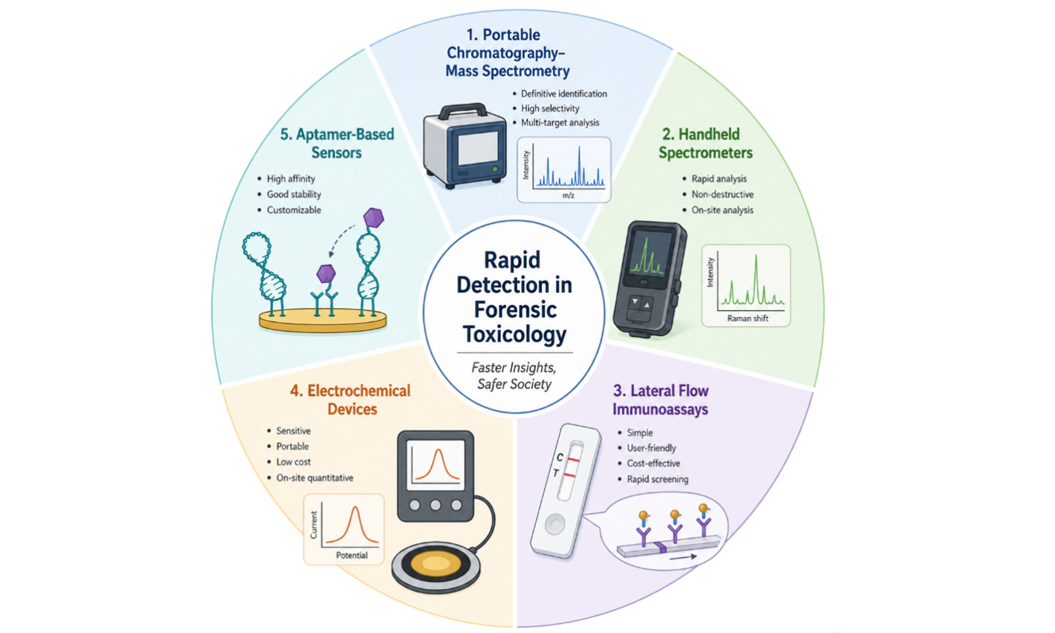
Abstract
The demand for timely, on-site toxicological evidence across poisoning investigations, drug enforcement, and public health emergencies has driven the development of rapid detection technologies in forensic toxicology. This review critically assesses the current landscape of rapid detection technologies, including portable chromatography-mass spectrometry, handheld spectrometers, lateral flow immunoassays, aptamer-based sensors, and electrochemical devices. Instrument-based methods offer definitive identification but face limitations in cost, complexity, and sensitivity for trace analysis. Sensor-based platforms prioritize simplicity and accessibility yet struggle with matrix interference, quantitative accuracy, and differentiation of new psychoactive substances (NPS) isomers. Despite significant advances, key translation barriers include a lack of standardization and limited field-relevant validation. We outline priority areas for future research, such as validation using authentic forensic specimens, integration of machine learning for adaptive data processing, and sustained collaboration to establish standardized protocols and evidentiary frameworks. Rapid detection technologies are not poised to replace laboratory analysis but to complement it, extending the reach of forensic toxicology into the field and enabling real-time decision-making for public health and safety.
Keywords


 Download PDF
INTRODUCTION
As an integral component of modern forensic science, forensic toxicology contributes critically to elucidating the cause and manner of death in suspected poisoning cases, assessing impairment in driving under the influence of drugs investigations, monitoring drug abuse, and providing critical evidence in criminal prosecutions. Over the past two decades, the scope of forensic toxicology has expanded substantially, driven by the escalating global drug crisis, the emergence of new psychoactive substances (NPS), and the increasing demand for timely toxicological evidence to support law enforcement and public health interventions[1,2]. This expansion has imposed increasing demands on detection methods, which must reconcile high sensitivity and specificity with the flexibility to address diverse forensic scenarios, ranging from crime scenes and emergency departments to public health incident responses[3,4].
Beyond traditional laboratory-based analysis, contemporary forensic toxicology requires analytical tools capable of meeting the immediate detection needs of multiple operational contexts[5]. In postmortem investigations, the concentrations of drugs and their metabolites undergo dynamic changes with increasing postmortem interval (PMI), which are influenced by complex factors including putrefactive degradation, tissue redistribution, and postmortem redistribution (PMR)[6,7]. These factors lead to discrepancies between measured drug concentrations and the original doses present at the time of death. In poisoning incidents, rapid identification of toxic substances provides immediate case characterization and enables prompt differentiation of toxicants to guide clinical treatment[8,9]. In traffic accident cases, on-site screening of blood for ethanol, drugs of abuse, and other addictive substances can significantly shorten detection time, thereby enhancing investigation efficiency[10]. Consequently, rapid detection technologies have emerged as a transformative force, not only within forensic toxicology but also across public health and public safety domains[11]. Traditional forensic toxicology workflows typically involve a sequential chain of operations, including sample collection, transportation, and preparation (separation, purification, and extraction), instrumental analysis, data processing, and final report issuance, which inevitably introduces time delays and increases turnaround times. On-site analytical methods transcend the spatial and temporal limitations inherent to conventional laboratory-based analysis, achieving “sample-in, result-out” capabilities and substantially shortening the decision cycle from sample acquisition to result interpretation[12,13]. Thus, the on-site analytical testing method is fundamentally reshaping the practice paradigm of forensic toxicology from a predominantly retrospective to a reactive model, an intelligence-generating framework embedded directly within law enforcement, emergency medical, and public health response workflows. In this context, rapid detection technologies are operationally defined as analytical methods that, by virtue of being deployable at the site of sample collection, can provide a qualitatively or quantitatively interpretable result significantly faster than traditional laboratory workflows, typically within 30 min of sample collection or sample introduction. This operational definition reflects the practical reality that rapid methods are not merely faster versions of laboratory techniques.
Over the past decade, the field of rapid detection has witnessed unprecedented growth, driven by parallel advances in multiple disciplines, including analytical chemistry, materials science, and nanotechnology[14-17]. In particular, the development of portable and field-deployable analytical devices has accelerated markedly, fueled by the miniaturization of optical and electronic components, the commercial availability of low-cost lasers and detectors, and the proliferation of smartphone-based readout systems[18,19]. Importantly, the progress in rapid detection methods for clinical diagnostics, food safety monitoring, environmental surveillance, and point-of-care medical testing has provided platform technologies readily adaptable for drug detection applications[20-22]. For forensic toxicology analysis, rapid detection methods can be classified into instrument-based field-deployable systems and sensor-based rapid screening platforms. The former category includes portable chromatographic-mass spectrometric instruments and handheld spectrometers, representing the direct translation of established laboratory gold standards into field-ready tools[23-25]. These instrument-based methods offer the significant advantage of identifying unknown substances through mass spectral interpretation or spectral library matching. In practice, however, instrument and consumable costs vary substantially depending on the manufacturer, model, and whether the device is domestically produced or imported. Operational costs are also influenced by sample preparation, maintenance, and operator training, which should be considered alongside analytical performance. Sensor-based platforms encompass a wide array of biosensors and chemical sensors, including lateral flow immunoassay (LFIA)-based kits, as well as emerging recognition-element platforms utilizing antibodies or aptamers coupled with colorimetric, fluorescent, or electrochemical signal transduction mechanisms[26-28]. These sensor-based approaches prioritize simplicity, low cost, and accessibility over instrumentation, enabling minimally trained personnel to obtain results rapidly within minutes.
Recently, the Shi group provided an overview of current rapid detection methods for toxicological analysis across diverse forensic scenarios, offering a valuable framework that connects emerging technologies to forensic toxicology practice[12]. Building upon this foundational review, the present review aims to extend the discussion by providing a more detailed examination from the perspective of sensor design principles and signal transduction mechanisms, critically evaluating the relative advantages, limitations, and suitability for specific forensic scenarios. We further summarize the key challenges facing practical deployment of these methods, including matrix effects, sensitivity limitations, standardization gaps, and outline future directions for advancing rapid detection technologies in forensic toxicology.
ADVANCES IN RAPID DETECTION METHODS
It is instructive to recognize that rapid detection methods in forensic toxicology can be broadly divided into instrument-based analytical systems [including portable chromatography-mass spectrometry, handheld Raman spectrometers, and portable near-infrared (NIR)/Fourier transform infrared (FTIR) spectrometers] and sensor-based detection platforms. These instruments offer the significant advantage of definitive or near-definitive identification of unknown substances through mass spectral interpretation or spectral library matching. Consequently, a relatively high level of technical maturity and regulatory acceptance can be achieved, particularly in applications such as drug crime scene investigation, customs inspection, and border enforcement actions.
Portable chromatography-mass spectrometry and handheld spectrometers
Portable gas chromatography-mass spectrometry (GC-MS) has matured substantially over the past decade, evolving from laboratory-bound instruments to field-deployable systems capable of delivering laboratory-grade performance at the point of need[29-31]. Recent advances have focused on expanding compound coverage and simplifying field operation through integration of advanced sample introduction techniques, including solid-phase microextraction (SPME), purge-and-trap, and thermal desorption, enabling trace-level detection in complex forensic matrices such as biological fluids and seized drug residues[32-35]. Recent efforts have concentrated predominantly on the identification of fentanyl and its synthetic opioids in seized powders[31,36-38]. Beyond direct sampling, innovations in sample introduction have expanded the application scope of portable mass spectrometry (MS). The coupling of automated solid-phase extraction (SPE) devices with portable MS has enabled rapid on-site screening of illicit drugs in wastewater, eliminating analyte degradation risks during transport[39]. For direct analysis of biological specimens, a portable solvent-assisted thermal desorption ionization source has been developed for hair analysis, achieving detection limits as low as 0.2 ng/mg for ketamine, cocaine, and morphine[35]. Complementary enrichment strategies, such as integrating SPME with nano-electrospray ionization in a single kit, allow on-site extractive sampling from complex liquid samples and living organisms[40]. Additionally, dedicated microcolumns featuring ionic liquid-based hybrid gel stationary phases have enabled effective separation of multiple narcotics prior to micro-GC detection, enhancing both resolution and sensitivity for complex samples[41]. Collectively, these advancements have transitioned portable chromatography-MS from a specialized tool for seized powder analysis to a versatile system capable of handling diverse forensic matrices, including wastewater and biological fluids.
Raman spectroscopy is a rapid, non-destructive, cost-effective technique that provides molecular fingerprint information, receiving widespread attention in forensic science[42,43]. Compared with portable chromatography-MS, handheld Raman spectroscopy offers distinct advantages in cost, operational simplicity, and speed, benefiting from significant advances in domestic manufacturing[44,45]. Currently, commercially available handheld Raman spectrometers, both domestic and international, have established spectral libraries covering virtually all traditional drugs of abuse, including cocaine, heroin, methamphetamine, 3,4-methylenedioxymethamphetamine (MDMA), ketamine, and cannabis, as well as a broad range of emerging NPS such as synthetic cannabinoids, synthetic cathinones, and fentanyl analogs[46-48]. The localization of Raman spectrometer production has achieved remarkable progress, with domestic instruments now delivering sensitivity and spectral resolution comparable to their international counterparts. However, the analytical performance of handheld Raman diminishes in complex biological matrices due to low analyte concentrations, background fluorescence, and spectral interference from matrix components[48]. To overcome sensitivity limitations, studies have explored the combination of handheld Raman with MS to achieve complementary analytical capabilities, making their integration particularly attractive for forensic applications where both speed and accuracy are required[49,50]. Besides, surface-enhanced Raman scattering (SERS) has emerged as a transformative approach in diverse forensic scenarios, including traditional drugs of abuse (cocaine, heroin, methamphetamine, MDMA, ketamine), NPS (which encompass a broad range of substance classes such as synthetic cannabinoids, synthetic cathinones, synthetic opioids including fentanyl analogs and nitazenes, phenylethylamines, piperazines, and tryptamines), pesticides, and even non-traditional forensic targets such as exosomal biomarkers for sudden cardiac death[51-55]. Given the continuous evolution of the NPS landscape, the integration of machine learning with SERS has become increasingly necessary. For instance, machine learning algorithms can learn subtle, nonlinear patterns from SERS data, enabling classification of trace-level NPS and their isomers despite spectral overlaps and signal variability, and recent research has focused on constructing machine learning models specifically for the differentiation and identification of diverse NPS[56,57]. Despite these promising applications, current machine learning models remain largely at the proof-of-concept stage, typically validated under controlled conditions with limited datasets. Their generalizability to new instruments, matrices, and emerging NPS classes, as well as their performance on authentic forensic specimens, requires further investigation.
As a complementary vibrational technique to Raman, FTIR spectroscopy has gained increasing attention for on-site forensic drug detection. Unlike Raman spectroscopy, which measures inelastic light scattering and can be severely hindered by sample fluorescence, FTIR measures the absorption of mid-infrared radiation (typically 4,000-400 cm-1) by molecular bonds, and is inherently immune to fluorescence interference, thereby offering superior molecular specificity and more reliable compound identification. However, field deployment of portable FTIR instruments is constrained by high manufacturing costs, stringent mechanical stability requirements, and inherently heavier, more expensive hardware. Although handheld products such as the Agilent 4300 (weighing ~2.2 kg) are commercially available, their adoption in routine field operations remains limited compared to NIR devices. By contrast, portable NIR spectrometers feature a substantially simpler hardware configuration. Broadband sources such as tungsten-halogen lamps and InGaAs detectors are inexpensive and readily miniaturized, enabling compact, lightweight devices, some of which are even available as smartphone-attachable modules. This cost-effectiveness and ease of integration have made NIR the dominant choice for portable spectroscopic screening, offering complementary information through infrared absorption with distinct advantages for on-site forensic drug detection[58-60]. Kranenburg et al. achieved > 97% true positive rates for cocaine, brown heroin, and colored MDMA tablets using a
Lateral flow immunoassays
LFIA, commonly implemented using immunochromatographic test strips, represents the most commercially mature and operationally accessible sensor-based platform for rapid drug screening in forensic toxicology[67,68]. These devices operate on the principle of capillary-driven fluid transport along a nitrocellulose membrane, where target analytes are captured by antibodies immobilized on test lines, generating a visual signal typically through gold nanoparticles (AuNPs)[68]. Owing to the small molecular size, commercial LFIA kits for drugs predominantly employ a competitive immunoassay format, in which target analytes compete with immobilized antigen conjugates for binding to limited labeled antibodies, resulting in an inverse relationship between analyte concentration and test line intensity. The analytical performance of LFIA in forensic applications has been extensively validated for common drugs, NPS, pesticides, benzodiazepines, and other emerging forensic toxicants[67,69-72].
In recent years, the proliferation of opioid analogs, particularly fentanyl and its derivatives, has driven increased development of LFIA kits targeting this class of substances. Numerous commercial test strips for fentanyl, norfentanyl, carfentanil, and furanylfentanyl are now available[73]. Despite their widespread use, LFIA kits face sensitivity limitations when applied to complex biological matrices such as whole blood, serum, or postmortem specimens. This sensitivity gap has motivated substantial research efforts focused on improving detection limits and expanding target coverage[74,75]. The most widely explored strategy involves replacing AuNPs with alternative signal reporters. Various fluorescent materials, including organic dyes, quantum dots, and upconversion nanoparticles, have been successfully integrated into LFIA platforms. Figure 1 illustrates the competitive immunoassay format commonly used in LFIA[71,76]. For example, fluorescent dye-labeled LFIA significantly enhances sensitivity for morphine and methamphetamine, enabling detection at concentrations substantially lower than AuNP-based counterparts[77]. Similarly, persistent luminescence nanoparticles that emit long-lived afterglow have been employed for methamphetamine detection, demonstrating markedly improved sensitivity by eliminating background autofluorescence through time-gated measurement[76]. Beyond fluorescence-based readouts, alternative signal transduction modalities have been integrated with LFIA to overcome the sensitivity limitations of colorimetric detection. LFIA coupled with SERS provides immunological specificity, high sensitivity, and molecular fingerprinting capability[78]. LFIA coupled with electrochemistry replaces visual readout with amperometric or impedimetric measurement using screen-printed electrodes (SPEs), offering quantitative capability and enhanced sensitivity at the cost of increased device complexity[71].
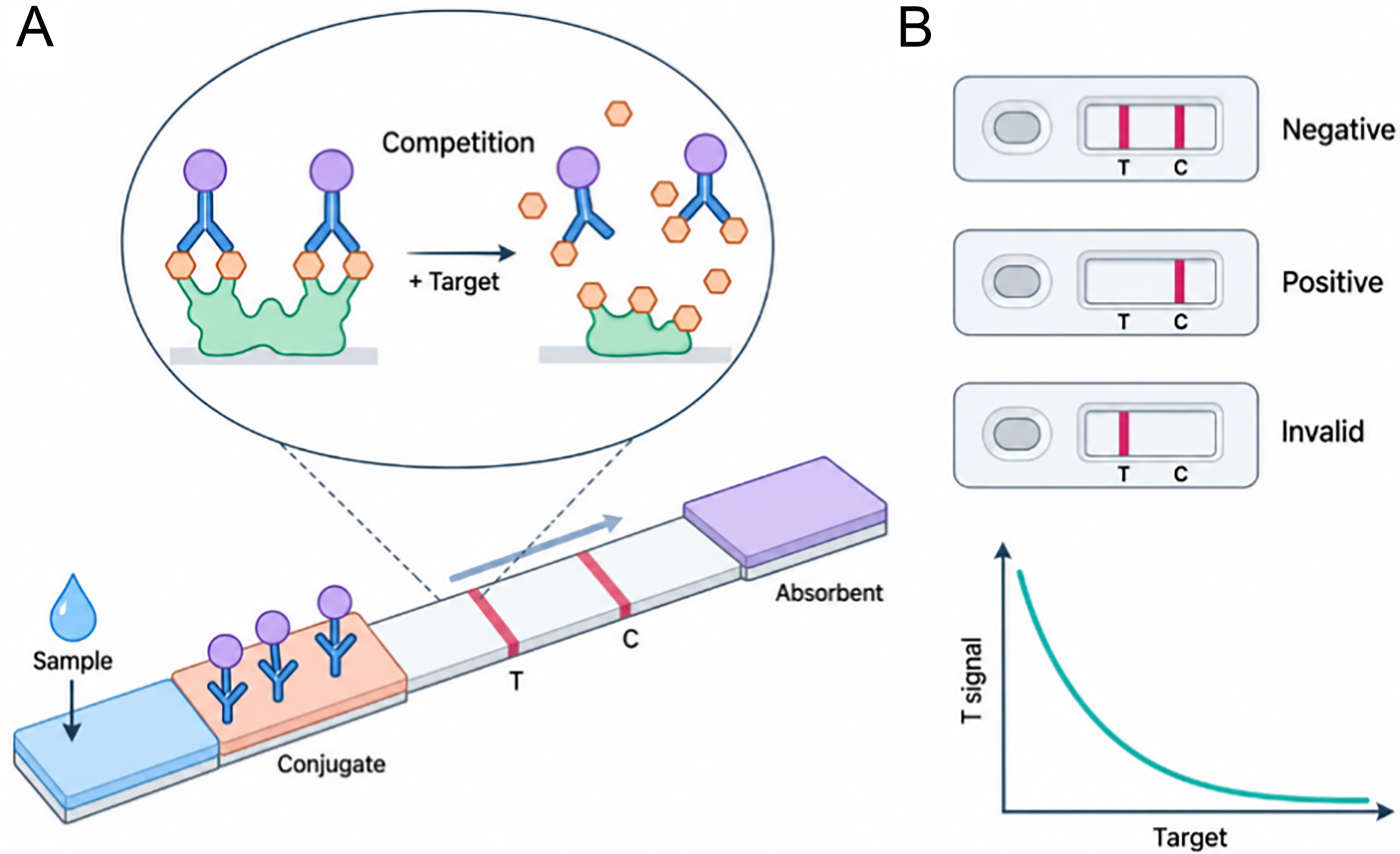
Figure 1. (A) Schematic illustration of the LFIA principle based on a competitive immunoassay format; (B) Signal output. LFIA: Lateral flow immunoassay.
A more fundamental innovation involves replacing antibodies with nucleic acid aptamers as recognition elements. Aptamers offer several distinct advantages over antibodies because they are chemically synthesized, eliminating batch-to-batch variability inherent to biological production, and they exhibit excellent thermal stability and ready modification[79,80]. While aptamer-LFIA has been more extensively developed for clinical diagnostics, its application to forensic toxicology remains relatively limited[69,81]. Nevertheless, proof-of-concept studies have demonstrated promise, with aptamer-based LFIA for methamphetamine and cocaine achieving detection limits comparable to or better than antibody-based counterparts while offering superior batch consistency and thermal stability[69]. However, the selection of high-affinity aptamers against small-molecule drug targets remains technically challenging and time-consuming, and aptamers may exhibit reduced affinity in complex biological matrices due to nuclease degradation or matrix-induced conformational changes.
LFIA technology has been successfully adapted for forensic toxicology targets outside the classical drug-of-abuse panel. Commercial and research-based test strips for pesticides, particularly paraquat and fluoroacetamide, have been reported, addressing the need for rapid screening in acute poisoning cases, occupational exposure incidents, and intentional contamination events[82,83]. These assays typically employ competitive formats analogous to LFIA and have been validated in urine and gastric contents. Notably, LFIA methods for pesticides generally achieve substantially higher sensitivity than their drug-detecting counterparts, with detection limits often reaching the low parts-per-billion (ng/mL) range, compared to the parts-per-million (μg/mL) range typical for traditional drugs of abuse[67]. This sensitivity advantage reflects the availability of high-affinity antibodies generated against pesticide immunogens, as well as the greater structural diversity of pesticide molecules, which present more epitopes for antibody recognition than the relatively small, compact drug molecules[68]. Nonetheless, sensitivity limitations in complex biological matrices, particularly blood and postmortem tissues, remain.
Specificity-engineered sensing methods
Unlike instrument-based methods, sensor platforms rely on molecular recognition between a recognition element and its target analyte, converting this binding event into a measurable signal[80]. The flexibility of synthetic recognition elements and tunable signal output modalities enable sensor performance tailored to specific forensic scenarios. The recognition part has expanded substantially beyond traditional antibodies to include nucleic acid aptamers, molecularly imprinted polymers (MIPs), and engineered functional materials[79,84]. Aptamers are single-stranded DNA or RNA oligonucleotides selected through systematic evolution of ligands by exponential enrichment (SELEX)[85,86]. They are chemically synthesized, eliminating batch-to-batch variability, exhibit excellent thermal stability, and can be readily modified with functional groups for surface immobilization or signal labeling[79,80]. MIPs, synthesized by polymerizing functional monomers around a template molecule, provide robust, low-cost recognition cavities with physical and chemical stability that exceeds most biological recognition elements, typically with lower affinity and selectivity[87]. Functional materials including metal-organic frameworks (MOFs), covalent organic frameworks (COFs), and graphene-based nanomaterials can interact with target analytes through non-specific or semi-specific mechanisms such as hydrogen bonding, π-π stacking, electrostatic interactions, and hydrophobic effects[88,89].
The signal transduction module encompasses a diverse array of readout modalities. Colorimetric sensors offer visual interpretation without specialized instrumentation, enabling rapid, cost-effective screening in resource-limited settings. Fluorescent sensors achieve significantly higher sensitivity through various signal modulation mechanisms, including quenching, enhancement, or ratiometric response, though they may be susceptible to photobleaching and autofluorescence interference[90]. Electrochemical sensors offer excellent sensitivity, quantitative capability, and miniaturization potential, making them well-suited for portable devices. Each modality presents distinct trade-offs between sensitivity, speed, cost, and operational complexity, with optimal choice depending critically on the specific forensic application. The following sections summarize each category, evaluating their working principles, practical advantages and limitations, and forensic toxicology applications.
Colorimetric sensors
Colorimetric sensors generate visually detectable color changes upon target analyte binding, enabling semi-quantitative or qualitative analysis. Colorimetric presumptive testing in forensic drug analysis has historically relied on well-established chemical spot tests, such as the Marquis test for opiates and amphetamines, the Scott test for cocaine, and the Prussian blue reaction for cyanide, which exploit characteristic color-producing reactions between specific reagents and functional groups of target analytes[91,92]. However, classical color tests suffer from poor specificity, producing false positives from structurally similar cutting agents and limited applicability to emerging novel psychoactive substances. These shortcomings have motivated the development of nanomaterial-based colorimetric sensors that offer improved sensitivity, selectivity, and adaptability[93,94]. AuNPs represent the most extensively employed nanomaterial for colorimetric sensing due to their distance-dependent optical properties. LFIA produces visible color signals through AuNP-labeled detection antibodies, enabling rapid on-site screening for drugs of abuse in urine, saliva, and hair matrices[70,83,95]. Various antibody-based colorimetric sensors have been widely developed for illicit drugs and NPS detection[96,97]. With the advances in aptamer selection, aptamer-functionalized AuNP sensors have been developed for cocaine, methamphetamine, and ketamine, achieving detection limits in the low micromolar range with response times under 10 min[69,98]. Notably, Xiao’s group has pioneered the development of aptamer-based colorimetric assays for rapid on-site detection of opioids such as heroin and oxycodone, achieving detection limits as low as the nM level[99]. Moreover, MIP-based colorimetric sensors offer an alternative recognition strategy with enhanced chemical and thermal stability[93]. By incorporating vinyl-functionalized chromophores into the polymer matrix during synthesis, analyte binding to imprinted cavities induces measurable color changes through conformational rearrangement or charge transfer. For example, a MIP-based sensor for methamphetamine employed a methacrylic acid-co-ethylene glycol dimethacrylate polymer with incorporated bromophenol blue, producing a blue-to-yellow color change upon analyte binding with a detection limit of 1.4 μM in biological samples[100]. The unique physicochemical properties of nanomaterials have spurred rapid advances in colorimetric sensing. Noble metal nanoparticles possess high extinction coefficients, size- and shape-dependent surface plasmon resonance (SPR) characteristics, and facile surface functionalization chemistry, enabling highly sensitive and selective detection[94,95,101]. Nanozymes, nanomaterials with intrinsic enzyme-like catalytic activity, have emerged as a powerful signal-amplification strategy. For instance, researchers developed a nanozyme-based lateral flow strip using 3,3’-diaminobenzidine (DAB) as a signal output for trace bloodstain detection in real criminal cases where commercial strips failed[102]. Figure 2 presents two representative colorimetric sensing strategies, each demonstrating a distinct transduction mechanism for different forensic targets as described above[98,99,103].
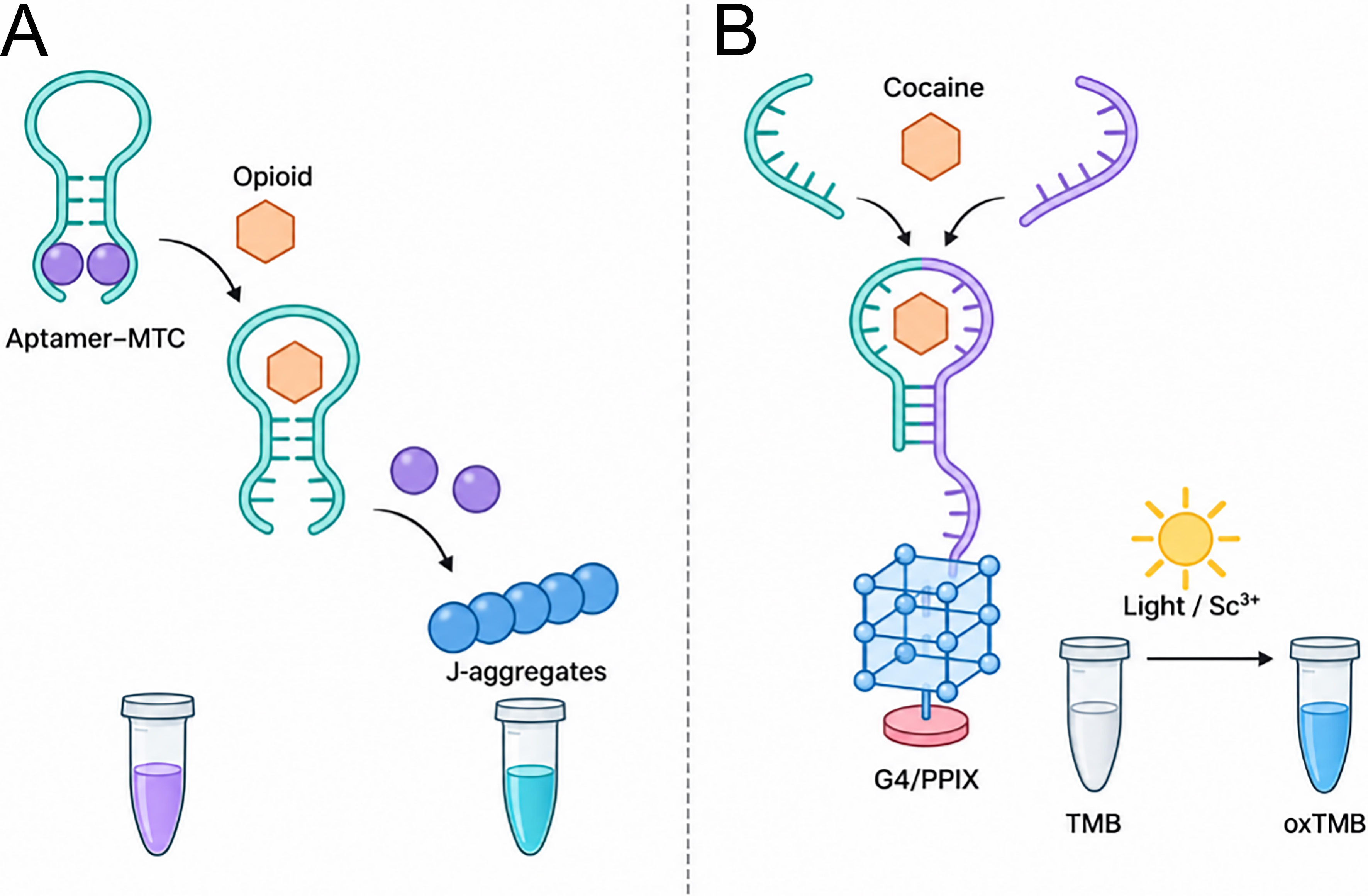
Figure 2. (A) Schematic of the aptamer-based MTC-displacement assay; (B) Principle of aptamer-based colorimetric cocaine tests. MTC: Methylene tripeptide complex; TMB: 3,3′,5,5′-Tetramethylbenzidine; G4: G-quadruplex; PPIX: protoporphyrin IX; oxTMB: oxidized 3,3′,5,5′-Tetramethylbenzidine.
Despite their operational simplicity, colorimetric sensors face several inherent limitations. Sensitivity is typically lower than that of fluorescent or electrochemical counterparts, with detection limits generally in the micromolar to sub-millimolar range, which is adequate for urine screening where drug concentrations are often elevated, but insufficient for trace analysis in hair or blood where nanomolar sensitivity may be required[28]. Quantitative accuracy is compromised by subjective visual color interpretation, though smartphone-based red-green-blue (RGB) analysis can mitigate this issue[104,105]. Matrix effects, particularly from colored or turbid biological specimens, can obscure color changes and produce false results[106]. Nonetheless, the exceptional simplicity and cost-effectiveness of colorimetric sensors position them as valuable frontline screening tools, particularly where immediate information suffices to guide subsequent action. From a practical standpoint, LFIA kits are among the most cost-effective and user-friendly rapid detection tools, requiring minimal operator training and no specialized instrumentation, which facilitates their adoption in routine field screening.
Fluorescent sensors
Fluorescent sensors achieve significantly higher sensitivity than colorimetric counterparts by exploiting efficient fluorescence signal transduction. While their construction, recognition element coupled with a signal reporter, remains analogous to colorimetric sensors, fluorescence-based methods offer distinct advantages in forensic toxicology[28]. Notably, the Dou group has made outstanding contributions to the direct fluorescence detection of synthetic cannabinoids by ingeniously designing intermolecular interactions, enabling highly sensitive and selective turn-on sensing of these compounds, such as JWH-018 and MDMB-type compounds with exceptional performance[107,108]. Building upon this foundation, aptamer-functionalized fluorescent sensors have been extended to a broader range of NPS and other forensic targets, including opioids (fentanyl, morphine, codeine, methadone), stimulants (cocaine, methamphetamine, amphetamine), and other emerging NPS such as synthetic cathinones, with detection limits in the low nanomolar to picomolar range, to which the Xiao group has made particularly significant contributions[98,109-111]. As shown in Figure 3, our group has advanced the field by developing a label-free, aptamer-based fluorescent sensor for rapid detection of THC and its metabolites[112,113]. Unlike conventional labeled strategies requiring fluorophore-modified aptamers, our approach eliminates covalent labeling, substantially reducing assay costs while maintaining low nanomolar sensitivity. Integrated with a portable fluorescence readout device, this platform enabled rapid on-site detection in saliva and urine, demonstrating potential for cost-effective, field-deployable screening[114-116].
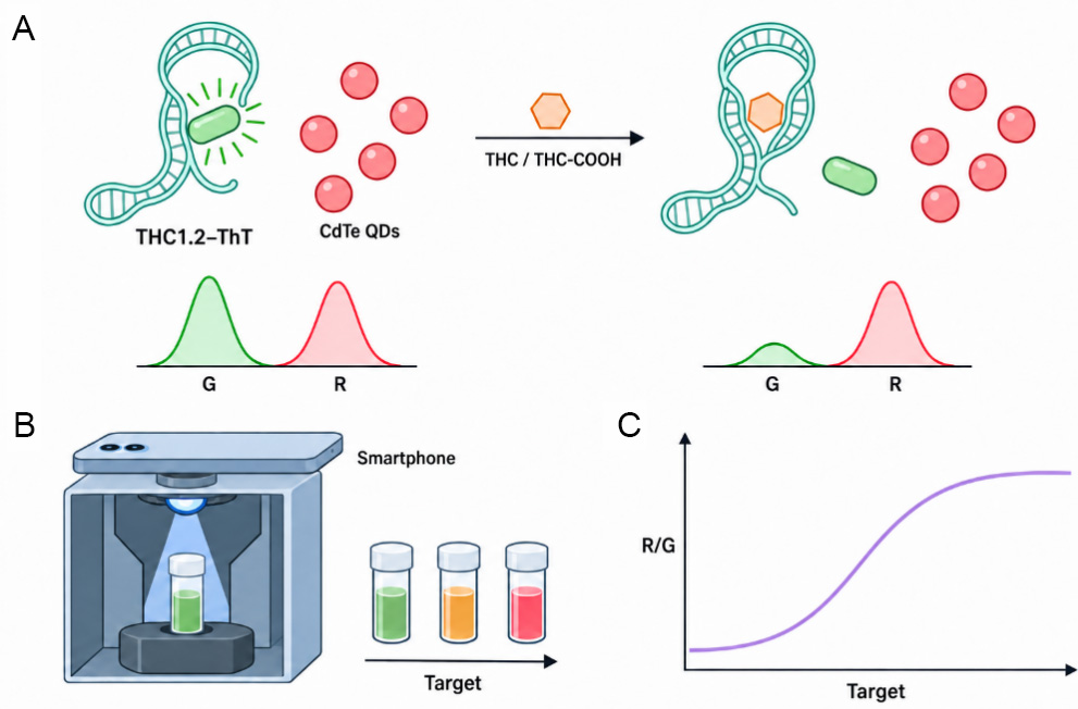
Figure 3. (A) Schematic diagram of ratiometric fluorescence for visualizing THC and its metabolite; (B) A smartphone-based portable fluorescence capture device; (C) Signal response of the sensor upon exposure to different target concentrations. THC: Tetrahydrocannabinol; THC-COOH: 11-nor-9-carboxy-tetrahydrocannabinol; QDs: quantum dots.
Beyond recognition elements, the diversity of fluorescent materials, including quantum dots, carbon dots, aggregation-induced emission (AIE) fluorescence, and MOFs provides a rich toolbox for sensor design, enabling tunable emission wavelengths, high quantum yields, and excellent photostability[117-120]. These innovations render fluorescent sensors suitable for trace analysis in challenging matrices such as hair extracts (pg/mg sensitivity), blood, and wastewater samples[120-122]. Like colorimetric sensors, fluorescent methods can be adapted for visual or semi-quantitative readout under portable UV-vis lamps, and miniaturized fluorescence readers (e.g., smartphone-based or handheld fluorometers) are increasingly available, facilitating on-site deployment. In recent years, the integration of machine learning with fluorescent sensing has emerged as a powerful strategy to address limitations in complex forensic matrices where spectral overlap, background autofluorescence, and environmental variability compromise analytical accuracy[119]. Despite their sensitivity advantages, fluorescent sensors face challenges that have slowed routine forensic practice. Photobleaching is the irreversible destruction of fluorophores under illumination, which complicates quantitative comparisons across different measurements[123]. Autofluorescence from endogenous fluorophores in biological matrices elevates backgrounds and reduces signal-to-noise ratios, particularly in blood and tissue homogenates. Nonetheless, the exceptional sensitivity and multiplexing capability of fluorescent sensors make them well-suited for trace-level detection applications where colorimetric methods prove inadequate. However, their performance in complex forensic matrices such as whole blood and decomposed tissue is substantially compromised by autofluorescence and light scattering. Sample pretreatment is often required, although this may partially negate the “minimal sample preparation” advantage. Time-gated or NIR detection has been explored to reduce autofluorescence, and ratiometric probes can correct for matrix-induced signal fluctuations[124]. Nevertheless, validation using authentic forensic specimens remains limited.
Electrochemical sensors
Electrochemical sensors transduce molecular recognition events into quantifiable electrical signals, including current, voltage, or impedance, using compact, portable potentiostats. Target analyte interactions at the working electrode surface generate electron transfer processes that are recorded as characteristic signals proportional to analyte concentration. This transduction mechanism offers exceptional advantages for forensic toxicology, such as detection limits that routinely reach low nanomolar to picomolar ranges and rapid response times (typically 30 s to 5 min)[125]. The working electrode, typically screen-printed carbon, gold, or platinum, serves as the platform for recognition element immobilization and signal generation. Modification of the electrode surface with nanomaterials (including graphene, carbon nanotubes, AuNPs, and metal oxides) enhances surface area, improves electron transfer kinetics, and provides anchoring sites for recognition elements, substantially improving sensitivity[126-128]. While direct electrochemical detection exploits the intrinsic redox activity of target analytes to generate measurable signals without the need for surface-immobilized recognition elements, this approach is fundamentally limited by poor specificity. The integration of MIPs with electrochemical transduction represents one of the most promising strategies for achieving selectivity without compromising robustness, offering a unique combination of selectivity, stability, and cost-effectiveness, making them particularly attractive for field-deployable forensic applications[129]. Complementing MIP-based approaches, aptamer-integrated electrochemical sensors exploit the conformational switching properties of synthetic oligonucleotides to achieve exceptional specificity. Target binding induces a conformational rearrangement that alters the proximity of the redox reporter to the electrode, producing a quantifiable change in faradaic current. This signal-on/signal-off transduction mechanism has been extensively exploited in recent years for the sensitive and selective detection of opioids[130,131]. For instance, electrochemical aptamer-based sensors have been successfully developed for fentanyl, achieving sub-nanomolar detection limits in undiluted serum and whole blood, with the ability to continuously monitor dynamic concentration fluctuations in real time[98,132]. These methods leverage the inherent advantages of aptamers, including SELEX selection against novel targets, excellent batch-to-batch reproducibility, and resilience to matrix effects, positioning aptamer-electrochemical hybrid technologies as a versatile and rapidly adaptable toolkit for combating the evolving landscape of illicit drug abuse. Despite their promising advantages, MIP- and aptamer-based electrochemical sensors present several critical limitations that warrant consideration. For MIP-based sensors, the major drawbacks include heterogeneous binding-site distribution, incomplete template removal, which leads to false-positive signals, and compromised reproducibility across batches. For aptamer-based sensors, the principal limitation is the intrinsic susceptibility of oligonucleotides to nuclease degradation in complex biological matrices. Additionally, aptamer conformational switching is highly sensitive to buffer composition and pH, making sensor performance vulnerable to environmental fluctuations in real-world forensic settings. Batch-to-batch variability in aptamer immobilization density and orientation further hinders reproducibility.
Beyond laboratory-based configurations, the translation of electrochemical sensors to forensic practice has been greatly accelerated by the development of diverse portable and wearable device formats. Among these, SPEs stand out as the most mature and widely adopted platform, with the advantages of single-use disposability, low cost, and plug-and-play operation without the need for electrode pretreatment or maintenance[133]. When coupled with handheld potentiostats, SPEs enable law enforcement personnel to perform on-site drug screening within minutes. As shown in Figure 4, 3D-printed electrochemical cells represent another innovative platform, allowing modular, customizable, and rapid prototyping of sensor housings tailored to specific forensic scenarios[134]. Fabricated from conductive filaments such as graphene-doped polylactic acid or carbon black composites, these 3D-printed devices integrate sample reservoirs, fluidic channels, and electrode alignment features into a single compact unit, facilitating field-deployable, point-of-need testing. USB-integrated smartphone-based detectors have further miniaturized electrochemical analysis by embedding potentiostat circuitry into USB-drive form factors that connect directly to mobile devices[135]. Powered by custom Android or iOS applications, these systems enable real-time voltammetric data acquisition, wireless transmission, and cloud-based storage, transforming smartphones into portable forensic laboratories[136]. In parallel, wearable electrochemical sensors have emerged as a transformative class of devices for direct evidence collection and real-time monitoring. Glove-based sensors integrate printed electrode arrays onto nitrile glove fingertips, enabling officers to swipe suspicious powders or liquids (e.g., fentanyl, cocaine, methamphetamine) and immediately perform electrochemical analysis without separate sampling tools[125,137]. Skin patches and temporary tattoo sensors conform to the skin surface for noninvasive monitoring of drugs excreted in sweat, including alcohol, nicotine, levodopa, and caffeine[137,138].
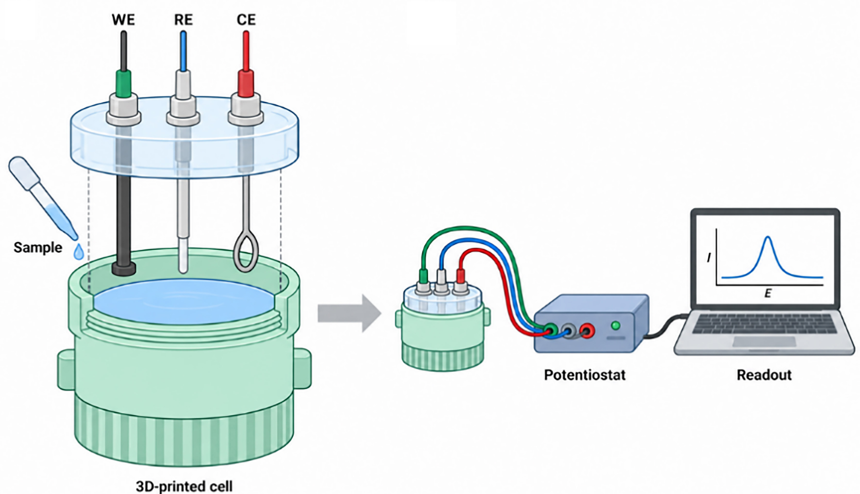
Figure 4. 3D-printed electrochemical cell and assembled system connected to a portable potentiostat via the custom cable developed and controlled by a laptop. WE: Working electrode; RE: reference electrode; CE: counter electrode.
Collectively, electrochemical sensors are transitioning from laboratory prototypes to practical field-deployable tools. Through the synergistic integration of nanomaterial-modified electrodes, MIP/aptamer-based recognition chemistries, flexible and wearable form factors, smartphone connectivity, and artificial intelligence-driven data analytics, these platforms enable rapid, sensitive, portable, and real-time detection of diverse illicit drugs. This technological convergence holds great promise for broad applications in crime scene investigation, roadside drug-impaired driving enforcement, wastewater-based epidemiology for community drug use surveillance, and clinical toxicology for emergency overdose diagnosis[137-139]. As portable potentiostats become increasingly affordable and regulatory frameworks adapt to accommodate these innovations, electrochemical sensors show considerable potential for future forensic applications, though further validation with real-world samples is needed.
Comparison of analytical performance
In conclusion, these methods can be broadly categorized into instrument-based systems (portable GC-MS, handheld Raman and NIR/FTIR spectrometers) and sensor-based platforms (LFIA, colorimetric, fluorescent, and electrochemical sensors). Instrument-based methods are commercially mature technologies that provide definitive or near-definitive identification of unknown substances through spectral library matching or mass spectral interpretation. They are well-established in forensic workflows, with regulatory acceptance and established evidentiary value. However, they are constrained by higher costs, greater operational complexity, and maintenance requirements, making them best suited for scenarios where definitive identification is paramount, such as customs inspection, crime scene triage, and seized drug analysis. In contrast, sensor-based platforms are generally more accessible, low-cost, and operationally simple, making them attractive for point-of-care testing, roadside screening, and resource-limited settings where instrument-based methods are impractical. However, most sensor platforms remain at the research and development stage, with limited commercial availability and insufficient field validation. Their performance is highly dependent on the specific target analyte and matrix, and they generally lack the standardization and regulatory acceptance of their instrument-based counterparts. Instrument-based methods such as portable GC-MS typically achieve limits of detection (LODs) in the ng/mL to μg/mL range, while handheld Raman and NIR/FTIR spectrometers are suitable for identifying pure substances or major components at the μg/mL level. For sensor-based platforms, the sensitivity follows a rough hierarchy: colorimetric sensors typically achieve LODs in the μg/mL to ng/mL range, fluorescent sensors offer higher sensitivity in the ng/mL to sub-ng/mL range, and electrochemical sensors provide the highest sensitivity, typically reaching ng/mL to pg/mL levels. It is noted that actual LODs vary considerably depending on the target analyte and matrix, and these ranges serve only as general references rather than absolute performance indicators. The typical time-to-result for all method categories is summarized and shown in Table 1, along with their suitable sample types and key factors affecting analysis time. In brief, handheld Raman and NIR/FTIR spectrometers provide results within seconds to a few minutes, suitable for seized powders and pills. Portable GC-MS requires 10-30 min depending on analyte complexity, applicable to biological fluids and wastewater, among sensor-based platforms, electrochemical sensors are the fastest (seconds) and compatible with complex matrices such as whole blood, while LFIA offers well-documented < 10 min results in urine and oral fluid; colorimetric and fluorescent sensors typically fall within 30 min, with time depending on assay design and target analyte.
Typical time-to-result of reported detection methods
| Method category | Time-to-result | Sample types | Key factors affecting analysis time | Ref. |
| Portable GC-MS | 10-30 min | Seized powders, biological fluids (blood, urine, hair extracts), wastewater | Number of target analytes; thermal desorption or SPME pre-concentration steps | [32-35] |
| Handheld Raman | Seconds to a few minutes | Seized powders, pills, liquids, drug paraphernalia; limited in colored/turbid biological fluids | No sample preparation required; spectral library matching is instantaneous | [44-48] |
| Portable NIR/FTIR | Seconds to a few minutes | Seized powders, pills, colored samples, packaging materials, beverages; biological fluids | No sample preparation required | [58-62] |
| LFIA | < 10 min | Urine, oral fluid, serum, whole blood, hair extracts | Antigen-antibody binding kinetics; analysis time is the most well-documented | [67-72] |
| Colorimetric Sensors | ~30 min | Urine, serum, water, seized samples; limited in colored/turbid biological fluids (hemoglobin, bilirubin interference) | Target analyte and assay format; incubation time; varies considerably across studies | [95-99] |
| Fluorescent Sensors | ~30 min | Urine, serum, hair extracts, wastewater; limited in blood due to autofluorescence from endogenous fluorophores | Depends on the design of the signal reporter probe; precise timing often not explicitly reported across studies | [107-113] |
| Electrochemical Sensors | Seconds to a few minutes | Whole blood, serum, urine, oral fluid, sweat; compatible with complex biological matrices | Fastest transduction mechanism; no separation or labeling required; instantaneous electrochemical readout | [125-129] |
PRACTICAL APPLICATIONS AND SCENARIOS IN FORENSIC TOXICOLOGY
Clinical emergency toxicology
In emergency settings, rapid identification of ingested or injected toxicants directly influences patient management decisions, including antidote administration, decontamination procedures, and disposition (admission and discharge). Overdose presentations, particularly involving opioids, stimulants, and sedative-hypnotics, require immediate intervention, yet clinical diagnosis based on signs and symptoms alone is frequently unreliable, particularly for mixed overdoses or atypical presentations[140]. Rapid detection technologies designed for clinical toxicology applications prioritize simplicity of operation (minimal hands-on time for busy clinicians), compatibility with whole blood or serum matrices (rather than urine, which does not reflect current impairment), and rapid turnaround (results within 15 min of sample collection). Point-of-care immunoassay platforms, similar to lateral flow devices but with integrated readers for quantitative or semi-quantitative output, have been implemented in many emergency departments for drugs of abuse screening[68,69,72]. Electrochemical sensors capable of detecting opioids and stimulants in whole blood within minutes are under development, offering the potential for true point-of-care therapeutic guidance[139]. The integration of portable mass spectrometry into emergency toxicology workflows remains limited by cost and complexity but may become feasible as devices become more user-friendly and affordable[34].
Poisoning-related death investigations
In forensic death investigations where poisoning is suspected, the accurate determination of toxicant identity and concentration is essential for establishing cause and manner of death. However, postmortem specimens present unique analytical challenges. Toxicant concentrations undergo dynamic changes during the PMI, the time between death and sample collection, due to putrefactive degradation, tissue redistribution, and PMR[6,14]. It is important to distinguish PMI from laboratory turnaround time: once samples are properly collected and preserved, PMR ceases, and subsequent reporting delays do not constitute continued PMR. However, prolonged storage may affect sample stability, particularly for unstable toxicants. Rapid detection technologies address this challenge by being deployed directly at the autopsy suite or immediately following specimen collection, thereby minimizing the PMI before analysis and enabling sample collection within the early postmortem window. For instance, portable GC-MS systems have been employed for on-site screening of gastric contents and liver homogenates for common poisons, providing preliminary results that guide subsequent targeted laboratory confirmation[6,14,30]. This rapid screening capability is particularly valuable for toxicants that are prone to postmortem degradation, such as synthetic cannabinoids (e.g., JWH-200, 5F-AB-PINACA) and certain antipsychotics (e.g., olanzapine), which have been shown to undergo significant concentration losses in stored blood samples due to instability and degradation[107,108]. Handheld spectrometers can rapidly screen suspicious materials recovered at death scenes, including drug paraphernalia, unidentified pills, or beverage residues, offering immediate investigative leads before autopsy findings are available[48,63]. Furthermore, quantitative rapid methods such as electrochemical sensors, when properly validated, can provide concentration estimates that, while less precise than laboratory liquid chromatography-tandem mass spectrometry (LC-MS/MS), may be sufficient to distinguish therapeutic use from lethal intoxication in acute poisoning cases. For instance, validated electrochemical sensors have enabled quantitative detection of THC-COOH in authentic urine, ADB-BUTINACA in seized samples, and ketamine in biological matrices[71,133,134].
Border inspection and customs enforcement
Border inspection and customs enforcement represent the frontline of efforts to intercept illicit drugs and emerging psychoactive substances before they enter the drug supply. Rapid detection technologies have been increasingly deployed in this domain to address the operational demands of high sample throughput, rapid decision-making, and non-destructive analysis that preserves evidentiary integrity. Handheld Raman and FTIR spectrometers can be deployed to screen suspicious powders, pills, and liquids directly through sealed containers without opening packages, minimizing both safety risks and chain-of-custody concerns. These instruments, equipped with onboard spectral libraries, can identify known controlled substances within seconds. For NPS not yet included in spectral libraries, transportable mass spectrometers provide valuable structural information through mass spectral interpretation. However, when an entirely unknown NPS emerges, definitive identification faces the same fundamental challenge as laboratory-based MS, requiring reference standards and a series of confirmatory experiments. Therefore, on-site analysis has inherent limitations for unknown NPS, and laboratory confirmation remains necessary for legally defensible results. The increasing prevalence of drug-laced products, including cannabis-infused edibles, vape cartridges containing synthetic cannabinoids, and blotter papers impregnated with potent psychedelics, has further driven the demand for versatile rapid screening tools capable of analyzing diverse sample matrices without extensive preparation.
Driving under the influence of drugs enforcement
Driving under the influence of drugs (DUID) has emerged as a major public safety concern, with prevalence approaching or exceeding that of alcohol-impaired driving in many jurisdictions. Unlike alcohol, which can be rapidly measured using portable breathalyzers, drug impairment requires the detection of a wide range of substances, including cannabis, stimulants, opioids, benzodiazepines, and dissociative anesthetics, presenting a more complex analytical challenge. Rapid detection technologies are characterized by noninvasive or minimally invasive sampling (typically oral fluid or sweat), analysis time under 10 min to avoid delaying traffic stops, and sufficient sensitivity to detect drugs at concentrations associated with impairment. Oral fluid LFIA have been explored for roadside screening, though practices vary considerably, with commercial devices enabling simultaneous detection of several drugs from a single swab. Confirmatory analysis of positive oral fluid samples by portable mass spectrometry has been explored in preliminary studies. Despite these advances, challenges persist, including variable correlation between oral fluid and blood drug concentrations, false positives from passive contamination, and the absence of universally accepted concentration thresholds for drug-impaired driving. Additionally, an analytical limitation arises when NPS or other emerging compounds are not represented in available spectral libraries. Although library-independent or suspect-screening approaches can partially mitigate this issue, definitive identification may remain challenging in the absence of appropriate reference spectra or authentic standards, which are often unavailable for newly emerging substances.
Additional emerging applications
Emerging applications include workplace drug testing, where rapid oral fluid or urine screening devices may enable on-site collection and analysis, eliminating the logistical burden and costs associated with sending specimens to centralized laboratories. In probation and parole supervision, rapid testing may support compliance monitoring with immediate results that inform supervision decisions. In wastewater-based epidemiology, rapid detection technologies are beginning to complement laboratory methods for near-real-time monitoring of community drug use. Finally, in disaster victim identification, rapid toxicological screening of postmortem specimens from mass casualty events (natural disasters, terrorist attacks, transportation accidents) may assist in prioritizing confirmatory analyses and guiding victim identification efforts. These expanding applications underscore the versatility of rapid detection technologies across diverse forensic and public health contexts, from routine compliance monitoring to emergency response scenarios.
CHALLENGES
Sensitivity limitations of portable instrumental methods
While portable chromatography-mass spectrometry systems have achieved detection limits approaching those of laboratory instruments for volatile and semi-volatile compounds, they generally remain less sensitive than their benchtop counterparts, particularly for trace analysis in complex biological matrices. This sensitivity gap would be particularly pronounced for miniaturized liquid chromatography-mass spectrometry systems, as the reduction in size of pumps, columns, and detectors inevitably compromises chromatographic resolution and signal-to-noise ratios, even though such compact systems remain less commercially mature than portable GC-MS. For analytes requiring sub-ng/mL detection limits, such as certain synthetic cannabinoids, fentanyl analogs at low-dose exposures, or drug concentrations in hair and wastewater samples, portable systems may fail to detect relevant concentrations, producing false negatives with significant legal and public health consequences. Furthermore, the sensitivity of handheld Raman and infrared spectrometers is inherently limited by the physics of photon scattering and absorption; these instruments reliably identify pure substances or major components but cannot detect trace adulterants or low-concentration analytes in mixtures. This limitation is particularly problematic given the increasing prevalence of potent synthetic opioids and the common practice of diluting drugs with inactive cutting agents, where the active pharmaceutical ingredient may constitute only a small fraction.
Matrix interference in sensor-based methods
Sensor-based platforms face substantial challenges when applied to real forensic specimens beyond simple buffer or urine matrices. Whole blood, serum, postmortem tissue, and putrefied biological samples contain diverse interferents (proteins, lipids, salts, cellular debris, and degradation products) that can significantly alter sensor performance. In colorimetric assays, hemoglobin and bilirubin mask target-induced color changes, while turbidity scatters incident light. In fluorescent sensors, endogenous fluorophores generate autofluorescence that compromises signal-to-noise ratios. Electrochemical sensors suffer from electrode fouling due to nonspecific protein adsorption, leading to signal drift and irreproducibility. While sample dilution, filtration, or SPE can mitigate these effects, such preprocessing steps negate the “minimal sample preparation” advantage of rapid detection methods. Furthermore, the selectivity of many sensor platforms, particularly those relying on functional materials or non-specific recognition mechanisms, may be insufficient to distinguish structurally similar NPS isomers that differ by a single substituent but carry vastly different legal statuses and toxicological profiles. Most published studies are method-development oriented, with insufficient characterization of specific samples and application scenarios. Besides, the reported analysis times converge within a narrow rapid window (typically < 10 min), and the limits of detection not only vary across different matrices but are also highly dependent on the specific target analyte and analytical methodology. Complex forensic matrices such as whole blood, decomposed tissue, and putrefied specimens contain endogenous electroactive species that can generate interfering faradaic signals overlapping with the target analyte’s redox potential. Nonspecific protein adsorption and degradation products may further foul the electrode surface. While antifouling coatings and sample dilution can partially mitigate these effects, they may compromise sensitivity or negate the advantage of minimal sample preparation. Most cited electrochemical studies were performed in buffer or spiked samples, and validation using authentic forensic specimens remains necessary. Accordingly, the choice of an optimal rapid detection method should be contextualized, with due consideration given to the specific matrix, intended use scenario, and target analyte.
Lack of standardization and quality assurance
Unlike established forensic methods (GC-MS, LC-MS/MS), which rely on validated protocols, calibrated instruments, certified reference materials, and routine proficiency testing, rapid detection technologies lack comparable standardization frameworks. Significant variability exists across different commercial devices and even across different lots of the same device, due to differences in antibody sourcing, nanoparticle synthesis, membrane composition, and manufacturing tolerances. For LFIA, cutoff concentrations vary substantially between manufacturers, leading to discrepant results that complicate result interpretation and cross-jurisdictional data comparison. For handheld spectrometers, onboard spectral libraries may lack reference spectra for emerging NPS, contain spectra of structurally similar but legally distinct compounds, or fail to account for batch-to-batch variability in illicit drug composition. The absence of external quality assessment schemes for field-deployable rapid methods means that end users, law enforcement officers, customs inspectors, or emergency physicians have no mechanism for verifying device performance before use, increasing the risk of uncorrected analytical errors.
Legal admissibility and evidentiary status
Perhaps the most fundamental barrier to broader adoption of rapid detection technologies in forensic toxicology is their uncertain legal status and limited evidentiary weight. In most jurisdictions, courts require analytical methods to satisfy established admissibility criteria, such as demonstrated error rates, peer-reviewed validation, and quality control procedures. Few rapid detection technologies have undergone the rigorous, multi-laboratory validation studies needed to meet these thresholds. Consequently, results from portable instruments and sensor-based devices are generally treated as screening information rather than definitive evidence, and confirmatory laboratory analysis is typically required. This “screen-and-confirm” paradigm diminishes the operational value of rapid detection technologies in time-sensitive legal contexts. While some jurisdictions have begun to explore pathways for admitting certain rapid test results under limited circumstances, a general legal framework for field-deployed rapid toxicological methods remains absent.
FUTURE PERSPECTIVE
The rapid detection technologies reviewed herein have collectively transformed forensic toxicology from a laboratory-centric, retrospective discipline into a field-deployable, intelligence-driven discipline. Yet, their full potential remains unrealized, constrained by persistent challenges in sensitivity, standardization, legal admissibility, and translation from research prototypes to operational tools. It is important to reiterate that rapid detection technologies are not intended to replace conventional laboratory-based confirmatory analysis, but rather to complement it. Rapid methods serve primarily as frontline screening and triage tools for timely field decision-making, while definitive identification, accurate quantification, and legally admissible interpretation still require laboratory-based methods such as GC-MS and LC-MS/MS. To translate this promise into routine forensic practice, future research efforts should focus on the following three priority areas. First, transitioning portable and miniaturized methods from proof-of-concept to practically deployable tools, rapid detection methods need to be validated in the complexity of real forensic specimens. Future work must prioritize the integration of miniaturized sample pretreatment modules. Second, the combination of machine learning and artificial intelligence into spectral and electrochemical data processing will be indispensable. While machine learning has shown promise in specific applications such as SERS-based NPS differentiation and NIR spectral interpretation, several critical limitations remain unresolved, including model generalizability, the need for representative training datasets, performance with authentic forensic specimens, robustness across instruments and matrices, and validation for routine forensic applications. Addressing these challenges is essential for translating current proof-of-concept applications into validated, routine forensic tools. These tools can compensate for the inherent limitations of miniaturized sensors by extracting subtle diagnostic features from noisy, low-resolution data, while also enabling adaptive algorithms that evolve with the dynamic NPS landscape. Finally, the successful translation of rapid detection technologies from academic prototypes to routine forensic practice requires sustained collaboration among forensic practitioners, analytical chemists, regulatory agencies, and instrument manufacturers. Field-relevant validation studies using authentic forensic specimens, rather than idealized laboratory samples, are essential to bridge the gap between proof-of-concept and operational deployment.
DECLARATIONS
Authors’ contributions
Conceptualization, methodology, investigation, funding acquisition, writing- original draft: Lin Y
Investigation, validation: Ye Y
Conceptualization, supervision, writing-review & editing: Liao L
Availability of data and materials
Not applicable.
AI and AI-assisted tools statement
During the preparation of this manuscript, the AI tool ChatGPT (version GPT-6 Astra, released 2026-09-25) was used to assist in generating graphical abstract elements and Figures 1-4, which were subsequently reviewed, edited, and incorporated into the final figures by the authors. The tool did not influence the study design, data collection, analysis, interpretation, or the scientific content of the work. All authors take full responsibility for the accuracy, integrity, and final content of the manuscript.
Financial support and sponsorship
The authors would like to gratefully acknowledge the National Natural Science Foundation of China (No. 22476135).
Conflicts of interest
Liao L is an Editorial Board Member of Legal Medicine Research. Liao L was not involved in any steps of editorial processing, including reviewer selection, manuscript handling, and decision-making. The other authors declare no conflicts of interest.
Ethical approval and consent to participate
Not applicable.
Consent for publication
Not applicable.
Copyright
© The Author(s) 2026.
REFERENCES
1. Weyermann C, Roux C. A different perspective on the forensic science crisis. Forensic Sci Int. 2021;323:110779.
2. Wang X, Yan H, Shi Y, Xiang P. China J Forensic Sci. 2025;5:23-32. Forefront technologies and global challenges: a review of the 61st TIAFT annual meeting 2024.
3. Zhang M. Forensic imaging: a powerful tool in modern forensic investigation. Forensic Sci Res. 2022;7:385-92.
4. Weyermann C, Willis S, Margot P, Roux C. Towards more relevance in forensic science research and development. Forensic Sci Int. 2023;348:111592.
5. Rosendo LM, Antunes M, Simão AY, et al. Sensors in the detection of abused substances in forensic contexts: a comprehensive review. Micromachines. 2023;14:2249.
6. Borra LCP, Van Der Hulst R, Elsinga PH, Boersma HH, Bosman IJ, Touw DJ. 50 Years of postmortem redistribution research: What have we learned? Forensic Sci Int. 2026;378:112699.
7. Wen Y, Han YZ, Gong D, et al. Postmortem distribution and postmortem redistribution of carbofuran-7-phenyl glucuronic acid in rabbits. Fa Yi Xue Za Zhi. 2022;38:601-5.
8. Basilicata P, Marisei M, Guadagni R, Sibilio M, Niola M, Pieri M. Pediatric poisoning management: How clinical practice can benefit from forensic approach. J Forensic Sci. 2024;69:1501-7.
9. Nuzhat Q, Nagesh SVSDNA, Neog P, Singh S, Bairagi KK. Analysis of acute poisoning cases presenting to the medical emergency department. J Forensic Sci Med. 2024;10:261-6.
10. Szeremeta M, Janica J, Jurkiewicz G, et al. Analysis of the prevalence of alcohol and psychoactive substances among drivers in the material from the Department of Forensic Medicine at the Medical University of Bialystok in Poland. Toxics. 2025;13:960.
11. Cozens AE, Johnson SD, Lee TC. A systematic review of sensors to combat crime and routes to further sensor development. Front Chem. 2025;13:1568867.
12. Tian YM, Yan YB, Wen D, Shi Y. Research progress on the application of novel functional materials for rapid detection of new psychoactive substances. Fa Yi Xue Za Zhi. 2025;41:314-25.
13. Morrison J, Watts G, Hobbs G, Dawnay N. Field-based detection of biological samples for forensic analysis: Established techniques, novel tools, and future innovations. Forensic Sci Int. 2018;285:147-60.
14. Abdelaal GMM, Hegazy NI, Etewa RL, Elmesallamy GEA. Postmortem redistribution of drugs: a literature review. Forensic Sci Med Pathol. 2024;20:1483-90.
15. Varghese B, Prasad L. Advances in nanotechnology for illicit drug detection: A systematic literature review using the PRISMA framework. Forensic Sci Int. 2025;374:112550.
16. Farook M, Sood A, Dogra V, Rathore L. Revolutionizing forensic science: the role of nanotechnology in crime detection and analysis. Curr Mater Sci. 2026;19:462-9.
17. Lavine B, Almirall J, Muehlethaler C, Neumann C, Workman J. Criteria for comparing infrared spectra - a review of the forensic and analytical chemistry literature. Forensic Chem. 2020;18:100224.
18. Manmana Y, Chetry B, Dutta S, Nath P, Citterio D. Integrating smartphone optical sensors with paper-based analytical devices: a future standalone platform for onsite detection and analysis. Trends Analyt Chem. 2026;196:118625.
19. Sharipov M, Uzokboev S, Nghia NN, et al. Recent progress in Arduino- and smartphone-based sensors for biochemical and environmental analysis. Trends Analyt Chem. 2025;183:118103.
21. Li Z, Wang P. Point-of-care drug of abuse testing in the opioid epidemic. Arch Pathol Lab Med. 2020;144:1325-34.
22. Yang SM, Lv S, Zhang W, Cui Y. Microfluidic point-of-care (POC) devices in early diagnosis: a review of opportunities and challenges. Sensors. 2022;22:1620.
23. Couch AN, Chang C, Davidson JT. Technical note: evaluation of a transportable linear ion trap mass spectrometer for seized drug screening operated under controlled laboratory conditions. Forensic Sci Int. 2025;367:112381.
24. Mohammed AA, Li B, Hong J, Xu W, Zhai Y. Rapid detection of surface chemical residues by miniature mass spectrometry. Talanta. 2026;297:128707.
25. Huang TY, Yu JCC. Development of crime scene intelligence using a hand-held raman spectrometer and transfer learning. Anal Chem. 2021;93:8889-96.
26. Dede M, van Dam A. Conjugation of visual enhancers in lateral flow immunoassay for rapid forensic analysis: a critical review. Anal Bioanal Chem. 2025;417:15-31.
27. Zouari M, Cetinkaya A, Ozkan SA. Innovative magnetic bead-based electrochemical platform for rapid and sensitive cocaine detection in biological fluids. Bioelectrochemistry. 2026;167:109072.
28. Rasheed S, Ikram M, Ahmad D, Naseer Abbas M, Shafique M. Advancements in colorimetric and fluorescent-based sensing approaches for point-of-care testing in forensic sample analysis. Microchem J. 2024;206:111438.
29. Frinculescu A, Shine T, Ramsey J, Couchman L, Frascione N, Abbate V. Analysis of drugs seized from amnesty bins at two major United Kingdom summer music festivals using two portable gas chromatography‐mass spectrometry (GC-MS) instruments. Drug Test Anal. 2024;16:1241-53.
30. Gozdzialski L, Aasen J, Larnder A, et al. Portable gas chromatography-mass spectrometry in drug checking: detection of carfentanil and etizolam in expected opioid samples. Int J Drug Policy. 2021;97:103409.
31. Fiorentin TR, Logan BK, Martin DM, Browne T, Rieders EF. Assessment of a portable quadrupole-based gas chromatography mass spectrometry for seized drug analysis. Forensic Sci Int. 2020;313:110342.
32. Capistran BA. Implementation of SPME and rapid GC-MS as a screening approach for forensic fire debris applications. Forensic Chem. 2024;38:100562.
33. Nicolodi C, Slominski WH, Parabocz GC, Pericolo S, Da-Col JA, Martendal E. Headspace solid-phase microextraction - gas chromatography - mass spectrometry qualitative screening method for active compounds, adulterants and impurities in ecstasy tablets seized in Northern Santa Catarina State, Brazil. Forensic Sci Int. 2024;355:111932.
34. Leary PE, Kammrath BW, Reffner JA. Portable gas chromatography-mass spectrometry: instrumentation and applications. In: Crocombe R, Leary P, Kammrath B, Editors. Portable Spectroscopy and Spectrometry. Wiley; 2021. pp. 367-89.
35. Wu S, Xu F, Wu Y, et al. Portable solvent-assisted thermal desorption ionization mass spectrometry for highly sensitive detection of illicit drugs in hair. Talanta. 2025;289:127769.
36. Crocombe RA, Giuntini G, Schiering DW, et al. Field‐portable detection of fentanyl and its analogs: a review. J Forensic Sci. 2023;68:1570-600.
37. Leary PE, Kizzire KL, Chan Chao R, Niedziejko M, Martineau N, Kammrath BW. Evaluation of portable gas chromatography-mass spectrometry (GC-MS) for the analysis of fentanyl, fentanyl analogs, and other synthetic opioids. J Forensic Sci. 2023;68:1601-14.
38. Zhang J, Zhang Y, Xu C, Huang Z, Hu B. Detection of abused drugs in human exhaled breath using mass spectrometry: a review. Rapid Commun Mass Spectrom. 2023;37:e9503.
39. Luo L, Wang C, Wu Y, et al. Rapid screening of illicit drugs in wastewater based on-site automated solid phase extraction and portable mass spectrometry techniques. J Chromatogr A. 2025;1753:466011.
40. Liu X, Wang B, Luo H, Zou J, Yang BC, Hu B. Portable miniature mass spectrometry for enhanced on-site detection of analytes in complex samples by integrating solid-phase microextraction and nano-electrospray ionization. Anal Chem. 2024;96:17471-5.
41. Bae SK, Kim SY, Kim YS, Myung S, Seo JH. Ionic liquid-based hybrid gel microcolumns for enhanced narcotic detection in portable micro-gas chromatography. ACS Appl Mater Interfaces. 2024;16:66402-10.
42. Chauhan S, Sharma S. Applications of Raman spectroscopy in the analysis of biological evidence. Forensic Sci Med Pathol. 2024;20:1066-90.
43. Ahmed MD, Maraz KM, Mazumder S. Unveiling molecular secrets: Raman spectroscopy as a versatile tool for advanced analysis and investigation in forensic science and pharmaceuticals. Mater Eng Res. 2023;5:291-305.
44. Kranenburg RF, Verduin J, de Ridder R, et al. Performance evaluation of handheld Raman spectroscopy for cocaine detection in forensic case samples. Drug Test Anal. 2021;13:1054-67.
45. Wang J, Liu K, Jin S, Jiang L, Liang P. A review of Chinese Raman spectroscopy research over the past twenty years. Appl Spectrosc. 2020;74:130-59.
46. Dong R, Wang J, Weng S, Yuan H, Yang L. Field determination of hazardous chemicals in public security by using a hand-held Raman spectrometer and a deep architecture-search network. Spectrochim Acta A Mol Biomol Spectrosc. 2021;258:119871.
47. Luo K, Huang R, Sun X, Hu W, Zeng L. Non-targeted identification of new Psychoactive substances in Raman spectra based on Global-Local multi-scale convolutional neural network. Microchem J. 2025;215:114215.
48. Santos LP, Nascimento MHC, Barros IHAS, et al. Portable Raman spectroscopy applied to the study of drugs of abuse. J Forensic Sci. 2022;67:1399-416.
49. Chang C, Monjardez G, Davidson JT. Assessment of a combined handheld Raman spectroscopy and transportable mass spectrometry approach for the analysis of seized drug mixtures. Forensic Sci Int. 2025;372:112512.
50. Cooman T, Ott CE, Dalzell KA, Burns A, Sisco E, Arroyo LE. Screening of seized drugs utilizing portable Raman spectroscopy and direct analysis in real time-mass spectrometry (DART-MS). Forensic Chem. 2021;25:100352.
51. Gao T, Dong Z, Xiong J, et al. Liquid-phase microextraction assisted surface-enhanced Raman spectroscopy: on-site quantification of amphetamine-type stimulants in complex samples from crime scenes. Anal Chim Acta. 2026;1404:345427.
52. Qiu T, Liu C, Ding Y, et al. SERS-based simplified analysis of paraquat in poisoning cases: bypassing complicated pretreatment with antioxidant sensor. Spectrochim Acta A Mol Biomol Spectrosc. 2025;329:125593.
53. Smith M, Logan M, Bazley M, et al. A Semi-quantitative method for the detection of fentanyl using surface-enhanced Raman scattering (SERS) with a handheld Raman instrument. J Forensic Sci. 2021;66:505-19.
54. Wang F, Wan H, Chen L, et al. Gemini SERS for cross-category biomarker detection and early warning of sudden cardiac death in acute coronary syndrome. ACS Nano. 2025;19:37802-14.
55. Wilson NG, Raveendran J, Docoslis A. Portable identification of fentanyl analogues in drugs using surface-enhanced Raman scattering. Sens Actuators B Chem. 2021;330:129303.
56. Jiang QY, Zhang Y, Sun Y, et al. On-site SERS analysis and intelligent multi-identification of fentanyl class substances by deep machine learning. Spectrochim Acta A Mol Biomol Spectrosc. 2025;325:125090.
57. Martens RR, Gozdzialski L, Newman E, Gill C, Wallace B, Hore DK. Trace detection of adulterants in illicit opioid samples using surface-enhanced raman scattering and random forest classification. Anal Chem. 2024;96:12277-85.
58. Williams SF, Stokes R, Tang PL, Blanco-Rodriguez AM. Detection & identification of hazardous narcotics and new psychoactive substances using Fourier transform infrared spectroscopy (FTIR). Anal Methods. 2023;15:3225-32.
59. Wermelinger M, Coppey F, Gasté L, Esseiva P. Exploring the added value of portable devices such as near infrared spectrometer in the field of illicit drugs analyses. Forensic Sci Int. 2023;348:111605.
60. Kranenburg RF, Ramaker HJ, Van Asten AC. On‐site forensic analysis of colored seized materials: detection of brown heroin and MDMA‐tablets by a portable NIR spectrometer. Drug Test Anal. 2022;14:1762-72.
61. Kranenburg RF, Ramaker H, van Asten AC. Portable near infrared spectroscopy for the isomeric differentiation of new psychoactive substances. Forensic Sci Int. 2022;341:111467.
62. Kranenburg RF, Ramaker HJ, Sap S, van Asten AC. A calibration friendly approach to identify drugs of abuse mixtures with a portable near‐infrared analyzer. Drug Test Anal. 2022;14:1089-101.
63. Van Beek A, Stuyver LI, Ramaker H, Mes EM, Van Asten AC, Kranenburg RF. Portable near-infrared detection to replace color tests in an analytical scheme for forensic drug identification. Forensic Chem. 2024;41:100615.
64. McKeown HE, Rook TJ, Pearson JR, Jones OA. Investigations into fentanyl precursors method classification by handheld Fourier transform infrared and Raman spectroscopy combined with multivariate statistical analysis. Forensic Chem. 2023;33:100476.
65. Kranenburg RF, Verduin J, Weesepoel Y, et al. Rapid and robust on‐scene detection of cocaine in street samples using a handheld near‐infrared spectrometer and machine learning algorithms. Drug Test Anal. 2020;12:1404-18.
66. Alpuché RM, Vyas B, Lednev IK. Flashforward: the current and future applications of vibrational spectroscopy for forensic purposes. J Raman Spectrosc. 2025;56:193-206.
67. Jara MDL, Alvarez LAC, Guimarães MCC, Antunes PWP, de Oliveira JP. Lateral flow assay applied to pesticides detection: recent trends and progress. Environ Sci Pollut Res Int. 2022;29:46487-508.
68. Bruijns B, Tiggelaar R, Knotter J, van Dam A. Use of lateral flow assays in forensics. Sensors. 2023;23:6201.
69. Erkocyigit B, Man E, Efecan E, et al. Non-invasive point-of-care detection of methamphetamine and cocaine via aptamer-based lateral flow test. Biosensors. 2025;15:31.
70. Li Z, Chen H, Feng S, Liu K, Wang P. Development and clinical validation of a sensitive lateral flow assay for rapid urine fentanyl screening in the emergency department. Clin Chem. 2020;66:324-32.
71. Deenin W, Wenninger N, Schmid MG, Kalcher K, Ortner A, Chaiyo S. Rapid electrochemical lateral flow device for the detection of Δ9-tetrahydrocannabinol. Anal Chim Acta. 2023;1279:341768.
72. Li W, Cheng X, Wang D, et al. A portable fluorescent lateral flow immunoassay system for ultrasensitive point-of-care detection of multiple illicit drugs. Anal Methods. 2025;17:9454-66.
73. Angelini DJ, Biggs TD, Maughan MN, Feasel MG, Sisco E, Sekowski JW. Evaluation of a lateral flow immunoassay for the detection of the synthetic opioid fentanyl. Forensic Sci Int. 2019;300:75-81.
74. de Campos EG, da Costa BRB, Dos Santos FS, et al. Alternative matrices in forensic toxicology: a critical review. Forensic Toxicol. 2021;40:1-18.
75. de Souza IAM, Dos Santos BP, do Nascimento SN, et al. Evaluation of the applicability of urine lateral flow immunochromatography tests for the detection of cocaine in plasma samples. Clin Biochem. 2025;135:110854.
76. Yuan H, Cheng J, Guo J, Li D, Guo J. Afterglow time-resolved lateral flow immunoassay platform with a low-cost handheld reader for on-site detection of methamphetamine. Microchem J. 2025;213:113615.
77. Fan L, Yang J, Wu J, et al. Deeply-dyed nanobead system for rapid lateral flow assay testing of drugs at point-of-care. Sens Actuators B Chem. 2022;362:131829.
78. Chen L, Wang F, Guo Z, Qiu T, Chen F, Cao Y. Anti-interference labeled SERS lateral flow immunoassay based on Prussian blue for ultra-sensitive quantitative detection of bloodstains in forensic field. Anal Bioanal Chem. 2025;418:597-607.
79. Alkhamis O, Canoura J, Yu H, Liu Y, Xiao Y. Innovative engineering and sensing strategies for aptamer-based small-molecule detection. Trends Analyt Chem. 2019;121:115699.
80. Li Y, Wei Y, Zhang J, Lin Y, Zheng C. From selection to sensing: recent advances in aptamer-based fluorescent and colorimetric platforms for illicit drug monitoring. Trends Analyt Chem. 2025;193:118457.
81. Majdinasab M, Badea M, Marty JL. Aptamer-based lateral flow assays: current trends in clinical diagnostic rapid tests. Pharmaceuticals. 2022;15:90.
82. Feng R, Wang M, Qian J, et al. Monoclonal antibody-based enzyme-linked immunosorbent assay and lateral flow immunoassay for the rapid screening of paraquat in adulterated herbicides. Microchem J. 2022;180:107644.
83. Li Q, Yang L, Duan C, Wang X, Yu X. Development of a lateral flow immunochromatography assay for the detection of fluoroacetamide in blood samples. One Health Adv. 2024;2:7.
84. Mohamed S, Santelli S, Giorgetti A, et al. The application of molecularly imprinted polymers in forensic toxicology: issues and perspectives. Chemosensors. 2024;12:279.
85. Zhao Y, Yavari K, Liu J. Critical evaluation of aptamer binding for biosensor designs. Trends Analyt Chem. 2022;146:116480.
86. Fang X, Tan W. Aptamers generated from cell-SELEX for molecular medicine: a chemical biology approach. Acc Chem Res. 2010;43:48-57.
87. Pan J, Chen W, Ma Y, Pan G. Molecularly imprinted polymers as receptor mimics for selective cell recognition. Chem Soc Rev. 2018;47:5574-87.
88. Jaid GM, AbdulRazak AA, Meskher H, Al-Saadi S, Alsalhy QF. Metal-organic frameworks (MOFs), covalent organic frameworks (COFs), and hydrogen-bonded organic frameworks (HOFs) in mixed matrix membranes. Mater Today Sustain. 2024;25:100672.
89. Georgakilas V, Tiwari JN, Kemp KC, et al. Noncovalent functionalization of graphene and graphene oxide for energy materials, biosensing, catalytic, and biomedical applications. Chem Rev. 2016;116:5464-519.
90. Gao M, Tang BZ. Fluorescent sensors based on aggregation-induced emission: recent advances and perspectives. ACS Sens. 2017;2:1382-99.
91. Philp M, Fu S. A review of chemical ‘spot’ tests: a presumptive illicit drug identification technique. Drug Test Anal. 2017;10:95-108.
92. de Souza DM, de Moura Messias PJ, Silva Santos ID, Ramalho ED, Ferrari Júnior E, de Oliveira Morais PA. Scott test associated with multivariate image analysis: a more selective alternative for cocaine research in forensic laboratories. Forensic Sci Int. 2022;335:111277.
93. Hossain MI, Yi DK, Kim S. Recent advances in nanomaterial-based and colorimetric technologies for detecting illicit drugs and environmental toxins. Appl Sci. 2026;16:693.
94. Chen CH, Wang CC, Ko PY, Chen YL. Nanomaterial-based adsorbents and optical sensors for illicit drug analysis. J Food Drug Anal. 2020;28:655-76.
95. Anudevi SD, Ebenezar KK, Narayan S. Colorimetric methods using gold nanoparticles for forensic investigations. Forensic Sci Med Pathol. 2025;21:1880-98.
96. Chen CA, Wang PW, Yen YC, et al. Fast analysis of ketamine using a colorimetric immunosorbent assay on a paper-based analytical device. Sens Actuators B Chem. 2019;282:251-8.
97. Zhang L, Li X, Li Y, Yu H. A colorimetric immuno-microarray for the quantitation and direct visualization of illicit drugs in body fluids. Analyst. 2021;146:538-46.
98. Canoura J, Liu Y, Perry J, Willis C, Xiao Y. Suite of aptamer-based sensors for the detection of fentanyl and its analogues. ACS Sens. 2023;8:1901-11.
99. Canoura J, Alkhamis O, Venzke M, Ly PT, Xiao Y. Developing aptamer-based colorimetric opioid tests. JACS Au. 2024;4:1059-72.
100. Akhoundian M, Alizadeh T. Enzyme-free colorimetric sensor based on molecularly imprinted polymer and ninhydrin for methamphetamine detection. Spectrochim Acta A Mol Biomol Spectrosc. 2023;285:121866.
101. Adegoke O, Nic Daeid N. Colorimetric optical nanosensors for trace explosive detection using metal nanoparticles: advances, pitfalls, and future perspective. Emerg Top Life Sci. 2021;5:367-79.
102. Hong J, Guo Z, Duan D, et al. Highly sensitive nanozyme strip: an effective tool for forensic material evidence identification. Nano Res. 2024;17:1785-91.
103. Lin Y, Chen X, Li Y, et al. Scandium-mediated photosensitization oxidation: a new strategy for fast and neutral pH colorimetric detection of cocaine by coupling split aptamer. Sens Actuators B Chem. 2023;380:133349.
104. Ki J, Kwon IH, Lee J, et al. A portable smartphone-based colorimetric sensor that utilizes dual amplification for the on-site detection of airborne bacteria. J Hazard Mater. 2023;460:132398.
105. Fyfe C, Yu S, Zhang J, Reid M. RGB color correction and gamut limitations in smartphone-based kinetic analysis of chemical reactions. Anal Bioanal Chem. 2025;417:5753-70.
106. Adrover-Jaume C, Clemente A, Rodríguez-Urretavizcaya B, et al. A paper biosensor for overcoming matrix effects interfering with the detection of sputum pyocyanin with competitive immunoassays. Microchim Acta. 2023;190:441.
107. Chen B, Li Y, Zhao C, et al. Accurate identification of MDMB-type synthetic cannabinoids through design of dual excited-state intramolecular proton transfer site probe and deep-learning. Anal Chem. 2025;97:11589-97.
108. Liu C, Xiao F, Li Y, et al. Intermolecular through‐space charge transfer enabled by bicomponent assembly for ultrasensitive detection of synthetic cannabinoid JWH‐018. Aggregate. 2023;4:e315.
109. Canoura J, Nguyen T, Byrd C, Hill R, Liu Y, Xiao Y. Generation of high-affinity aptamers for indazole synthetic cannabinoids. Anal Chem. 2024;96:11488-97.
110. Yang K, Alkhamis O, Canoura J, et al. Exploring the landscape of aptamers: from cross-reactive to selective to specific, high-affinity receptors for cocaine. JACS Au. 2024;4:760-70.
111. Yu H, Alkhamis O, Canoura J, Liu Y, Xiao Y. Advances and challenges in small‐molecule DNA aptamer isolation, characterization, and sensor development. Angew Chem Int Ed Engl. 2021;60:16800-23.
112. Lin Y, Li Y, Chang H, et al. Rapid testing of Δ9-tetrahydrocannabinol and its metabolite on-site using a label-free ratiometric fluorescence assay on a smartphone. Anal Chem. 2023;95:7363-71.
113. Wei Y, Yang L, Ye Y, et al. A simple aptamer-dye fluorescence sensor for detecting Δ9-tetrahydrocannabinol and its metabolite in urban sewage. Chem Commun. 2024;60:5205-8.
114. Wang Y, Wang Z, Tong Y, et al. Aptamer‐based fluorescent sensor for highly sensitive detection of methamphetamine. Luminescence. 2024;39:e4687.
115. Wang Z, Wang Y, Tong Y, Niu W. Enzyme-aided amplification strategy for sensitive detection of methamphetamine based on fluorescence aptamer sensor. Anal Sci. 2024;40:2125-32.
116. Wei Y, Zhang Z, Zhang J, Lin Y, Zheng C. Plasma-assisted rapid synthesis of Y-MOFs for highly sensitive and robust phosphate fluorescence sensing in urine. Sens Actuators B Chem. 2026;446:138706.
117. Elmizadeh H, Bardajee GR, Moaddeli A. Ultrasensitive and rapid detection of methamphetamine in forensic biological fluids using fluorescent apta-nanobiosensors based on CdTe quantum dots. Microchem J. 2023;189:108519.
118. Beshahwored SS. Carbon quantum dots (CQDs) in forensic investigations: a review of current applications and future perspectives. RSC Adv. 2025;15:34718-32.
119. Huang R, Zhou Y, Hu J, Peng A, Hu W. Deep learning-assisted multicolor fluorescent probes for image and spectral dual-modal identification of illicit drugs. Sens Actuators B Chem. 2023;394:134348.
120. Liu L, He G, Ye L, et al. Development of smartphone-based AIE fluorescence-quenching immunochromatographic sensors for the detection of illicit drugs in various complex sample matrices. Anal Bioanal Chem. 2025;417:6165-77.
121. Yang M, Wen W, Yang H, Di B, Chen J, Su M. High-sensitivity immunochromatographic assay based on time-resolved fluorescence microspheres for the simultaneous detection of mescaline and LSD in hair. Anal Bioanal Chem. 2026;418:7091-102.
122. Ding Y, Li X, Guo Y, et al. Rapid and sensitive detection of ketamine in blood using novel fluorescence genosensor. Anal Bioanal Chem. 2017;409:7027-34.
123. Luo J, Zhu Z. Sensitive and high-throughput time-resolved luminescence detection of tetracycline in milk for eliminating background fluorescence on a miniaturized apparatus. Anal Chem. 2024;96:11115-20.
124. Gopalan D, Yoshikawa AM, Young BE, Kwak JW, Eisenstein M, Soh HT. Time-gated fluorescent aptamer sensors eliminate autofluorescence during continuous molecular detection in complex biological matrices. ACS Sens. 2026;11:4477-88.
125. Bilge S, Dogan-Topal B, Gürbüz MM, Yücel A, Sınağ A, Ozkan SA. Recent advances in electrochemical sensing of cocaine: A review. Trends Anal Chem. 2022;157:116768.
126. Parrilla M, Joosten F, De Wael K. Enhanced electrochemical detection of illicit drugs in oral fluid by the use of surfactant-mediated solution. Sens Actuators B Chem. 2021;348:130659.
127. Drăgan AM, Parrilla M, Cambré S, et al. Microneedle array-based electrochemical sensor functionalized with SWCNTs for the highly sensitive monitoring of MDMA in interstitial fluid. Microchem J. 2023;193:109257.
128. Rui H, Ting Y, Yan MY. Advances in the application of novel carbon nanomaterials in illicit drug detection. New J Chem. 2023;47:2161-72.
129. Almabadi MH, Truta FM, Adamu G, et al. Integration of smart nanomaterials for highly selective disposable sensors and their forensic applications in amphetamine determination. Electrochim Acta. 2023;446:142009.
130. Yu H, Chen Z, Liu Y, Alkhamis O, Song Z, Xiao Y. Fabrication of aptamer‐modified paper electrochemical devices for on‐site biosensing. Angew Chem Int Ed Engl. 2020;60:2993-3000.
131. Liu Y, Canoura J, Alkhamis O, Xiao Y. Immobilization strategies for enhancing sensitivity of electrochemical aptamer-based sensors. ACS Appl Mater Interfaces. 2021;13:9491-9.
132. Canoura J, Liu Y, Alkhamis O, Xiao Y. Aptamer-based fentanyl detection in biological fluids. Anal Chem. 2023;95:18258-67.
133. Arrieiro MO, Arantes LC, Moreira DA, et al. Electrochemical detection of eutylone using screen-printed electrodes: Rapid and simple screening method for application in forensic samples. Electrochim Acta. 2022;412:140106.
134. Novais ADS, Ribeiro DG, Magalhães de Almeida Melo L, et al. Simple, miniaturized, adaptable, robust and transportable (SMART) 3D-printed electrochemical cell: A friendly tool for on-site and forensic analysis. Sens Actuators B Chem. 2024;398:134667.
135. Conrado TT, Pedão ER, Ferreira VS, et al. Sensitive, integrated, mass‐produced, portable and low‐cost electrochemical 3D-printed sensing set (SIMPLE-3D-SenS): a promising analytical tool for forensic applications. Sens Actuators B Chem. 2025;427:137215.
136. Ganesan M, Devi RK, Wang C, et al. From lab to body: advanced electrochemical biosensors for illicit drug detection via nanomaterials, AI, and wearable tech. ACS Sens. 2025;10:9153-82.
137. Gao T, Tang W, Liu Y, et al. Wearable chemical sensors in forensic science: Advances and perspectives. Trends Analyt Chem. 2026;195:118555.
138. Teymourian H, Parrilla M, Sempionatto JR, et al. Wearable electrochemical sensors for the monitoring and screening of drugs. ACS Sens. 2020;5:2679-700.
139. Montiel NF, Mazurków J, van Echelpoel R, et al. Evaluation of an innovative portable heroin electrochemical sensor for empowering forensic laboratories. Drug Test Anal. 2026;18:260-9.
Cite This Article
 Open Access
How to Cite
Lin Y, Ye Y, Liao L. Rapid detection in forensic toxicology: current advances, applications, and challenges. Leg Med Res. 2026;1:7. https://dx.doi.org/10.20517/lmr.2026.09
Download Citation
If you have the appropriate software installed, you can download article citation data to the citation manager of your choice. Simply select your manager software from the list below and click on download.
Export Citation File
Type of Import
Tips on Downloading Citation
Citation Manager File Format
Type of Import
Direct Import: When the Direct Import option is selected (the default state), a dialogue box will give you the option to Save or Open the downloaded citation data. Choosing Open will either launch your citation manager or give you a choice of applications with which to use the metadata. The Save option saves the file locally for later use.
Indirect Import: When the Indirect Import option is selected, the metadata is displayed and may be copied and pasted as needed.
About This Article
Copyright

Data & Comments
Data
0








Comments
Comments must be written in English. Spam, offensive content, impersonation, and private information will not be permitted. If any comment is reported and identified as inappropriate content by OAE staff, the comment will be removed without notice. If you have any queries or need any help, please contact us at [email protected].