



Epigenetic dysregulation of gastrointestinal tumors
 0
0  ,
, Abstract
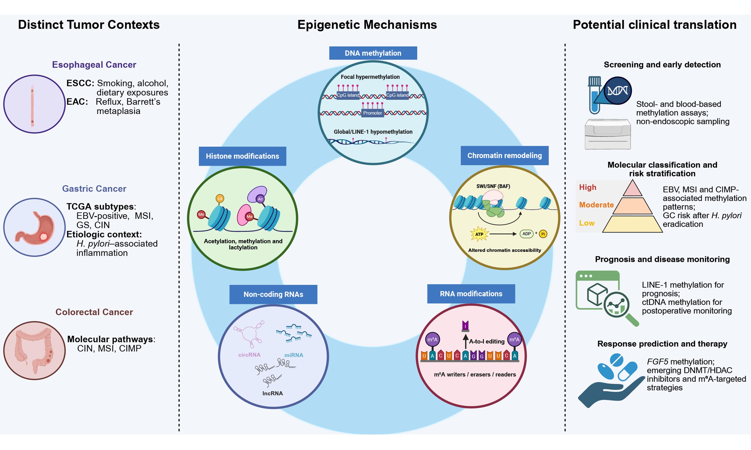
Gastrointestinal cancers mainly include esophageal cancer, gastric cancer, and colorectal cancer. These tumor types account for a huge number of new cases and deaths across the world. Although numerous genetic studies have shown the importance of gene mutations in their pathology, they are still insufficient to capture all layers of cancer development. Recently, epigenetic dysregulation has drawn attention because of its potential in the diagnosis and treatment of gastrointestinal cancers. Among diverse epigenetic pathways, DNA methylation is the best-studied and has displayed clinical value. Additionally, research on histone modifications, chromatin remodeling and RNA-mediated regulation is rapidly accumulating. In this review, we systematically illustrate the epigenetic dysregulation in gastrointestinal cancers to provide hints for clinical transformation.
Keywords
INTRODUCTION
Gastrointestinal cancers are among the cancer types with the highest incidence and mortality rates worldwide. According to the 2022 global cancer statistics (GLOBOCAN), esophageal cancer (EC), gastric cancer (GC), and colorectal cancer (CRC) rank as the most prevalent and lethal gastrointestinal malignancies, raising a major challenge to global public health[1,2]. Notably, gastrointestinal malignancies also encompass liver cancer, pancreatic cancer, and biliary tract cancer. However, in this review, the terms EC, GC, and CRC are used to refer to the gastrointestinal tract malignancies that share a common mucosal epithelial origin.
In 2022, there were 4.906 million new cases of gastrointestinal tumors, accounting for 24.6% of all new cancer cases worldwide[1]. Among them, CRC is the most common tumor, which ranks third in incidence and second in mortality globally, leading to approximately 1.9 million new cases and 0.9 million deaths[3]. Additionally, GC accounts for about 0.97 million new cases and 0.66 million deaths annually, ranking as the fifth most common cancer and the fourth leading cause of cancer death worldwide[4]. Meanwhile, EC has approximately 0.511 million new cases and 0.445 million deaths annually, with five-year survival rates as low as 20%[5]. Moreover, the disease burden of gastrointestinal tumors is expected to increase further by 2050, with approximately 9.06 million new cases and 6.42 million deaths annually, showing a rapidly increasing trend[6]. Strikingly, these tumors are especially prominent in Asia, and particularly in East Asia. There were approximately 0.517 million new CRC cases and 0.24 million CRC deaths reported in China in 2022[3]. Additionally, new cases and deaths of EC in China accounted for 44% and 42% of the global number, respectively[7].
To develop effective therapies for these gastrointestinal cancers, it is critical to understand their genetic basis. However, considerable evidence indicates that genetic models based solely on DNA sequence alterations cannot fully explain the initiation, progression, and heterogeneity of gastrointestinal tumors. Instead, epigenetic alterations function as another layer of cancer regulation. Epigenetic regulation refers to the mechanisms by which gene expression is modulated without altering DNA sequence. The main layers include DNA methylation, post-translational histone modifications, Adenosine Triphosphate (ATP)-dependent chromatin remodeling, and RNA-mediated regulatory mechanisms. Recent studies revealed that these mechanisms play important roles in gastrointestinal tumor development, shifting the research focus from a purely genetic perspective toward the broader field of epigenetics. This review introduces the epigenetic alterations associated with gastrointestinal cancers and discusses their potential applications in clinical diagnosis and treatment.
EPIGENETIC REGULATION SHAPES TUMOR BIOLOGY
Epigenetic regulation shapes transcriptional states and cell identity without changing the primary DNA sequence. Epigenetic dysregulation is associated with various diseases, such as osteoporosis[8], nervous system diseases[9], and cancers[10]. Importantly, these changes are often mitotically heritable, but many are also potentially reversible. This reversibility forms the basis for their potential in cancer treatment. While genetic lesions classically initiate oncogenesis by activating oncogenes or repressing tumor suppressors, the epigenetic landscape contributes to tumorigenesis either dependent or independent of genetic alteration. Additionally, epigenetic dysregulation also impacts how malignant cells acquire lineage plasticity, evade microenvironmental stress, and resist therapeutic pressure. Importantly, these epigenetic mechanisms rarely act independently. Instead, changes in one epigenetic layer often influence other layers, collectively shaping cancer-associated transcriptional states. In gastrointestinal malignancies, this epigenetic network operates within tissues impacted by epithelial renewal, inflammation, microbiota, diet-related exposures, and toxic injury, resulting in epithelial-mesenchymal transition (EMT), metastatic dissemination, and therapy resistance[11,12].
DNA methylation is the most extensively characterized epigenetic layer in cancer biology. In malignant transformation, the methylation landscape is usually disrupted in two directions. On the one hand, promoter CpG island hypermethylation can silence tumor suppressor genes, DNA repair components, and differentiation programs. On the other hand, global DNA hypomethylation can destabilize repetitive elements and pericentromeric regions, contributing to chromosomal instability, structural variation, and aberrant activation of normally silenced retrotransposons. The clinical relevance of these changes is clearest in selected settings. For instance, MLH1 promoter hypermethylation is a major mechanism of mismatch-repair deficiency and microsatellite instability in sporadic CRC[13]. Also, MGMT promoter hypermethylation impairs DNA repair and is associated with benefit from temozolomide in glioblastoma[14]. These examples explain why DNA methylation is useful both as a disease mechanism and as a stratification marker. Therefore, DNA methylation should not be regarded as a uniform process, since focal and global methylation changes can have different biological consequences.
Histone modifications impact chromatin condensation and recruit effector proteins that determine whether regulatory elements are active, poised, or repressed. Active promoters are often marked by H3K4me3, while polycomb-repressed regions are commonly associated with H3K27me3[15]. Cancer studies have shown that this layer is not merely correlative. Recurrent EZH2 Tyr641 mutations in germinal-center-derived follicular lymphoma and diffuse large B-cell lymphoma alter Polycomb Repressive Complex 2 (PRC2) activity. As a result, H3K27me3-associated chromatin states and malignant differentiation programs are reshaped[16].
Histone modifications also interact with ATP-dependent chromatin remodeling complexes. The SWItch/Sucrose NonFermentable (SWI/SNF) (also called BAF) machinery can slide, evict, or restructure nucleosomes and change transcription-factor access to enhancers and promoters. Inactivation of core subunits can therefore have broad regulatory effects. For example, frequent ARID1A mutations in ovarian clear cell carcinoma rewire enhancer landscapes, disrupt three-dimensional regulatory loops, and allow malignant cells to change lineage identity or acquire phenotypic plasticity[17,18].
Non-coding RNAs add another route through which epigenetic programs are regulated. Long non-coding RNAs (lncRNAs) can act as molecular scaffolds that guide chromatin regulators to specific genomic loci[19]. Their dysregulation is associated with tumor development, metastasis, and drug resistance. HOTAIR is a well-known example that recruits PRC2, reprograms the chromatin state, and promotes metastatic dissemination in breast cancer[20]. Other RNA classes act through different mechanisms. MicroRNAs fine-tune oncogenic or tumor-suppressive networks after transcription, whereas circular RNAs can sequester microRNAs or modulate nuclear protein interactions[21].
Covalent RNA modifications, especially N6-methyladenosine (m6A), can influence transcript stability, processing, and translation efficiency. In acute myeloid leukemia, promoter-bound METTL3 deposits m6A marks on oncogenic transcripts and enhances their translation[10]. This example illustrates how RNA-centered regulation can cooperate with chromatin states to sustain malignant phenotypes. Some epigenetic regulators may create druggable vulnerabilities in selected tumor contexts given their roles as enzymes or specialized protein-interaction hubs. Overall, epigenetic pathways synergistically impact tumor progression, plasticity, immune evasion and therapeutic resistance. Figure 1 summarizes the major epigenetic mechanisms and their functional links to tumor progression, plasticity, immune evasion and therapeutic resistance.
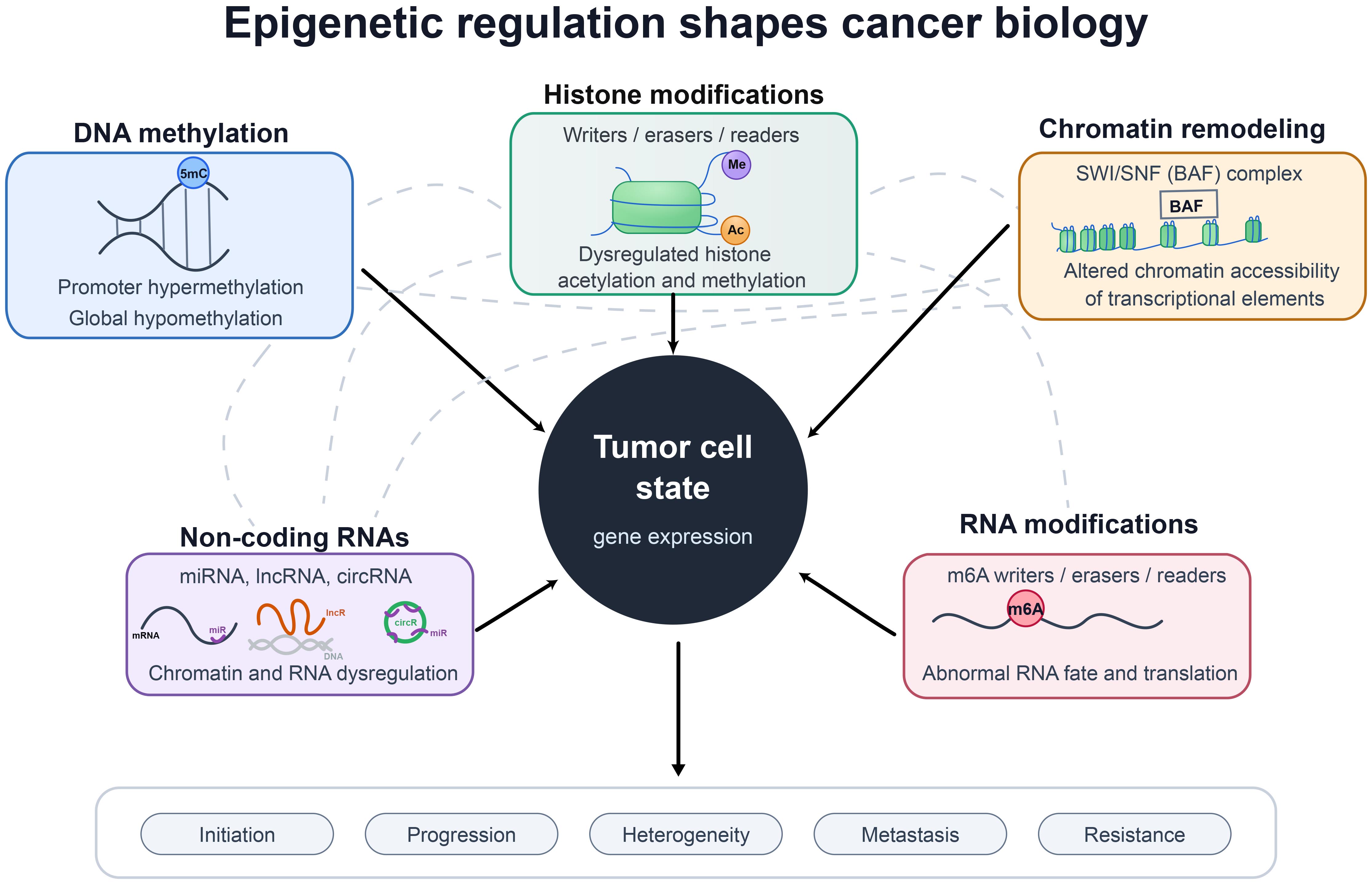
Figure 1. Epigenetic dysregulation shapes tumor biology. Five major epigenetic layers (DNA methylation, histone modifications, chromatin remodeling, non-coding RNAs, and RNA modifications) collectively influence tumor cell state and gene expression. Solid arrows indicate direct contributions to clinical trajectories (e.g., progression, metastasis, and resistance). Gray dashed lines highlight the extensive cross-talk among these layers, illustrating how alterations in one mechanism actively reshape others to drive oncogenesis. miRNA: MicroRNA; lncRNA: long non-coding RNA; circRNA: circular RNA; BAF: BRG-/BRM-associated factor.
EPIGENETIC REGULATION IN ESOPHAGEAL CANCER
Esophageal mucosa is repeatedly exposed to mechanical trauma, dietary carcinogens, microbial populations, and chronic inflammatory reflux. Therefore, EC is a useful setting for examining how environmental injury interacts with epigenetic reprogramming. EC includes two major histological and etiological subtypes: esophageal squamous cell carcinoma (ESCC) and esophageal adenocarcinoma (EAC). They differ in epidemiology, risk factors, precursor lesions, and molecular background[22]. They share overlapping classes of epigenetic machinery, but within distinct cellular and developmental trajectories.
DNA methylation abnormalities and biomarkers in ESCC
In ESCC, aberrant DNA methylation is the most mature epigenetic hallmark. Large-scale epigenomic studies have identified widespread methylation alterations that correlate with patient outcomes[23-25]. Genome-wide hypomethylation often affects repetitive elements, including long interspersed nucleotide sequences (LINE-1). This pattern has been associated with chromosomal instability and poor prognosis after curative tumor resection[24,26]. Moreover, global methylation loss coexists with localized promoter CpG island hypermethylation, which can silence regulatory loci. One example is hypermethylation-mediated silencing of the tumor suppressor SEMA3B and its antisense lncRNA SEMA3B-AS1, which has been associated with ESCC progression and unfavorable survival[27].In addition, multi-region methylation profiling has revealed both intertumor and intratumor methylation heterogeneity in ESCC. This heterogeneity correlates with lymph node metastasis, suggesting that subclonal epigenetic diversification may accompany selection of more aggressive tumor-cell populations[28].
Research on DNA methylation biomarkers in ESCC is also expanding rapidly. For instance, CDH1 promoter hypermethylation is observed in 43% of ESCC patients and is associated with recurrence of early-stage ESCC[29]. Also, the association between ESCC and promoter hypermethylation of DNA damage repair genes, such as MGMT and MLH1, has been well studied[30,31]. Additionally, targeted bisulfite sequencing has generated multi-gene methylation panels for ESCC detection and identified several genomic regions that were able to act as biomarkers, including regions covering STK3 and ZNF418[32]. Locus-specific hypermethylation, including FGF5 promoter methylation, has been reported as a candidate marker of sensitivity to definitive chemoradiotherapy[33]. These findings highlight their clinical potential, but careful validation across independent cohorts, sample types, and treatment settings remains necessary.
Histone modification, chromatin remodeling, and RNA-mediated regulation in ESCC
Histone-modifying enzymes and structural chromatin states provide further mechanistic insight into ESCC pathogenesis. The available evidence is more pathway-specific than that for DNA methylation, but several studies support its functional relevance. The histone demethylase JMJD3 promotes ESCC pathogenesis through epigenetic regulation of MYC proto-oncogene (MYC)[34]. Histone modifiers can also cooperate with transcriptional regulators to repress expression of non-coding RNAs. For example, SOX4 recruits EZH2 and Histone deacetylase 3 (HDAC3) to the miR-31 promoter, thereby silencing miR-31 expression and promoting an invasive cellular phenotype[35]. In addition, super-enhancer remodeling contributes to oncogenic transcription in ESCC. Active H3K27 acetylation (H3K27ac) can activate the lncRNA CCAT1, which coordinates SPRY4 and HOXB13 expression, resulting in ESCC migration and proliferation[36].
At the architectural level, chromatin remodeling complexes appear to be disrupted early during squamous carcinogenesis. Somatic mutations in SWI/SNF complex subunits have been reported as early events in esophageal squamous carcinogenesis[37]. Furthermore, loss or downregulation of the core SWI/SNF subunit ARID1A is associated with infiltrative growth in established ESCC tumors[38]. These structural changes usually intersect with lineage-specific transcriptional programs. The squamous transcription factors TP63, SOX2, and KLF5 form a core regulatory circuitry that binds open, hyper-accessible chromatin regions and reshapes enhancer usage[39]. Chromatin accessibility profiling has also identified hyper-accessible enhancers and enhancer RNAs in ESCC cells, including a putative enhancer-derived enhancer RNA (eRNA) for Epidermal Growth Factor Receptor (EGFR)[40].
Evidence for non-coding RNAs and m6A RNA modifications in ESCC is growing, but it remains immature. Interestingly, some ncRNAs not only act as downstream effectors, but also influence chromatin-modifying complexes or DNA methylation directly. The lncRNA POU3F3, for example, promotes DNA methylation in ESCC cells[41]. Similarly, circIMMP2L promotes malignant progression by securing nuclear retention of the transcriptional corepressor CtBP1 and modulating downstream epigenetic modifications[42]. Moreover, m6A RNA modification extends another epigenetic layer to ESCC cell-state control. The key point is not simply the presence of m6A marks, but their dynamic deposition and removal on mRNA transcripts. The m6A demethylase ALKBH5 suppresses esophageal cancer malignancy by regulating primary microRNA biogenesis and RAI1 expression[43]. METTL14, a critical m6A methylase, participates in a positive feedback loop with miR-99a-5p and TRIB2 in radioresistant ESCC cell populations. This circuit recruits HDAC2, sustains a cancer stem cell phenotype, and promotes radioresistance[44]. These molecule-specific mechanisms broaden the biological view of ESCC plasticity. However, the breadth of clinical validation available for RNA methylation biomarkers remains inadequate.
Epigenetic alterations in EAC and Barrett’s-associated progression
EAC has a different epigenetic context from ESCC. Its epigenetic landscape is closely linked to the inflammatory and metaplastic trajectory of Barrett’s esophagus. In this glandular lineage, widespread DNA hypomethylation occurs early in the premalignant metaplastic stage. It may also synergize with focal gene amplification during progression toward adenocarcinoma[45]. Importantly, DNA methylation profiling has delineated molecular subclasses across Barrett’s esophagus and EAC[46]. This classification becomes more informative when methylation profiles are integrated with transcriptomic and genomic data[47]. One subtype-associated event is hypermethylation-mediated downregulation of NDRG4, a tumor-suppressive gene in EAC[48]. Meanwhile, chromatin-level studies suggest that global accessibility shifts can establish a hybrid stomach-intestinal chromatin state that underlies metaplastic cellular reprogramming in human Barrett’s tissue[49].
Overall, DNA methylation and localized chromatin states provide the strongest epigenetic evidence base for EAC progression. Direct evidence for EAC-specific histone modifiers, lncRNA scaffolds, and m6A RNA-modification pathways remains comparatively sparse. This imbalance matters for interpretation. ESCC and EAC share broad epigenetic mechanisms, but they deploy them within distinct lineage and developmental contexts. This distinct lineage context underscores the need to evaluate epigenetic mechanisms in ESCC and EAC independently, despite their shared molecular machinery.
Clinical translation of epigenetic markers in esophageal cancer treatment
Clinical translation remains limited and differs by histological subtype. No epigenetic biomarker or epigenetic drug is established in the routine management of ESCC. The DNA methylation panels, LINE-1 methylation, and FGF5 methylation remain candidate tissue markers that require prospective assay validation. In Barrett’s esophagus and EAC, non-endoscopic cell collection coupled to methylated VIM and CCNA1 testing has progressed. The EsoGuard assay has been analytically validated as a CLIA/CAP laboratory-developed test using samples collected with the FDA-cleared EsoCheck device[50]. However, analytical validation alone does not establish population-level clinical utility. The 2022 American College of Gastroenterology (ACG) guideline found insufficient evidence to recommend routine biomarker panels. Therefore, positive molecular tests still require a confirmatory endoscopy[51]. In addition, azacitidine priming has entered early-phase evaluation in resectable gastric and esophageal adenocarcinoma. However, neither DNA methyltransferase nor histone deacetylase inhibitor is used as standard treatment for ESCC or EAC[52]. Future studies should prioritize prospective validation in well-defined risk populations, particularly for ESCC-specific methylation markers, where tumor heterogeneity and the lack of standardized assays remain major barriers to clinical implementation.
EPIGENETIC DYSREGULATION IN GASTRIC CANCER
GC remains a major cause of cancer morbidity and mortality worldwide. Its development reflects long-standing mucosal injury, exposure to microbes and viruses, genetic changes that are either inherited or acquired, and changes in the regulation of chromatin and RNA[53,54]. Epigenetic alterations are especially relevant in the stomach because they are not confined to established tumors. They can be detected in non-neoplastic mucosa and can accumulate during metaplasia and dysplasia, thus helping define tumor subgroups. In the Cancer Genome Atlas Program (TCGA) classification, GC is divided into Epstein-Barr virus (EBV)-positive, microsatellite instability (MSI), genomically stable, and chromosomal instability subtypes. Among them, EBV-positive tumors show extensive CpG island hypermethylation, whereas MSI tumors often involve methylation-associated mismatch-repair deficiency[55].
DNA methylation abnormalities in GC
DNA methylation is the best-characterized epigenetic alteration in GC. Two broad patterns are repeatedly described: focal CpG island promoter hypermethylation and global hypomethylation. As observed in EC, promoter hypermethylation can silence tumor-suppressive or DNA-repair genes, whereas global hypomethylation often affects repetitive elements and may contribute to chromosomal instability and retrotransposon activity[56-58]. Notably, genome-wide methylation studies have identified methylation-defined GC subgroups, indicating that DNA methylation functions both as a gene-specific regulatory event and as a tumor-classification feature[55,59].
The EBV-positive and MSI subtypes illustrate two clinically recognizable methylation patterns. EBV-positive GC shows extensive CpG island hypermethylation, a feature that helps distinguish it from other molecular subtypes[55]. In MSI tumors, promoter methylation of mismatch-repair genes, particularly MLH1, leads to mismatch-repair deficiency and microsatellite instability[55]. These subtype-linked methylation patterns are useful because they link epigenetic alterations to established molecular classifications rather than treating GC as a single disease.
Helicobacter pylori infection provides another gastric cancer-specific link between inflammation and DNA methylation. Aberrant methylation can be detected in H. pylori-infected noncancerous gastric mucosa, supporting an epigenetic defect model in which histologically non-malignant tissue already carries cancer-associated methylation changes[60]. This effect has practical implications. Methylation-based markers have been tested to stratify the risk of primary GC after H. pylori eradication, including in high-risk individuals who remain under endoscopic surveillance[61]. This epigenetic defect model separates GC-specific DNA methylation biology from a purely tumor-centered view. The methylation signal in noncancerous mucosa may reflect previous inflammatory injury, persistent epithelial remodeling, or selection of altered epithelial clones. This explains why risk remains measurable after H. pylori clearance: although eradication removes the inflammatory stimulus, it does not necessarily erase the accumulated methylation changes[60,61].
At the gene level, promoter hypermethylation has been reported in cell-cycle regulators, DNA-repair genes, adhesion-related genes, and apoptosis-associated genes, including CDKN2A/p16INK4a, MLH1, MGMT, CDH1, and CASP8[62-66]. LINE-1 hypomethylation in upper gastrointestinal cancers has been associated with increased retrotransposition and tumor-specific insertions, providing a plausible connection between global hypomethylation and genomic instability[56]. In GC models, DNA hypomethylation can also promote invasive behavior by regulating SP1 binding at the CDCA3 promoter[67]. Taken together, abnormal DNA methylation patterns in GC and noncancerous gastric mucosa shed light on its clinical applications.
Histone modification and chromatin remodeling in GC
Compared with DNA methylation, research on histone modifications in GC has focused on specific mechanisms. For instance, EZH2, the catalytic subunit of PRC2, mediates H3K27 trimethylation and drives gastric tumorigenesis by silencing tumor suppressor genes[68-71]. Furthermore, EZH2 can interact with the DNA methylation machinery, suggesting that polycomb-mediated repression and promoter methylation often cooperate to silence target genes[69].
Other chromatin regulators appear to act in more restricted settings. Inhibition of the lysine demethylase KDM4C induces senescence in TP53-mutant GC cells, suggesting that dependency on histone demethylases may vary by genetic background[72]. Chromatin remodeling defects show a similar subtype bias. The BAF subunit ARID1A is frequently altered in subsets of GC, especially MSI and EBV-associated tumors, and TCGA identified ARID1A among the most recurrent chromatin-regulatory alterations[55,73]. These findings indicate that chromatin remodeling in GC is highly subtype-specific, rather than a general process across all tumors.
Non-coding RNAs, RNA editing and m6A RNA modification in GC
Non-coding RNA studies in GC cover many molecules and phenotypes, so the evidence is less uniform than that for DNA methylation. Several miRNAs have been linked to drug response, tumor progression, or biomarker potential. For example, miR-27a has been associated with HIF-1α-related multidrug resistance, whereas miR-215/192 and miR-148a-3p have been reported in GC progression and biomarker studies[74-76]. LncRNAs and circRNAs have also been widely studied. For instance, HOTAIR is linked to cisplatin resistance and histone-mark switching during the epithelial-mesenchymal transition[77]. Additionally, MALAT1 promotes tumor progression via a miRNA-associated axis, while circAKT3 and circPVT1 are implicated in cisplatin resistance, proliferation, and prognostic stratification[78-81]. However, compared with DNA methylation alterations, evidence supporting ncRNA-based mechanisms in GC remains less consistent.
RNA editing provides a distinct post-transcriptional mechanism. ADAR-mediated A-to-I RNA editing is altered in GC and has been associated with disease progression and prognosis, with ADAR1 overexpression and ADAR2 downregulation contributing to an editing imbalance in tumor tissues[82]. CPEB3 has also been reported to suppress GC progression partly through regulation of ADAR1 mRNA localization and ADAR1-mediated RNA editing[83].
m6A RNA modification is an emerging area in GC research. METTL3 has been reported to promote GC proliferation, migration, epithelial-mesenchymal transition, and MYC-associated transcriptional programs[84,85]. The m6A reader YTHDF1 can promote gastric carcinogenesis by enhancing translation of FZD7 and activating Wnt/β-catenin signaling[86]. These findings support a role for m6A-mediated post-transcriptional control, but most data still come from mechanistic studies rather than prospective clinical cohorts.
Clinical translation of epigenetic markers in GC treatment
Clinical use of epigenetic information in GC remains indirect. MSI status is incorporated into clinical biomarker assessment and treatment selection, whereas EBV status is mainly used for molecular or pathological classification in selected settings. These features are generally assessed by immunohistochemistry, MSI testing, sequencing, or Epstein-Barr Virus Early RNA (EBER) in situ hybridization rather than by methylation assay[87]. The extensive CpG island hypermethylation of EBV-positive tumors and MLH1-associated methylation in MSI tumors are therefore biologically and taxonomically important but are not routine stand-alone clinical tests. RIMS1 methylation measured in non-neoplastic antral and body biopsies is undergoing prospective validation for risk stratification after H. pylori eradication[61]. However, external validation, assay standardization, and incorporation into surveillance algorithms are still required. Overall, DNA methylation currently represents the most clinically advanced epigenetic approach in GC, whereas histone modifications, ncRNAs, and m6A-related biomarkers remain largely investigational. Further prospective studies with standardized assays and clinically relevant endpoints are required before these emerging markers can be incorporated into routine clinical practice.
EPIGENETIC REGULATION IN COLORECTAL CANCER
CRC is probably the most well-documented gastrointestinal tumor. Based on its Consensus Molecular Subtypes (CMS), it is divided into four subtypes (CMS1-4). On the other hand, CRC pathogenesis is driven by three major molecular mechanisms: chromosomal instability (CIN, ~85% of cases), microsatellite instability (MSI, ~15% of cases), and the CpG island methylator phenotype (CIMP, ~20% of cases). Notably, CIMP-high tumors frequently overlap with MSI-high or Recombinant B-Raf Proto Oncogene Serine/Threonine Protein Kinase (BRAF)-mutant tumors. These pathway-based features are distinct from, yet complementary to, the CMS classification. The classic adenoma-carcinoma sequence model proposed by Fearon and Vogelstein emphasized the sequential accumulation of genetic mutations in APC, KRAS, and TP53[88]. However, genetic alterations alone cannot fully account for the heterogeneity, plasticity, and clinical behavior of CRC. Over the past two decades, growing evidence has demonstrated that epigenetic dysregulation plays a critical driving role in CRC initiation, progression, metastasis, and therapeutic resistance.
DNA methylation in CRC
Aberrant DNA methylation is a hallmark of CRC, manifesting both as genome-wide hypomethylation and as locus-specific hypermethylation of promoter CpG islands. The CIMP defines a distinct subset of CRCs characterized by simultaneous hypermethylation of multiple loci. In a large cohort analysis of 295 sporadic CRCs, Toyota and colleagues first identified CIMP-high tumors, accounting for approximately 20% of cases[89]. Subsequent studies have established the clinical relevance of CIMP. For instance, CIMP-high tumors are associated with BRAF mutations, proximal colon location, and high-level MSI[90]. In terms of prognosis, CIMP-high status generally predicts worse overall survival[91,92]. Notably, this prognostic effect is possibly context-dependent. Some studies reported that CIMP-high confers higher mortality among microsatellite-stable cancers, but not MSI-high cancers[93].
Beyond global CIMP status, hypermethylation of individual tumor suppressor gene promoters occurs early in colorectal tumorigenesis. A comprehensive analysis including colorectal adenomas and carcinomas showed that promoters of APC, p16/CDKN2A, and RASSF1A are already methylated in adenomas at frequencies comparable to those seen in carcinomas[94]. Even in aberrant crypt foci, the earliest histologically identifiable lesions, hypermethylation of APC can be detected and correlates with reduced gene expression[95]. Similarly to other gastrointestinal tumors, methylation of DNA repair gene promoters, including MGMT, MLH1, and MSH2, has been linked to CRC carcinogenesis[94,96]. Genome-wide methylation sequencing has further expanded the list of frequently methylated genes in CRC, including NDRG4, BMP3, MFAP2, APOL3, RNASEL, SFRP1, and SFRP2, all of which distinguish cancer from normal tissue with high sensitivity[97,98]. To clarify their clinical use, combining machine learning and integrated analysis of TCGA DNA methylation signatures with comorbidity patterns revealed that a three-gene panel of ADHFE1, ADAMTS5, and MIR129-2 achieved the best CRC classification performance among ten candidate genes[99]. In terms of prognosis, p16/CDKN2A promoter hypermethylation is present in about one-third of tumors and is associated with loss of p16 expression, thus correlating with shorter patient survival[95,100]. Collectively, these studies establish DNA methylation as an early and heterogeneous feature of colorectal tumorigenesis and provide the biological basis for molecular classification and biomarker development.
The clinical application of DNA methylation biomarkers has been extensively validated in non-invasive settings. For plasma-based screening, a prediction model including five blood leukocyte DNA methylation markers for detecting colorectal neoplasms reached excellent performance with an area under the curve (AUC) of 0.85, which can further improve to 0.89 by combining with a lifestyle risk score[101]. More recently, circulating tumor DNA (ctDNA) methylation haplotypes have emerged as powerful prognostic tools. The ColonES assay, developed by a multicenter study, detects methylation haplotypes across 191 genomic regions, predicting CRC with 86% sensitivity and advanced adenoma with 79% sensitivity[102]. Importantly, patients with high preoperative ctDNA methylation scores had significantly shorter relapse-free and overall survival[102]. Additionally, methylation of BCAT1 and IKZF1 in plasma ctDNA frequently occurred in CRC and rapidly reversed after surgical resection, thus can inform adequacy of surgical resection[103]. Using a single-tube multiplex qMSP assay for six methylation markers, Zhu and colleagues reported a specificity of 98.2% for CRC detection with sensitivity of 67.5 %[104]. Strikingly, highly methylated ctDNA levels were significantly associated with shorter relapse-free and overall survival[104]. Collectively, these findings suggest DNA methylation is critical to CRC generation and development and already displays remarkable potential in clinical diagnosis.
Histone modifications and chromatin accessibility in CRC: from initiation to therapy resistance
Although the roles of histone modifications in CRC are not as well developed as DNA methylation, their dysregulation contributes to every stage of CRC pathogenesis. Regarding tumor initiation, the H3K36 methyltransferase SETD2 acts as a tumor suppressor, whose mutation is frequent in CRC[105]. In a conditional knockout mouse model crossed with ApcMin/+ mice, SETD2 loss increased the number of intestinal polyps by 3.5-fold and promoted progression to adenocarcinoma[106]. For CRC progression, the H3K9 methyltransferase SUV39H2 is significantly elevated in tumor tissues, and its high expression correlates with distant metastasis and poor survival[107]. Conversely, the H3K9 demethylase JMJD2C is overexpressed in liver metastases compared to primary tumors[108]. Mechanistically, JMJD2C binds the MALAT1 promoter, reduces repressive H3K9me3 and H3K36me3 marks, and therefore upregulates MALAT1 expression, which in turn enhances β-catenin signaling and promotes metastasis in mouse models[108]. An emerging modification, histone lactylation, links metabolism to epigenetics. In KRAS-mutant CRC tissues, lactate accumulation drives histone H3 lysine 9 lactylation (H3K9la). ChIP-seq revealed that H3K9la is enriched at the GRAMD1A promoter, a cholesterol transporter gene. Knockdown of GRAMD1A suppresses proliferation and migration in KRAS-mutant cells, identifying H3K9la as a key mediator of KRAS-driven tumorigenesis[109].
Chemoresistance is a major clinical challenge, and histone acetylation plays a central role. HDAC3 mediates 5-fluorouracil resistance by directly deacetylating p21, thereby promoting its ubiquitination and degradation[110]. Treatment with the HDAC3 inhibitor restored p21 acetylation and stability and re-sensitized resistant cells to 5-FU[110]. Similarly, HDAC6 is overexpressed in chemoresistant CRC cells, and its selective inhibitor ACY1215 synergizes with chemotherapy to increase apoptosis and inhibit proliferation[111,112]. Besides HDACs, histone acetylase p300 is elevated in a substantial proportion of CRCs, acting as an oncogene through enhancing H3K27 acetylation and activating β-catenin[113].
Chromatin accessibility dynamics have been mapped at single-cell resolution. Using single-cell ATAC-seq on 48 polyps, 27 normal tissues, and 6 CRCs, researchers identified a continuum of chromatin remodeling from normal stem cells through polyps to cancer, characterized by progressive loss of differentiation-associated transcription factor motifs and gain of proliferation-associated motifs[114]. In the context of metastasis, CRC cells lose colon-specific accessible sites and instead gain accessibility at HNF4A binding sites[115]. Overall, histone modification and chromatin accessibility are critical epigenetic pathways in CRC, spanning tumorigenesis, proliferation, metastasis, and drug resistance. Although these findings indicate the clinical potential of therapy targeting histone modifications, there is a lack of solid evidence to support the application of enzyme- or pathway-specific inhibitors.
RNA-mediated regulation in CRC
LncRNAs and circRNAs function as key regulators of gene expression in CRC. HOTAIR is one of the most extensively studied lncRNAs in CRC. HOTAIR expression was higher in tumors from patients who developed liver metastasis, and its overexpression increased cell invasion significantly[116]. Mechanistically, HOTAIR binds to PRC2 and promotes H3K27me3-mediated silencing of metastasis suppressor genes[116]. Another lncRNA, MALAT1, is elevated in metastatic CRC and facilitates tumor progression by upregulating PI3K/Akt/mTOR and Wnt pathways[117,118]. In addition, circular RNAs, characterized by their covalently closed structure and high stability, often act as microRNA sponges. CircCCDC66 is upregulated more than 8-fold in CRC tissues; its knockdown reduces tumor proliferation, migration, and invasion[119]. In the context of drug resistance, hsa_circ_0020095 is markedly elevated in cisplatin-resistant CRC cells, and it sponges miR-487a-3p to upregulate SOX9, accelerating cell proliferation and drug resistance[120].
In terms of RNA modification, an integrative analysis of 583 CRC patients from TCGA and Gene Expression Omnibus (GEO) cohorts, examining 20 m6A regulators, identified two distinct clusters with significantly different overall survival[121]. Furthermore, m6A-associated gene signatures were constructed and validated as independent prognostic predictors[121]. Additionally, METTL3 is elevated in nearly two-thirds of tumors and predicts poor survival[122]. Mechanistically, METTL3 methylates the 3’UTR of MYC mRNA, increasing its stability and protein levels[123,124]. These findings support a role for m6A-associated RNA regulation in CRC biology, but therapeutic translation remains at an early stage.
Overall, epigenetic dysregulation is a driving force in colorectal cancer pathogenesis. DNA methylation signatures provide the most abundant insights in tumor pathology, serving as robust biomarkers for early detection, prognosis, and treatment prediction. Moreover, they have been applied in clinical settings with acceptable accuracy and sensitivity. However, more cautious validation across multiple centers and distinct molecular subtypes is still necessary. Histone modifications and chromatin accessibility orchestrate critical processes underlying tumor initiation, progression, metastasis, and acquired drug resistance. Importantly, although some histone modification-based therapies have shown potential in CRC clinical treatment, they still need large-scale validation in patients across tumor stages and subtypes before clinical implementation. Additionally, it is not entirely clear how individual pathways function and how they form a network in this process. Finally, ncRNAs, including HOTAIR, MALAT1, and circCCDC66, function as key regulators of chromatin modification and miRNA sponging. Emerging RNA modifications m6A add post-transcriptional control layers that influence prognosis and metabolism.
Clinical translation of epigenetic markers in CRC treatment
CRC has the most established clinical use of epigenetic testing among the cancers reviewed. For stool-based screening, multitarget DNA/Fecal Immunochemical Test (FIT) assays incorporate methylated DNA markers and are included among recommended screening strategies, while positive results require diagnostic colonoscopy[125,126]. For blood-based screening, the methylated SEPT9 test achieved ~75% sensitivity at 87% specificity for CRC detection[127]. Nonetheless, it has a restricted US regulatory indication for average-risk adults aged 50 years or older who have not completed other recommended screening, and it should not be presented as a replacement for first-line screening or colonoscopy[128]. In MLH1/PMS2-deficient tumors, MLH1 promoter methylation is used as a cascade test, along with BRAF V600E, to support a sporadic origin and triage germline evaluation for Lynch syndrome[129]. A cell-free DNA assay incorporating genomic and epigenomic signals received US approval in 2024, although its sensitivity for advanced precancerous lesions was substantially lower than for CRC[130,131]. By contrast, ctDNA methylation assays for postoperative monitoring and inhibitors of DNA methyltransferases or histone deacetylases still require validation. A phase II trial of pembrolizumab plus azacitidine showed limited activity in refractory metastatic CRC[132]. Overall, DNA methylation represents the most clinically mature epigenetic layer in CRC, with established applications in screening and molecular classification, whereas histone modifications, chromatin remodeling, ncRNAs, and RNA modifications remain primarily at the investigational stage.
CONTEXT-DEPENDENT EPIGENETIC DYSREGULATIONS IN GASTROINTESTINAL CANCERS
Esophageal, gastric, and colorectal cancers involve the same broad classes of epigenetic regulation, but the biological consequences of these alterations depend on tissue lineage, etiological exposure, and precursor state. Figure 2 illustrates this cross-cancer epigenetic framework. ESCC develops in a squamous lineage associated with smoking, alcohol, and dietary carcinogens, whereas EAC is linked to reflux, Barrett’s metaplasia, and glandular reprogramming. GC includes inflammation-associated methylation in non-neoplastic mucosa, distinct EBV-positive and MSI molecular subtypes. CRC develops through conventional adenoma-carcinoma pathways, and has the most established framework linking CIMP, mismatch-repair deficiency, and non-invasive molecular screening[55,88,133]. Table 1 compares the representative epigenetic patterns, cancer-specific contexts, and translational status across these settings.
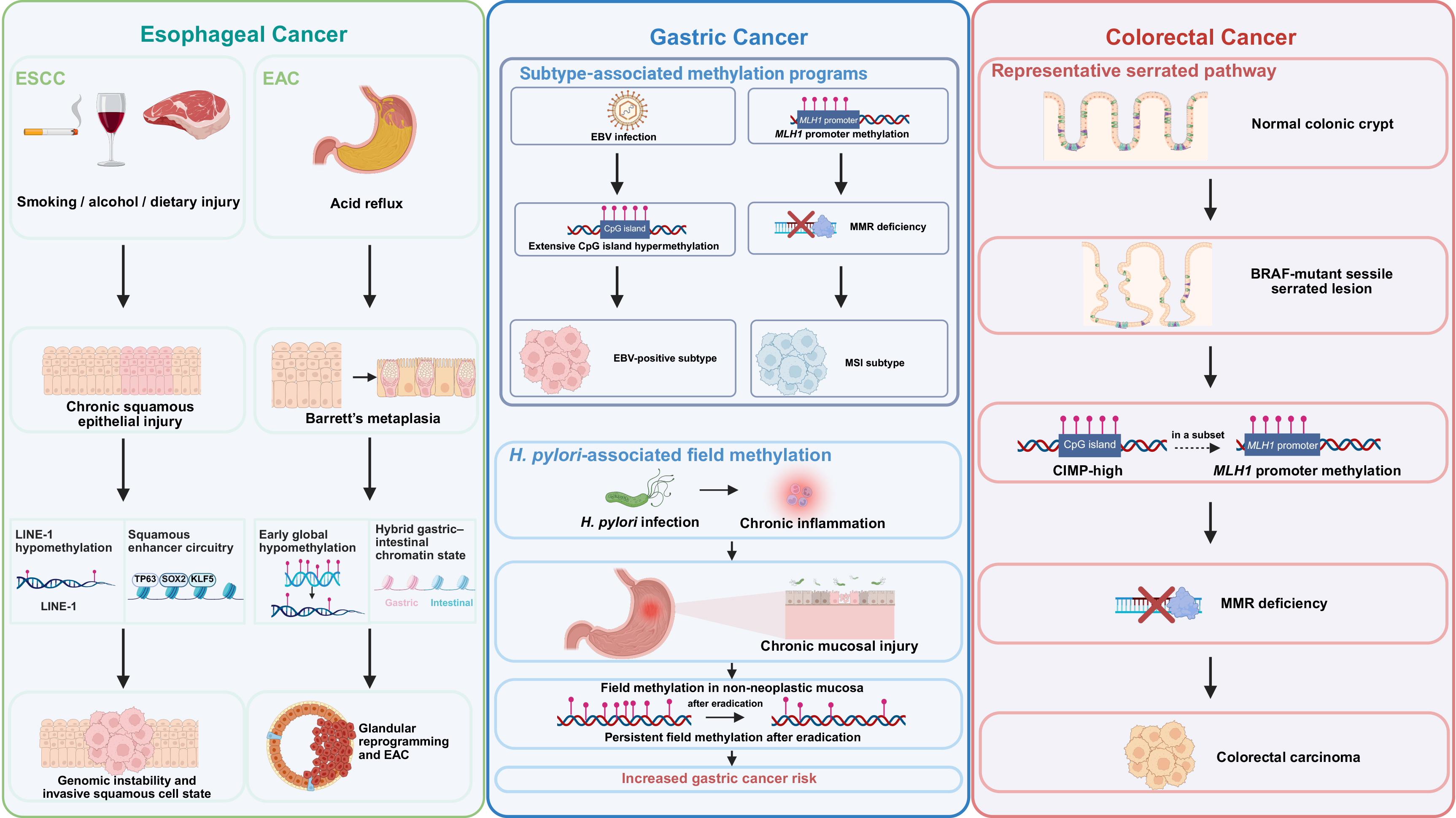
Figure 2. Tumor-specific epigenetic programs in gastrointestinal carcinogenesis. Representative epigenetic alterations are shown for ESCC, Barrett’s-associated EAC, gastric cancer and the colorectal serrated pathway. These include LINE-1 or global hypomethylation, lineage-specific enhancer and chromatin states, EBV- and Helicobacter pylori-associated methylation, CIMP-high, MLH1 promoter methylation and mismatch-repair deficiency. Created in BioRender.com. ESCC: Esophageal squamous cell carcinoma; EAC: esophageal adenocarcinoma; EBV: Epstein-Barr virus; MMR: DNA mismatch repair; CIMP:CpG island methylator phenotype.
Tumor-specific epigenetic patterns and translational status across esophageal, gastric, and colorectal cancers
| Tumor type or subtype | Representative epigenetic alteration | Cancer-specific context | Translational status | References |
| ESCC | Promoter CpG island hypermethylation panels, including STK3- and ZNF418-containing regions | Squamous lineage; exposure-associated carcinogenesis; substantial inter- and intratumor methylation heterogeneity | Retrospective biomarker evidence | Pu et al., 2017[32]; Talukdar et al., 2021[23] |
| ESCC | Global DNA hypomethylation and LINE-1 methylation loss | Genomic instability associated with squamous carcinogenesis; prognostic effects vary by cohort and disease context | Retrospective biomarker evidence | Iwagami et al., 2013[26]; Kawano et al., 2014[24] |
| Barrett's esophagus/EAC | Early global hypomethylation and methylation-defined molecular patterns | Reflux-Barrett’s-EAC sequence and glandular lineage reprogramming | Retrospective biomarker evidence | Alvarez et al., 2011[45]; Jammula et al., 2020[47] |
| EBV-positive and MSI gastric cancer | Subtype-associated CpG island hypermethylation and MLH1-associated methylation in MSI tumors | Virus- and mismatch-repair-associated molecular subtypes; distinct from CRC CIMP | Molecular classification | Cancer Genome Atlas Research Network, 2014[55]; Zouridis et al., 2012[59]; Ajani et al., 2025[87] |
| H. pylori-associated gastric cancer risk | Field methylation in non-neoplastic mucosa, including RIMS1 methylation | Persistent epimutation burden after inflammation-associated gastric injury and H. pylori eradication | Clinical validation | Maekita et al., 2006[60]; Yamada et al., 2025[61] |
| CRC, CIMP context | CIMP-associated promoter CpG island hypermethylation | Serrated pathway, BRAF mutation and MSI-associated tumor evolution; distinct from CMS classification | Molecular classification | Toyota et al., 1999[89]; Ogino et al., 2009[90]; Guinney et al., 2015[134] |
| CRC, MLH1/PMS2-deficient tumors | MLH1 promoter methylation | Distinguishes many sporadic MLH1-deficient tumors from cases requiring further Lynch syndrome evaluation | Clinical implementation | Kuismanen et al., 2000[13]; Aronson et al., 2025[129] |
| CRC screening | Multitarget stool DNA methylation markers combined with fecal hemoglobin | Non-invasive screening in average-risk populations | Clinical implementation | Imperiale et al., 2014[135]; Imperiale et al., 2024[125]; Davidson et al., 2021[126] |
| CRC screening, adults not completing recommended screening | Plasma methylated SEPT9 | Blood-based screening for a defined average-risk population that has not completed other recommended screening | Clinical implementation | Jin et al., 2015[127]; US Food and Drug Administration, 2016[128] |
| CRC blood-based screening | Combined cfDNA genomic and epigenomic signals, including methylation and fragmentation patterns | Average-risk population screening; performance differs between CRC and advanced precancerous lesions | Clinical implementation | Chung et al., 2024[130]; US Food and Drug Administration, 2024[131] |
| CRC postoperative monitoring | ctDNA methylation markers and methylation haplotypes | Post-resection molecular monitoring and recurrence-risk assessment | Clinical validation | Symonds et al., 2018[103]; Mo et al., 2023[102] |
DNA methylation illustrates why apparently similar epigenetic phenotypes should not be treated as equivalent across tumor types. In EBV-positive GC, extensive CpG island hypermethylation occurs in a virus-associated gastric epithelial context, whereas MLH1-associated methylation is more characteristic of the MSI GC subtype. CRC CIMP is instead commonly associated with the serrated pathway, BRAF mutation, proximal tumor location, MLH1 methylation, and MSI[55,89,90]. CRC CIMP may overlap with features enriched in CMS1, but it is not equivalent to the transcriptome-based CMS classification[134]. Thus, widespread CpG island hypermethylation represents a shared molecular pattern whose biological origin and clinical interpretation remain tumor- and subtype-specific. Similarly, the prognostic significance of global or LINE-1 hypomethylation is context-dependent. LINE-1 hypomethylation has been associated with poor outcome after curative resection in ESCC and with adverse prognosis in selected GC and CRC
Notably, the greater volume of HDAC-related research in CRC should also be interpreted as an asymmetry in the evidence base rather than as evidence that HDAC dysregulation is intrinsically more important in CRC. The reviewed CRC literature includes extensive preclinical work on fluoropyrimidine resistance and selective HDAC inhibition. By contrast, ESCC studies remain concentrated on specific regulatory circuits. This imbalance reflects differences in model availability, historical drug-development priorities, and the larger CRC literature on chemotherapy resistance.
Translational development is also uneven. CRC currently has the clearest clinical applications, including multitarget stool DNA/FIT screening and MLH1 promoter methylation testing. In Barrett’s esophagus and EAC, non-endoscopic methylation testing has progressed from analytically validated laboratory-developed testing to clinical evaluation, but it is not established as routine biomarker testing. In GC, RIMS1 methylation is under prospective evaluation for risk stratification after H. pylori eradication. But it still requires implementation-level validation. Histone modifiers, chromatin-remodeling defects, ncRNAs, RNA editing, and m6A regulators remain predominantly mechanistic, retrospective, or early-clinical-stage findings across the three cancers. Therefore, shared epigenetic mechanisms do not imply uniform biological or clinical effects, as their significance is shaped by lineage, etiological context, precursor state, molecular subtype, and the availability of clinically validated assays.
CONCLUSION
In summary, epigenetic dysregulation is a feature of and contributor to esophageal, gastric, and colorectal cancers. While aberrant DNA methylation yields the most clinically advanced biomarkers for early detection, prognosis, and treatment response across all three tumor types, implementation differs markedly by tumor type, and robust evidence remains limited for many proposed markers. However, the relative contribution of histone modifiers, chromatin remodelers, and RNA-mediated mechanisms varies by tumor subtype and anatomical context.
Looking forward, several priorities emerge in this field. First, functional dissection of epigenetic crosstalk within specific genetic backgrounds (e.g., KRAS-mutant CRC, EBV-positive gastric cancer) is necessary to identify context-dependent vulnerabilities. Second, single-cell multi-omic technologies will facilitate tracking epigenetic heterogeneity during therapy resistance and metastasis. Third, prospective validation of multi-omic epigenetic panels in large, well-annotated cohorts is urgently required. This is particularly important because many epigenetic alterations are dynamic and may also arise in inflamed, aging, or premalignant tissues, making it difficult to distinguish true cancer-specific signals from background epigenetic remodeling or field effects.
For clinical translation, blood- or stool-based assays that capture dynamic DNA methylation changes have potential for integration into screening programs and minimal residual disease monitoring, but proposed roles in the population still require indication-specific prospective validation. Moreover, epigenetic drugs [HDAC inhibitors, DNA methyltransferase (DNMT) inhibitors, and emerging m6A modulators] should be evaluated in rationally designed combination trials guided by tumor subtype and resistance mechanisms. Ultimately, incorporating epigenetic biomarkers into routine clinical workflows would enable precision risk stratification and real-time monitoring of treatment response across gastrointestinal cancers.
DECLARATIONS
Acknowledgments
We thank Andrew Hutchins for comments on the manuscript. The Graphical Abstract was created with BioRender.com.
Authors’ contributions
Designed the project and drafted the manuscript: Huang Z, Ma R
Drafted the manuscript: Xiao Z
Supervised and designed the study, acquired funding, and wrote the manuscript: Zhou X
All authors revised the manuscript.
Availability of data and materials
Not applicable.
AI and AI-assisted tools statement
During the preparation of this manuscript, the AI tool ChatGPT (version 5.6-sol, released 2026-06-26) was used solely for language editing. The tool did not influence the study design, data collection, analysis, interpretation, or the scientific content of the work. All authors take full responsibility for the accuracy, integrity, and final content of the manuscript.
Financial support and sponsorship
Funding was from the Shenzhen Innovation Committee of Science and Technology (RCBS20221008093109033 to Zhou X) and the Guangdong Basic and Applied Basic Research Foundation (2023A1515111170 to Zhou X).
Conflict of interest
All authors declared that there are no conflicts of interest.
Ethical approval and consent to participate
Not applicable.
Consent for publication
Not applicable.
Copyright
© The Author(s) 2026.
REFERENCES
1. Bray F, Laversanne M, Sung H, et al. Global cancer statistics 2022: GLOBOCAN estimates of incidence and mortality worldwide for 36 cancers in 185 countries. CA Cancer J Clin. 2024;74:229-63.
2. Singh A. Global burden of five major types of gastrointestinal cancer. Prz Gastroenterol. 2024;19:236-54.
3. Wu S, Zhang Y, Lin Z, Wei M. Global burden of colorectal cancer in 2022 and projections to 2050: incidence and mortality estimates from GLOBOCAN. BMC Cancer. 2025;25:1770.
4. Grantham T, Ramachandran R, Parvataneni S, Budh D, Gollapalli S, Gaduputi V. Epidemiology of gastric cancer: global trends, risk factors and premalignant conditions. J Community Hosp Intern Med Perspect. 2023;13:100-6.
5. Sheikh M, Roshandel G, McCormack V, Malekzadeh R. Current status and future prospects for esophageal cancer. Cancers. 2023;15:765.
6. Danpanichkul P, Pang Y, Tothanarungroj P, et al. Gastrointestinal cancer statistics in 2022 and projection to 2050: GLOBOCAN estimates across 185 countries. Cancer. 2026;132:e70245.
7. Qi L, Sun M, Liu W, et al. Global esophageal cancer epidemiology in 2022 and predictions for 2050: a comprehensive analysis and projections based on GLOBOCAN data. Chin Med J. 2024;137:3108-16.
8. Reppe S, Noer A, Grimholt RM, et al. Methylation of bone SOST, its mRNA, and serum sclerostin levels correlate strongly with fracture risk in postmenopausal women. J Bone Miner Res. 2015;30:249-56.
9. Karayol R, Borroto MC, Haghshenas S, et al. MSL2 variants lead to a neurodevelopmental syndrome with lack of coordination, epilepsy, specific dysmorphisms, and a distinct episignature. Am J Hum Genet. 2024;111:1330-51.
10. Barbieri I, Tzelepis K, Pandolfini L, et al. Promoter-bound METTL3 maintains myeloid leukaemia by m6A-dependent translation control. Nature. 2017;552:126-31.
11. Quante M, Varga J, Wang TC, Greten FR. The gastrointestinal tumor microenvironment. Gastroenterology. 2013;145:63-78.
12. Tan T, Shi P, Abbas MN, et al. Epigenetic modification regulates tumor progression and metastasis through EMT (Review). Int J Oncol. 2022;60:70.
13. Kuismanen SA, Holmberg MT, Salovaara R, de la Chapelle A, Peltomäki P. Genetic and epigenetic modification of MLH1 accounts for a major share of microsatellite-unstable colorectal cancers. Am J Pathol. 2000;156:1773-9.
14. Hegi ME, Diserens AC, Gorlia T, et al. MGMT gene silencing and benefit from temozolomide in glioblastoma. N Engl J Med. 2005;352:997-1003.
15. Hahn MA, Li AX, Wu X, et al. Loss of the polycomb mark from bivalent promoters leads to activation of cancer-promoting genes in colorectal tumors. Cancer Res. 2014;74:3617-29.
16. Morin RD, Johnson NA, Severson TM, et al. Somatic mutations altering EZH2 (Tyr641) in follicular and diffuse large B-cell lymphomas of germinal-center origin. Nat Genet. 2010;42:181-5.
17. Jones S, Wang TL, Shih IeM, et al. Frequent mutations of chromatin remodeling gene ARID1A in ovarian clear cell carcinoma. Science. 2010;330:228-31.
19. Tsai MC, Manor O, Wan Y, et al. Long noncoding RNA as modular scaffold of histone modification complexes. Science. 2010;329:689-93.
20. Gupta RA, Shah N, Wang KC, et al. Long non-coding RNA HOTAIR reprograms chromatin state to promote cancer metastasis. Nature. 2010;464:1071-6.
21. Sawamura H, Taniguchi K, Ikeda Y, Tsuji A, Kitagishi Y, Matsuda S. Roles of gut dysbiosis, anti-proliferative proteins, and post-transcriptional regulation in carcinogenesis. J Transl Genet Genom. 2022;6:157-68.
22. Genome Atlas Research Network. Integrated genomic characterization of oesophageal carcinoma. Nature. 2017;541:169.
23. Talukdar FR, Soares Lima SC, Khoueiry R, et al. Genome-wide DNA methylation profiling of esophageal squamous cell carcinoma from global high-incidence regions identifies crucial genes and potential cancer markers. Cancer Res. 2021;81:2612-24.
24. Kawano H, Saeki H, Kitao H, et al. Chromosomal instability associated with global DNA hypomethylation is associated with the initiation and progression of esophageal squamous cell carcinoma. Ann Surg Oncol. 2014;21 Suppl 4:S696-702.
25. Hoshimoto S, Takeuchi H, Ono S, et al. Genome-wide hypomethylation and specific tumor-related gene hypermethylation are associated with esophageal squamous cell carcinoma outcome. J Thorac Oncol. 2015;10:509-17.
26. Iwagami S, Baba Y, Watanabe M, et al. LINE-1 hypomethylation is associated with a poor prognosis among patients with curatively resected esophageal squamous cell carcinoma. Ann Surg. 2013;257:449-55.
27. Dong Z, Liang X, Wu X, et al. Promoter hypermethylation-mediated downregulation of tumor suppressor gene SEMA3B and lncRNA SEMA3B-AS1 correlates with progression and prognosis of esophageal squamous cell carcinoma. Clin Exp Metastasis. 2019;36:225-41.
28. Teng H, Xue M, Liang J, et al. Inter- and intratumor DNA methylation heterogeneity associated with lymph node metastasis and prognosis of esophageal squamous cell carcinoma. Theranostics. 2020;10:3035-48.
29. Lee EJ, Lee BB, Han J, et al. CpG island hypermethylation of E-cadherin (CDH1) and integrin alpha4 is associated with recurrence of early stage esophageal squamous cell carcinoma. Int J Cancer. 2008;123:2073-9.
30. Ishii T, Murakami J, Notohara K, et al. Oesophageal squamous cell carcinoma may develop within a background of accumulating DNA methylation in normal and dysplastic mucosa. Gut. 2007;56:13-9.
31. Roth M, Abnet C, Hu N, et al. p16, MGMT, RARβ2, CLDN3, CRBP and MT1G gene methylation in esophageal squamous cell carcinoma and its precursor lesions. Oncol Rep. 2006; 15:1591-7.
32. Pu W, Wang C, Chen S, et al. Targeted bisulfite sequencing identified a panel of DNA methylation-based biomarkers for esophageal squamous cell carcinoma (ESCC). Clin Epigenetics. 2017;9:129.
33. Iwabu J, Yamashita S, Takeshima H, et al. FGF5 methylation is a sensitivity marker of esophageal squamous cell carcinoma to definitive chemoradiotherapy. Sci Rep. 2019;9:13347.
34. Li SM, He LR, Chen JW, et al. JMJD3 promotes esophageal squamous cell carcinoma pathogenesis through epigenetic regulation of MYC. Signal Transduct Target Ther. 2020;5:165.
35. Koumangoye RB, Andl T, Taubenslag KJ, et al. SOX4 interacts with EZH2 and HDAC3 to suppress microRNA-31 in invasive esophageal cancer cells. Mol Cancer. 2015;14:24.
36. Zhang E, Han L, Yin D, et al. H3K27 acetylation activated-long non-coding RNA CCAT1 affects cell proliferation and migration by regulating SPRY4 and HOXB13 expression in esophageal squamous cell carcinoma. Nucleic Acids Res. 2017;45:3086-101.
37. Nakazato H, Takeshima H, Kishino T, et al. Early-stage induction of SWI/SNF mutations during esophageal squamous cell carcinogenesis. PLoS One. 2016;11:e0147372.
38. Ozawa Y, Nakamura Y, Fujishima F, et al. Decreased expression of ARID1A contributes to infiltrative growth of esophageal squamous cell carcinoma. Tohoku J Exp Med. 2015;235:185-91.
39. Jiang YY, Jiang Y, Li CQ, et al. TP63, SOX2, and KLF5 establish a core regulatory circuitry that controls epigenetic and transcription patterns in esophageal squamous cell carcinoma cell lines. Gastroenterology. 2020;159:1311-1327.e19.
40. Choi S, Sathe A, Mathé E, Xing C, Pan Z. Identification of a putative enhancer RNA for EGFR in hyper-accessible regions in esophageal squamous cell carcinoma cells by analysis of chromatin accessibility landscapes. Front Oncol. 2021;11:724687.
41. Li W, Zheng J, Deng J, et al. Increased levels of the long intergenic non-protein coding RNA POU3F3 promote DNA methylation in esophageal squamous cell carcinoma cells. Gastroenterology. 2014;146:1714-26.e5.
42. Liang Y, Mao Q, Wang L, et al. CircIMMP2L promotes esophageal squamous cell carcinoma malignant progression via CtBP1 nuclear retention dependent epigenetic modification. Clin Transl Med. 2021;11:e519.
43. Chen P, Li S, Zhang K, et al. N6-methyladenosine demethylase ALKBH5 suppresses malignancy of esophageal cancer by regulating microRNA biogenesis and RAI1 expression. Oncogene. 2021;40:5600-12.
44. Liu Z, Wu K, Gu S, et al. A methyltransferase-like 14/miR-99a-5p/tribble 2 positive feedback circuit promotes cancer stem cell persistence and radioresistance via histone deacetylase 2-mediated epigenetic modulation in esophageal squamous cell carcinoma. Clin Transl Med. 2021;11:e545.
45. Alvarez H, Opalinska J, Zhou L, et al. Widespread hypomethylation occurs early and synergizes with gene amplification during esophageal carcinogenesis. PLoS Genet. 2011;7:e1001356.
46. Kaz AM, Wong CJ, Luo Y, et al. DNA methylation profiling in Barrett’s esophagus and esophageal adenocarcinoma reveals unique methylation signatures and molecular subclasses. Epigenetics. 2011;6:1403-12.
47. Jammula S, Katz-Summercorn AC, Li X, et al.; Oesophageal Cancer Clinical and Molecular Stratification (OCCAMS) consortium. Identification of subtypes of Barrett’s esophagus and esophageal adenocarcinoma based on DNA methylation profiles and integration of transcriptome and genome data. Gastroenterology. 2020;158:1682-1697.e1.
48. Cao L, Hu T, Lu H, Peng D. N-MYC downstream regulated gene 4 (NDRG4), a frequent downregulated gene through DNA hypermethylation, plays a tumor suppressive role in esophageal adenocarcinoma. Cancers. 2020;12:2573.
49. Singh H, Ha K, Hornick JL, et al. Hybrid stomach-intestinal chromatin states underlie human Barrett’s metaplasia. Gastroenterology. 2021;161:924-939.e11.
50. Ghosal A, Verma S, Le IT, Lee VT, de Guzman BJ, Aklog L. Analytical validation of a DNA methylation biomarker test for the diagnosis of Barrett’s esophagus and esophageal adenocarcinoma from samples collected using EsoCheck®, a non-endoscopic esophageal cell collection device. Diagnostics. 2024;14:1784.
51. Shaheen NJ, Falk GW, Iyer PG, et al. Diagnosis and management of Barrett’s esophagus: an updated ACG guideline. Am J Gastroenterol. 2022;117:559-87.
52. Schneider BJ, Shah MA, Klute K, et al. Phase I study of epigenetic priming with azacitidine prior to standard neoadjuvant chemotherapy for patients with resectable gastric and esophageal adenocarcinoma: evidence of tumor hypomethylation as an indicator of major histopathologic response. Clin Cancer Res. 2017;23:2673-80.
53. Zhao S, Allis CD, Wang GG. The language of chromatin modification in human cancers. Nat Rev Cancer. 2021;21:413-30.
54. Jassim A, Rahrmann EP, Simons BD, Gilbertson RJ. Cancers make their own luck: theories of cancer origins. Nat Rev Cancer. 2023;23:710-24.
55. Genome Atlas Research Network. Comprehensive molecular characterization of gastric adenocarcinoma. Nature. 2014;513:202-9.
56. Baba Y, Yasuda N, Bundo M, et al. LINE-1 hypomethylation, increased retrotransposition and tumor-specific insertion in upper gastrointestinal cancer. Cancer Sci. 2024;115:247-56.
57. Lin R, Qian Y, Zhang J, et al. Genome-wide DNA methylation profiling of gastric cardia cancer. J Gastroenterol Hepatol. 2023;38:290-300.
58. Suzuki K, Suzuki I, Leodolter A, et al. Global DNA demethylation in gastrointestinal cancer is age dependent and precedes genomic damage. Cancer Cell. 2006;9:199-207.
59. Zouridis H, Deng N, Ivanova T, et al. Methylation subtypes and large-scale epigenetic alterations in gastric cancer. Sci Transl Med. 2012;4:156ra140.
60. Maekita T, Nakazawa K, Mihara M, et al. High levels of aberrant DNA methylation in Helicobacter pylori-infected gastric mucosae and its possible association with gastric cancer risk. Clin Cancer Res. 2006;12:989-95.
61. Yamada H, Abe S, Charvat H, et al. Precision risk stratification of primary gastric cancer after eradication of H. pylori by a DNA methylation marker: a multicentre prospective study. Gut. 2025;74:1410-8.
62. Azarkhazin F, Tehrani GA. Detecting promoter methylation pattern of apoptotic genes Apaf1 and Caspase8 in gastric carcinoma patients undergoing chemotherapy. J Gastrointest Oncol. 2018;9:295-302.
63. Park TJ, Han S, Cho Y, Paik WK, Kim YB, Lim IK. Methylation of O6-methylguanine-DNA methyltransferase gene is associated significantly with K-ras mutation, lymph node invasion, tumor staging, and disease free survival in patients with gastric carcinoma. Cancer. 2001;92:2760-8.
64. Oue N, Sentani K, Yokozaki H, Kitadai Y, Ito R, Yasui W. Promoter methylation status of the DNA repair genes hMLH1 and MGMT in gastric carcinoma and metaplastic mucosa. Pathobiology. 2001;69:143-9.
65. Kang GH, Lee S, Kim JS, Jung HY. Profile of aberrant CpG island methylation along the multistep pathway of gastric carcinogenesis. Lab Invest. 2003;83:635-41.
66. Song SH, Jong H, Choi HH, et al. Methylation of specific CpG sites in the promoter region could significantly down-regulate p16INK4a expression in gastric adenocarcinoma. Int J Cancer. 2000;87:236-40.
67. Yu J, Hua R, Zhang Y, Tao R, Wang Q, Ni Q. DNA hypomethylation promotes invasion and metastasis of gastric cancer cells by regulating the binding of SP1 to the CDCA3 promoter. J Cell Biochem. 2020;121:142-51.
68. Jie B, Weilong C, Ming C, et al. Enhancer of zeste homolog 2 depletion induces cellular senescence via histone demethylation along the INK4/ARF locus. Int J Biochem Cell Biol. 2015;65:104-12.
69. Rezaei S, Hosseinpourfeizi MA, Moaddab Y, Safaralizadeh R. Contribution of DNA methylation and EZH2 in SRBC down-regulation in gastric cancer. Mol Biol Rep. 2020;47:5721-7.
70. Fujii S, Ochiai A. Enhancer of zeste homolog 2 downregulates E-cadherin by mediating histone H3 methylation in gastric cancer cells. Cancer Sci. 2008;99:738-46.
71. Guo L, Yang TF, Liang SC, Guo JX, Wang Q. Role of EZH2 protein expression in gastric carcinogenesis among Asians: a meta-analysis. Tumour Biol. 2014;35:6649-56.
72. Wang K, Gong Z, Chen Y, et al. KDM4C-mediated senescence defense is a targetable vulnerability in gastric cancer harboring TP53 mutations. Clin Epigenetics. 2023;15:163.
73. Wang K, Kan J, Yuen ST, et al. Exome sequencing identifies frequent mutation of ARID1A in molecular subtypes of gastric cancer. Nat Genet. 2011;43:1219-23.
74. Bao C, Guo L. MicroRNA-148a-3p inhibits cancer progression and is a novel screening biomarker for gastric cancer. J Clin Lab Anal. 2020;34:e23454.
75. Xu YJ, Fan Y. MiR-215/192 participates in gastric cancer progression. Clin Transl Oncol. 2015;17:34-40.
76. Zhao Q, Li Y, Tan BB, Fan LQ, Yang PG, Tian Y. HIF-1α induces multidrug resistance in gastric cancer cells by inducing MiR-27a. PLoS One. 2015;10:e0132746.
77. Song Y, Wang R, Li LW, et al. Long non-coding RNA HOTAIR mediates the switching of histone H3 lysine 27 acetylation to methylation to promote epithelial-to-mesenchymal transition in gastric cancer. Int J Oncol. 2019;54:77-86.
78. Chen J, Li Y, Zheng Q, et al. Circular RNA profile identifies circPVT1 as a proliferative factor and prognostic marker in gastric cancer. Cancer Lett. 2017;388:208-19.
79. Huang X, Li Z, Zhang Q, et al. Circular RNA AKT3 upregulates PIK3R1 to enhance cisplatin resistance in gastric cancer via miR-198 suppression. Mol Cancer. 2019;18:71.
80. Li J, Gao J, Tian W, Li Y, Zhang J. Long non-coding RNA MALAT1 drives gastric cancer progression by regulating HMGB2 modulating the miR-1297. Cancer Cell Int. 2017;17:44.
81. Yan J, Dang Y, Liu S, Zhang Y, Zhang G. LncRNA HOTAIR promotes cisplatin resistance in gastric cancer by targeting miR-126 to activate the PI3K/AKT/MRP1 genes. Tumour Biol. 2016;37:16345-16355.
82. Chan TH, Qamra A, Tan KT, et al. ADAR-mediated RNA editing predicts progression and prognosis of gastric cancer. Gastroenterology. 2016;151:637-650.e10.
83. Chen J, Li L, Liu TY, et al. CPEB3 suppresses gastric cancer progression by inhibiting ADAR1-mediated RNA editing via localizing ADAR1 mRNA to P bodies. Oncogene. 2022;41:4591-605.
84. Liu T, Yang S, Sui J, et al. Dysregulated N6-methyladenosine methylation writer METTL3 contributes to the proliferation and migration of gastric cancer. J Cell Physiol. 2020;235:548-62.
85. Yang DD, Chen ZH, Yu K, et al. METTL3 promotes the progression of gastric cancer via targeting the MYC pathway. Front Oncol. 2020;10:115.
86. Pi J, Wang W, Ji M, et al. YTHDF1 promotes gastric carcinogenesis by controlling translation of FZD7. Cancer Res. 2021;81:2651-65.
87. Ajani JA, D'Amico TA, Bentrem DJ, et al. Gastric cancer, version 2.2025, NCCN clinical practice guidelines in oncology. J Natl Compr Canc Netw. 2025;23:169-91.
89. Toyota M, Ahuja N, Ohe-Toyota M, Herman JG, Baylin SB, Issa JP. CpG island methylator phenotype in colorectal cancer. Proc Natl Acad Sci U S A. 1999;96:8681-6.
90. Ogino S, Nosho K, Kirkner GJ, et al. CpG island methylator phenotype, microsatellite instability, BRAF mutation and clinical outcome in colon cancer. Gut. 2009;58:90-6.
91. Shiovitz S, Bertagnolli MM, Renfro LA, et al.; Alliance for clinical trials in oncology. CpG island methylator phenotype is associated with response to adjuvant irinotecan-based therapy for stage III colon cancer. Gastroenterology. 2014;147:637-45.
92. Kang KJ, Min BH, Ryu KJ, et al. The role of the CpG island methylator phenotype on survival outcome in colon cancer. Gut Liver. 2015;9:202-7.
93. Dahlin AM, Palmqvist R, Henriksson ML, et al. The role of the CpG island methylator phenotype in colorectal cancer prognosis depends on microsatellite instability screening status. Clin Cancer Res. 2010;16:1845-55.
94. Derks S, Postma C, Moerkerk PT, et al. Promoter methylation precedes chromosomal alterations in colorectal cancer development. Cell Oncol. 2006;28:247-57.
95. Chan AO, Broaddus RR, Houlihan PS, Issa JP, Hamilton SR, Rashid A. CpG island methylation in aberrant crypt foci of the colorectum. Am J Pathol. 2002;160:1823-30.
96. Lee KH, Lee JS, Nam JH, et al. Promoter methylation status of hMLH1, hMSH2, and MGMT genes in colorectal cancer associated with adenoma-carcinoma sequence. Langenbecks Arch Surg. 2011;396:1017-26.
97. Li JS, Riggins K, Yang L, et al. DNA methylation profiling at base-pair resolution reveals unique epigenetic features of early-onset colorectal cancer in underrepresented populations. Clin Epigenetics. 2025;17:11.
98. Park SK, Baek HL, Yu J, et al. Is methylation analysis of SFRP2, TFPI2, NDRG4, and BMP3 promoters suitable for colorectal cancer screening in the Korean population? Intest Res. 2017;15:495-501.
99. Tsai YH, Lai YH, Chen SJ, Cheng YC, Pai TW. DNA methylation biomarker discovery for colorectal cancer diagnosis assistance through integrated analysis. Cancer Inform. 2025;24:11769351251324545.
100. Mitomi H, Fukui N, Tanaka N, et al. Aberrant p16INK4a methylation is a frequent event in colorectal cancers: prognostic value and relation to mRNA expression and immunoreactivity. J Cancer Res Clin Oncol. 2010;136:323-31.
101. Li N, Luo C, Chen Y, et al. Identification and validation of blood leukocyte DNA methylation biomarkers for early detection of colorectal neoplasm. Chin Med J. 2026;139:2334-45.
102. Mo S, Dai W, Wang H, et al. Early detection and prognosis prediction for colorectal cancer by circulating tumour DNA methylation haplotypes: a multicentre cohort study. EClinicalMedicine. 2023;55:101717.
103. Symonds EL, Pedersen SK, Murray DH, et al. Circulating tumour DNA for monitoring colorectal cancer-a prospective cohort study to assess relationship to tissue methylation, cancer characteristics and surgical resection. Clin Epigenetics. 2018;10:63.
104. Zhu D, Li J, Zhang W, et al. Highly specific multiplex DNA methylation detection for liquid biopsy of colorectal cancer. Clin Chim Acta. 2025;565:120026.
105. Bushara O, Wester JR, Jacobsen D, et al. Clinical and histopathologic characterization of SETD2-mutated colorectal cancer. Hum Pathol. 2023;131:9-16.
106. Yuan H, Li N, Fu D, et al. Histone methyltransferase SETD2 modulates alternative splicing to inhibit intestinal tumorigenesis. J Clin Invest. 2017;127:3375-91.
107. Shuai W, Wu J, Chen S, et al. SUV39H2 promotes colorectal cancer proliferation and metastasis via tri-methylation of the SLIT1 promoter. Cancer Lett. 2018;422:56-69.
108. Wu X, Li R, Song Q, et al. JMJD2C promotes colorectal cancer metastasis via regulating histone methylation of MALAT1 promoter and enhancing β-catenin signaling pathway. J Exp Clin Cancer Res. 2019;38:435.
109. Zhang C, Yu R, Li S, et al. KRAS mutation increases histone H3 lysine 9 lactylation (H3K9la) to promote colorectal cancer progression by facilitating cholesterol transporter GRAMD1A expression. Cell Death Differ. 2025;32:2225-38.
110. Jin W, Wang J, Feng Y, Chen B, Hu Z. HDAC3-mediated deacetylation of p21 stabilizes protein levels and promotes 5-FU resistance in colorectal cancer cells. Adv Cancer Biol Metastasis. 2025;14:100136.
111. Tan Y, Zhang S, Zhu H, et al. Histone deacetylase 6 selective inhibitor ACY1215 inhibits cell proliferation and enhances the chemotherapeutic effect of 5-fluorouracil in HCT116 cells. Ann Transl Med. 2019;7:2.
112. Lee DH, Won HR, Ryu HW, Han JM, Kwon SH. The HDAC6 inhibitor ACY‑1215 enhances the anticancer activity of oxaliplatin in colorectal cancer cells. Int J Oncol. 2018;53:844-54.
113. Zhang Y, Wang S, Kang W, et al. CREPT facilitates colorectal cancer growth through inducing Wnt/β-catenin pathway by enhancing p300-mediated β-catenin acetylation. Oncogene. 2018;37:3485-500.
114. Becker WR, Nevins SA, Chen DC, et al. Single-cell analyses define a continuum of cell state and composition changes in the malignant transformation of polyps to colorectal cancer. Nat Genet. 2022;54:985-95.
115. Li S, Yang M, Teng S, et al. Chromatin accessibility dynamics in colorectal cancer liver metastasis: uncovering the liver tropism at single cell resolution. Pharmacol Res. 2023;195:106896.
116. Kogo R, Shimamura T, Mimori K, et al. Long noncoding RNA HOTAIR regulates polycomb-dependent chromatin modification and is associated with poor prognosis in colorectal cancers. Cancer Res. 2011;71:6320-6.
117. Zhang J, Li Q, Xue B, He R. MALAT1 inhibits the Wnt/β-catenin signaling pathway in colon cancer cells and affects cell proliferation and apoptosis. Bosn J Basic Med Sci. 2020;20:357-64.
118. Xu J, Xiao Y, Liu B, et al. Exosomal MALAT1 sponges miR-26a/26b to promote the invasion and metastasis of colorectal cancer via FUT4 enhanced fucosylation and PI3K/Akt pathway. J Exp Clin Cancer Res. 2020;39:54.
119. Hsiao KY, Lin YC, Gupta SK, et al. Noncoding effects of circular RNA CCDC66 promote colon cancer growth and metastasis. Cancer Res. 2017;77:2339-50.
120. Sun Y, Cao Z, Shan J, et al. Hsa_circ_0020095 promotes oncogenesis and cisplatin resistance in colon cancer by sponging miR-487a-3p and modulating SOX9. Front Cell Dev Biol. 2020;8:604869.
121. Sun W, Su Y, Zhang Z. Characterizing m6A modification factors and their interactions in colorectal cancer: implications for tumor subtypes and clinical outcomes. Discov Oncol. 2024;15:457.
122. Chen H, Gao S, Liu W, et al. RNA N6-methyladenosine methyltransferase METTL3 facilitates colorectal cancer by activating the m6A-GLUT1-mTORC1 axis and is a therapeutic target. Gastroenterology. 2021;160:1284-1300.e16.
123. Li T, Hu PS, Zuo Z, et al. METTL3 facilitates tumor progression via an m6A-IGF2BP2-dependent mechanism in colorectal carcinoma. Mol Cancer. 2019;18:112.
124. Xiang S, Liang X, Yin S, Liu J, Xiang Z. N6-methyladenosine methyltransferase METTL3 promotes colorectal cancer cell proliferation through enhancing MYC expression. Am J Transl Res. 2020;12:1789-806.
125. Imperiale TF, Porter K, Zella J, et al.; BLUE-C Study Investigators. Next-generation multitarget stool DNA test for colorectal cancer screening. N Engl J Med. 2024;390:984-93.
126. Davidson KW, Barry MJ, Mangione CM, et al.; US Preventive Services Task Force. Screening for colorectal cancer: US preventive services task force recommendation statement. JAMA. 2021;325:1965-77.
127. Jin P, Kang Q, Wang X, et al. Performance of a second-generation methylated SEPT9 test in detecting colorectal neoplasm. J Gastroenterol Hepatol. 2015;30:830-3.
128. US Food and Drug Administration. PMA P130001: FDA summary of safety and effectiveness data. 2016. Available from: https://www.accessdata.fda.gov/cdrh_docs/pdf13/p130001b.pdf [Last accessed on 14 Jul 2026].
129. Aronson M, Palma L, Semotiuk K, et al. Canadian consensus for the assessment and testing of Lynch syndrome. J Med Genet. 2025;62:326-34.
130. Chung DC, Gray DM 2nd, Singh H, et al. A cell-free DNA blood-based test for colorectal cancer screening. N Engl J Med. 2024;390:973-83.
131. US Food and Drug Administration. Shield-P230009. Available from: https://www.fda.gov/medical-devices/recently-approved-devices/shield-p230009 [Last accessed on 14 Jul 2026].
132. Kuang C, Park Y, Augustin RC, et al. Pembrolizumab plus azacitidine in patients with chemotherapy refractory metastatic colorectal cancer: a single-arm phase 2 trial and correlative biomarker analysis. Clin Epigenetics. 2022;14:3.
133. Smyth EC, Lagergren J, Fitzgerald RC, et al. Oesophageal cancer. Nat Rev Dis Primers. 2017;3:17048.
134. Guinney J, Dienstmann R, Wang X, et al. The consensus molecular subtypes of colorectal cancer. Nat Med. 2015;21:1350-6.
135. Imperiale TF, Ransohoff DF, Itzkowitz SH, et al. Multitarget stool DNA testing for colorectal-cancer screening. N Engl J Med. 2014;370:1287-97.
136. Shigaki H, Baba Y, Watanabe M, et al. LINE-1 hypomethylation in gastric cancer, detected by bisulfite pyrosequencing, is associated with poor prognosis. Gastric Cancer. 2013;16:480-7.
137. Inamura K, Yamauchi M, Nishihara R, et al. Tumor LINE-1 methylation level and microsatellite instability in relation to colorectal cancer prognosis. J Natl Cancer Inst. 2014;106:dju195.
Cite This Article
 Open Access
How to Cite
Download Citation
Export Citation File:
Type of Import
Tips on Downloading Citation
Citation Manager File Format
Type of Import
Direct Import: When the Direct Import option is selected (the default state), a dialogue box will give you the option to Save or Open the downloaded citation data. Choosing Open will either launch your citation manager or give you a choice of applications with which to use the metadata. The Save option saves the file locally for later use.
Indirect Import: When the Indirect Import option is selected, the metadata is displayed and may be copied and pasted as needed.
About This Article
Special Topic
Copyright

Data & Comments
Data
0









Comments
Comments must be written in English. Spam, offensive content, impersonation, and private information will not be permitted. If any comment is reported and identified as inappropriate content by OAE staff, the comment will be removed without notice. If you have any queries or need any help, please contact us at [email protected].