


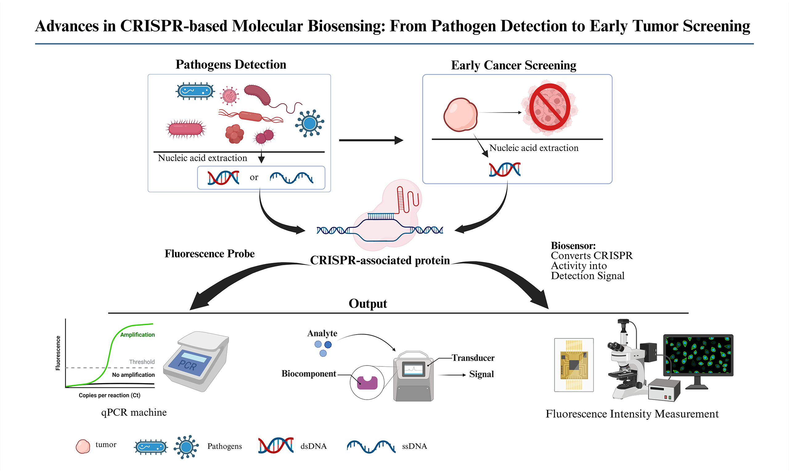
Advances in CRISPR-based molecular biosensing: from pathogen detection to early tumor screening
 0
0 Abstract
The Clustered Regularly Interspaced Short Palindromic Repeats/CRISPR-associated (CRISPR/Cas) system, with its programmable nucleic acid recognition and trans-cleavage-mediated signal amplification capabilities, has evolved from a gene-editing tool into a core technology for next-generation molecular biosensing. This review systematically elucidates the molecular mechanisms of this technology, focusing on the trans-cleavage activity of key proteins such as Cas12, Cas13, and Cas14, in conjunction with the functional characteristics of tool enzymes like Cas9, and the fluorescence, electrochemical, and colorimetric signal readout strategies they employ. Furthermore, this article provides a detailed summary of the application advancements of CRISPR biosensors in the rapid test of pathogens, including viruses, bacteria, and parasites, and early tumor screening, which encompasses the highly sensitive detection of various nucleic acid biomarkers, including gene mutation, methylation, fusion gene, and microRNA. Furthermore, this review summarizes the technological evolution of device integration, from fully automated, closed microfluidics to portable systems, to achieve point-of-care testing. Finally, the article proposes future development directions, including the integration of this technology with nanomaterials and artificial intelligence. This review aims to provide a systematic reference for the further development and application transformation of CRISPR molecular biosensing technology.
Keywords
INTRODUCTION
In contemporary society, pathogen infection and tumor remain two major challenges to human health, together constituting a significant portion of the global disease burden[1,2]. According to the World Health Organization, non-communicable diseases account for more than 70% of all deaths worldwide, with cardiovascular diseases and tumor being the main representatives, while infectious diseases remain a critical factor in mortality in many regions, especially for low-income and vulnerable populations[3]. Globally, the number of tumor-related deaths and the proportion of tumor-related deaths to total deaths are slowly increasing with population aging, consistently ranking among the leading causes of mortality[4].
Although China's infectious disease prevention and control system has achieved remarkable results, effectively controlling a variety of major infectious diseases, public health emergencies can still pose a severe threat to public health[5]. For instance, the COVID-19 pandemic witnessed a dramatic surge in infection-related mortality, inflicting irreparable suffering on individuals and families while also imposing substantial socioeconomic burdens[6]. These facts underscore the critical strategic importance of developing early, rapid, and accurate screening technology for both persistent tumor threats and unpredictable emerging epidemics.
As a cornerstone of Precision Medicine, Molecular Diagnostics offers an unprecedented molecular perspective for disease prevention, diagnosis, treatment, and prognosis management by analyzing information from biomacromolecules such as Nucleic Acids and Proteins. Thanks to the deep integration of Multi-omics Technologies, modern Molecular Diagnostics has achieved a qualitative leap in the depth and breadth of detection. However, traditional technology paradigms represented by PCR, Sequencing, and Immunoassay still appear inadequate in dealing with complex biological matrices, capturing trace Molecular Markers, and realizing Point-of-Care Applications[7]. These technical bottlenecks not only limit their effectiveness in Difficult Disease Diagnosis and Dynamic Efficacy Monitoring but also hinder their widespread adoption in scenarios such as Primary Healthcare and Emergency Response. Therefore, overcoming the limitations of existing technologies and developing a new generation of highly sensitive, integrated, and automated Molecular Diagnostic Platforms has become an urgent need to drive modern medicine to a higher level of development.
The Clustered Regularly Interspaced Short Palindromic Repeats/CRISPR-associated (CRISPR/Cas) system, as a revolutionary gene editing tool, has garnered widespread attention in the Biomedical Field due to its specificity and efficiency. Initially discovered as an Adaptive Immune Mechanism in bacteria and archaea, this system defends against Virus and Plasmid invasion by recognizing and cleaving Exogenous Nucleic Acid Sequences[8,9]. Through its unique Nucleic Acid Binding and Cleavage Activity, the CRISPR/Cas System enables highly specific recognition of Nucleic Acid Targets and significantly enhances detection sensitivity via Signal Amplification Mechanisms. Currently, Nucleic Acid Detection Methods constructed with the CRISPR/Cas System have undergone Clinical Validation, and the preliminary results are in line with expectations, demonstrating broad application prospects[10-12]. However, before achieving large-scale clinical application, the CRISPR/Cas System still faces a series of challenges that need to be addressed, including the risk of Off-target Editing, the need for further enhancement of Detection Specificity, improved ability to handle Complex Samples, and refinement of the Stability of Portable Devices[13-16].
Rapid point-of-care testing technologies are crucial for the early diagnosis and control of infectious diseases[17]. Although traditional polymerase chain reaction offers high sensitivity and specificity, its reliance on laboratory settings and trained personnel limits its application in resource-constrained environments[18,19]. In recent years, the CRISPR/Cas System has been established as a core technology for next-generation molecular diagnostics due to its exceptional sequence-specific recognition capability and trans-cleavage activity[20,21]. To realize its potential for on-site applications, cutting-edge research has focused on the deep integration of CRISPR detection with isothermal amplification, microfluidic technologies, and intelligent terminals, leading to the development of a series of automated and integrated portable detection devices[22,23]. Currently, the research focus in this field is shifting from validating the detection performance of CRISPR/Cas Systems to integrating multiple steps, such as sample processing, nucleic acid amplification, CRISPR/Cas reaction, and signal readout, into a closed, portable, and even disposable miniaturized device[24,25]. This integration process aims to minimize manual operation, reduce the risk of cross-contamination, and simplify device dependence, thereby promoting the translation of CRISPR diagnostics from the laboratory to point-of-care settings.
In summary, this review summarizes the research and latest advances in CRISPR biosensors for pathogen detection and early Tumor screening, systematically elucidates the application of CRISPR/Cas System-based point-of-care testing devices in integration, and proposes the challenges and prospects faced by CRISPR biosensors, aiming to provide references and basis for the further development of this method.
DIAGNOSTIC MECHANISMS OF VARIOUS CAS ENZYMES
The molecular diagnostic applications of the CRISPR/Cas system primarily rely on Class II effector proteins. These single-polypeptide effectors, guided by a single guide RNA (sgRNA) or CRISPR RNA (crRNA), can specifically recognize and cleave target nucleic acids, forming the mechanistic core of CRISPR biosensing. Based on their target molecules and cleavage activities, Cas enzymes used in diagnostics can be mainly categorized into several classes, including Cas9, Cas12, Cas13, and Cas14[26-28].
Cas9: recognition mechanism based on specific cleavage
Cas9 is the earliest and most extensively studied and applied effector protein of the CRISPR system. It recognizes specific protospacer adjacent motifs [(PAM), e.g., NGG)] on double-stranded DNA (dsDNA) guided by (sgRNA, a single RNA molecule formed by the fusion of crRNA and trans-activating crRNA (tracrRNA)), and then the HNH and RuvC nuclease domains of Cas9 cleave the DNA strand complementary to the sgRNA and the opposite strand, respectively, generating a site-specific double-strand break[29]. In diagnostics, the "gene scissors" function of Cas9 does not directly generate an amplifiable reporter signal; instead, it creates conditions for subsequent signal readout through precise cleavage of the target DNA.However, recent structural work by Smith has revealed that DNA supercoiling - a common topological state in live cells - can induce Cas9 off-target activity by lowering the activation energy for R-loop formation and allowing HNH domain movement even with mismatched sequences. This finding is critical for diagnostic applications that rely on Cas9’s specificity, as it implies that supercoiled DNA templates (e.g., from isothermal amplification) may produce false-positive signals if the sgRNA tolerates partial mismatches. Therefore, careful control of DNA topology or the use of high-fidelity Cas9 variants is recommended when implementing Cas9-based diagnostic readouts[30].
A representative example is a technique that integrates Nucleic Acid Sequence-Based Amplification (NASBA) with CRISPR/Cas9 cleavage. This approach first employs NASBA to isothermally amplify the original RNA target, generating a large quantity of dsDNA amplicons. Subsequently, Cas9 is utilized to specifically recognize and cleave these DNA amplification products. Only when the DNA amplicon sequence perfectly matches the target RNA will Cas9 cleave and disrupt a trigger sequence designed to activate a "Toehold" sensor, thereby indicating the detection result through a color change in the solution. This method successfully achieved single-base discrimination among different Zika virus strains, with detection sensitivity reaching the femtomolar level[31]. However, due to the lack of trans-cleavage activity in Cas9, its signal amplification capability is limited. The detection process often requires complex signal transduction designs, which to some extent restricts its application in point-of-care testing[32].
Cas12 and Cas13: signal amplification mechanisms based on the trans-cleavage effect
The discovery of Cas12 and Cas13 represents a revolutionary breakthrough in the field of CRISPR diagnostics. They share a key characteristic: trans-cleavage activity (also known as the "trans-cleavage effect"). After the Cas enzyme, guided solely by crRNA (independent of tracrRNA), recognizes and cleaves the target nucleic acid (cis-cleavage), it becomes activated and subsequently non-specifically cleaves reporter molecules in the surrounding environment, achieving cascade amplification of the signal[33,34]. Integrating this programmable reaction module of the CRISPR/Cas system into DNA functional circuits offers novel strategies and prospects for constructing complex and intelligent biosensing systems[35].
The Cas12 family primarily targets dsDNA or single-stranded DNA (ssDNA). Guided by crRNA (for Cas12a) or sgRNA (for Cas12b), upon recognition of a dsDNA target containing a specific PAM sequence (e.g., TTTV, TTN, TTTN), its trans-cleavage activity is activated, leading to indiscriminate degradation of single-stranded DNA reporters in the system. It should be noted that when the target is ssDNA, both Cas12a and Cas12b recognize and cleave the target in a PAMindependent manner, and can still trigger trans-cleavage activity. In terms of activation efficiency, Cas12a responds more efficiently to dsDNA targets than to ssDNA targets, whereas Cas12b exhibits higher activation efficiency with ssDNA targets than with dsDNA targets[36,37] (Figure 1 mainly illustrates the mechanisms by which Cas12a triggers transcleavage activity using dsDNA as the target, and Cas12b using ssDNA as the target). Based on this mechanism, the DETECTR platform primarily employs Cas12a. When combined with pre-amplification techniques such as recombinase polymerase amplification (RPA), it achieves detection sensitivity at the attomolar (aM) level and can accurately distinguish between Human Papillomavirus (HPV) types 16 and 18[38]. Similarly, the HOLMES platform utilizes the trans-cleavage activity of Cas12a to enable highly sensitive detection and single-nucleotide polymorphism (SNP) discrimination of specific DNA targets[39,40]. The subsequently improved HOLMESv2 platform adopts Cas12b, which retains activity at higher temperatures, further enhancing reaction speed and broadening the scope of application[41].
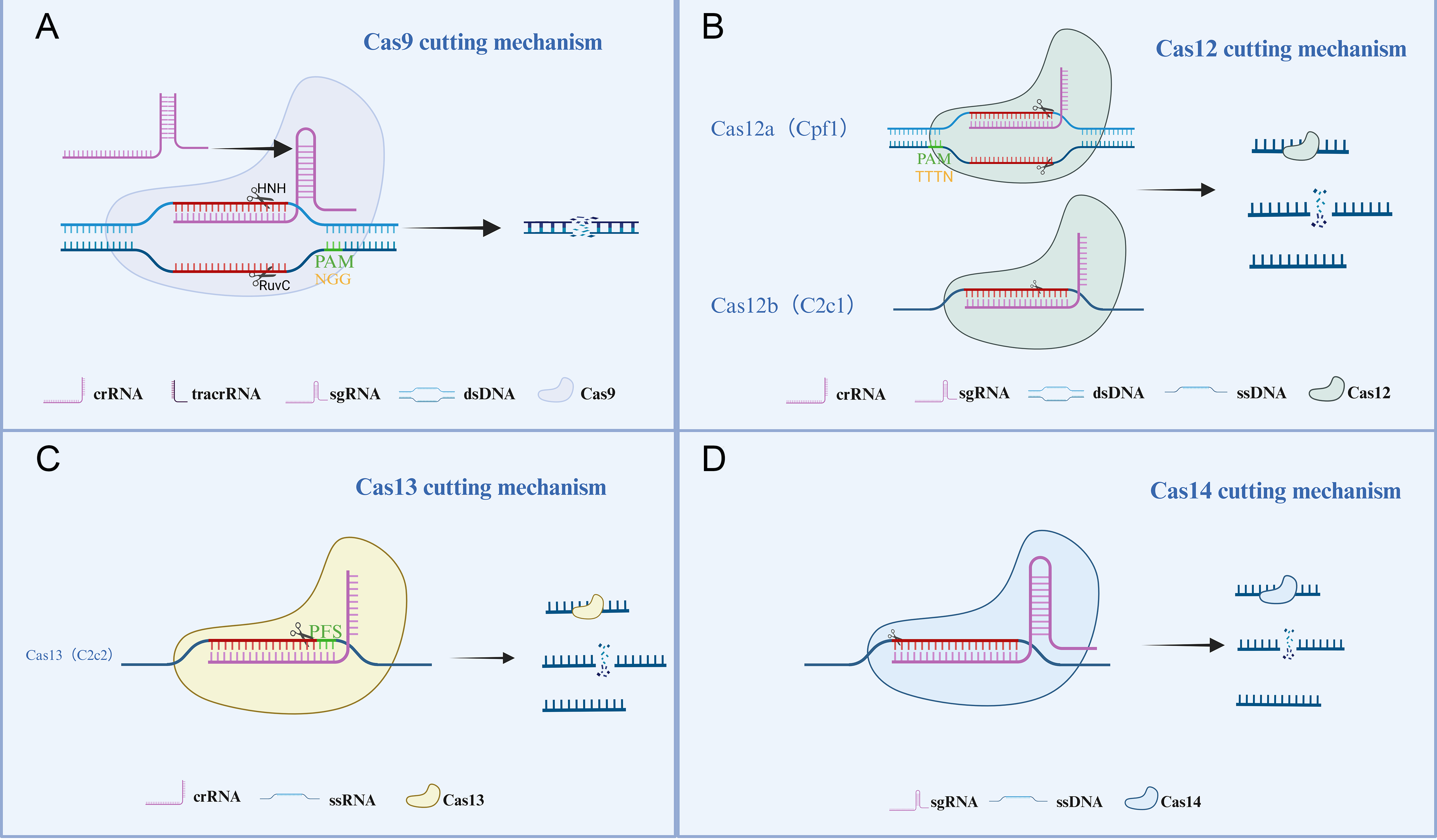
Figure 1. Mechanisms of cleavage action of various Cas enzymes. (A) Cas9 recognizes the PAM sequence (NGG) on dsDNA via sgRNA (crRNA:tracrRNA complex). The HNH and RuvC nuclease domains of Cas9 cleave the DNA strand complementary to the sgRNA and the opposite DNA strand, respectively, generating a site-specific dsDNA break. (B) Cas12a recognizes the PAM sequence (TTTV/TTTN) on dsDNA via crRNA; Cas12b targets ssDNA via sgRNA without requiring a PAM sequence. Their trans-cleavage activity is then activated, leading to the degradation of ssDNA reporters in the system. (C) Cas13 recognizes the 3'protospacer flanking site (PFS) on single-stranded RNA (ssRNA) via crRNA, which activates its trans-cleavage activity and subsequently degrades ssRNA reporters. (D) Cas14 targets ssDNA via sgRNA independently of a PAM sequence; its trans-cleavage activity is activated, resulting in degradation of ssDNA reporters.(Created in BioRender. Yan, S. (2026) https://BioRender.com/k9vp6hk).
Notably, a recent study by Scholz introduced Cas12a2, a distinct Cas12 variant that, upon RNA-target recognition, unleashes indiscriminate dsDNA degradation. While Cas12a2 was originally developed for cell killing, its promiscuous DNase activation after a specific RNA trigger provides a conceptually new signal transduction mechanism: the target RNA (e.g., a viral or cancer-marker transcript) activates Cas12a2 to cleave nonspecific DNA reporters. This principle could be adapted for ultra-sensitive RNA detection without separate reverse transcription (RT) or pre-amplification steps, as the transcleavage activity is amplified by the enzyme itself. The Scholz paper[42] thus expands the diagnostic toolbox beyond canonical Cas12a and Cas12b, offering a onestep, RNAdirected DNA cleavage reporter system.
The Cas13 family (exemplified by Cas13a) primarily targets RNA. Guided by crRNA, upon recognition of a single-stranded RNA (ssRNA) target that carries a Protospacer Flanking Site (PFS, typically requiring a non-G base) at its 3' end, its trans-cleavage activity is activated, leading to the degradation of single-stranded RNA reporters in the system[43,44]. The renowned SHERLOCK platform leverages this principle, converting DNA or RNA targets into RNA recognizable by Cas13 through pre-amplification via Recombinase RPA and T7 in vitro transcription, thereby activating the reporter system to generate a fluorescent signal. This technology has been successfully applied to detect Zika virus, Dengue virus, and cancer-related mutations, achieving a sensitivity at the attomolar (aM) level[45]. Subsequently developed SHERLOCKv2 enabled parallel detection of multiple targets by employing multiple Cas13/Cas12a homologs, further expanding its application scope[46].
Cas14: PAM-independent ssDNA detection
Cas14 is a more compact Cas protein (typically composed of 400-700 amino acids), with the unique advantage of targeting ssDNA without requiring a PAM sequence. Similar to Cas12, after specific ssDNA targets are recognized and cis-cleaved by Cas14 guided by sgRNA, it further exhibits trans-cleavage activity on ssDNA reporters. This property confers unique advantages for detecting point mutations, single-nucleotide polymorphisms (SNPs), and ssDNA viruses[47]. For example, integrating Cas14 with the DETECTR platform (Cas14-DETECTR) successfully achieved PAM-unrestricted, highly specific detection of SNPs in the human genome associated with eye color-related genes[48,49].This strategy of integrating different CRISPR systems with specific detection platforms aligns with the research approach of achieving precise temporal and spatial control over CRISPR/Cas9 gene manipulation through the design of pH-responsive RNA triplexes. Together, they exemplify the latest trend of optimizing CRISPR performance via engineering the systems themselves or their operational environment[50].
Mechanism comparison and application summary
In summary, the cleavage mechanisms of various Cas enzymes are illustrated in Figure 1. The core diagnostic mechanism of different Cas enzymes lies in their specific recognition of the target nucleic acid (cis-cleavage) and whether they possess signal amplification capability (trans-cleavage). Cas9 relies on precise cis-cleavage for target recognition but requires additional signal transduction steps. In contrast, Cas12, Cas13, and Cas14 employ a cascade amplification mechanism of "cis-recognition and trans-cleavage, " enabling in-situ signal amplification, which significantly enhances detection sensitivity and operational convenience[27]. Table 1 summarizes their key characteristics.
Main functional characteristics of various Cas enzymes
| Protein | Target acid | Guide RNA | Recognition sequence | Non-specific reporter molecule for trans-cleavage |
| Cas9[29] | dsDNA | sgRNA | PAM (such as 5'-NGG-3') | No |
| Cas12a/b[36] | dsDNA/ssDNA | crRNA/sgRNA | PAM (such as TTTV/5'-TTTN-3')/No | ssDNA |
| Cas13[43] | ssRNA | crRNA | PFS(Non-G nucleotide at the 3' proto-spacer flanking site) | ssRNA |
| Cas14[47] | ssDNA | sgRNA | No | ssDNA |
Next, this review will focus on the signal amplification mechanisms and readout methods based on the aforementioned Cas enzymes, especially Cas12/Cas13 and others with trans-cleavage activity.
COLLATERAL EFFECT SIGNAL AMPLIFICATION MECHANISM
Fluorophore-quencher reporter mechanism
The core principle of the Fluorophore-Quencher Reporter (FQ reporter) mechanism lies in the activation of Cas proteins upon recognition and binding to specific targets, which subsequently triggers their nonspecific cleavage activity. This activity leads to the degradation of ssDNA or single-stranded RNA reporter probes present in the system. These probes are typically labeled with a fluorophore at one end and a quencher at the other. When the probe remains intact, fluorescence is quenched due to Förster Resonance Energy Transfer (FRET). Upon cleavage by the Cas protein, the fluorophore and quencher are separated, restoring fluorescence and generating a detectable signal[51,52]. Currently, research on fluorescent probes is rapidly evolving, encompassing the development of novel probe materials and investigations into their diverse applications[53,54].
As illustrated in Figure 2A typical Immuno-CRISPR detection strategy integrates antibody-DNA barcode conjugates, a Cas12a/crRNA complex, and a collateral FQ reporter probe to construct the detection system. In this design, an antigen-antibody interaction forms a sandwich complex comprising “capture antibody-target protein-antibody-streptavidin-DNA barcode.” Subsequently, the Cas12a/crRNA complex binds to the DNA barcode and becomes activated, thereby exerting its trans-cleavage activity to cleave the FQ reporter probe. The resulting fluorescence signal enables sensitive detection of the target protein.
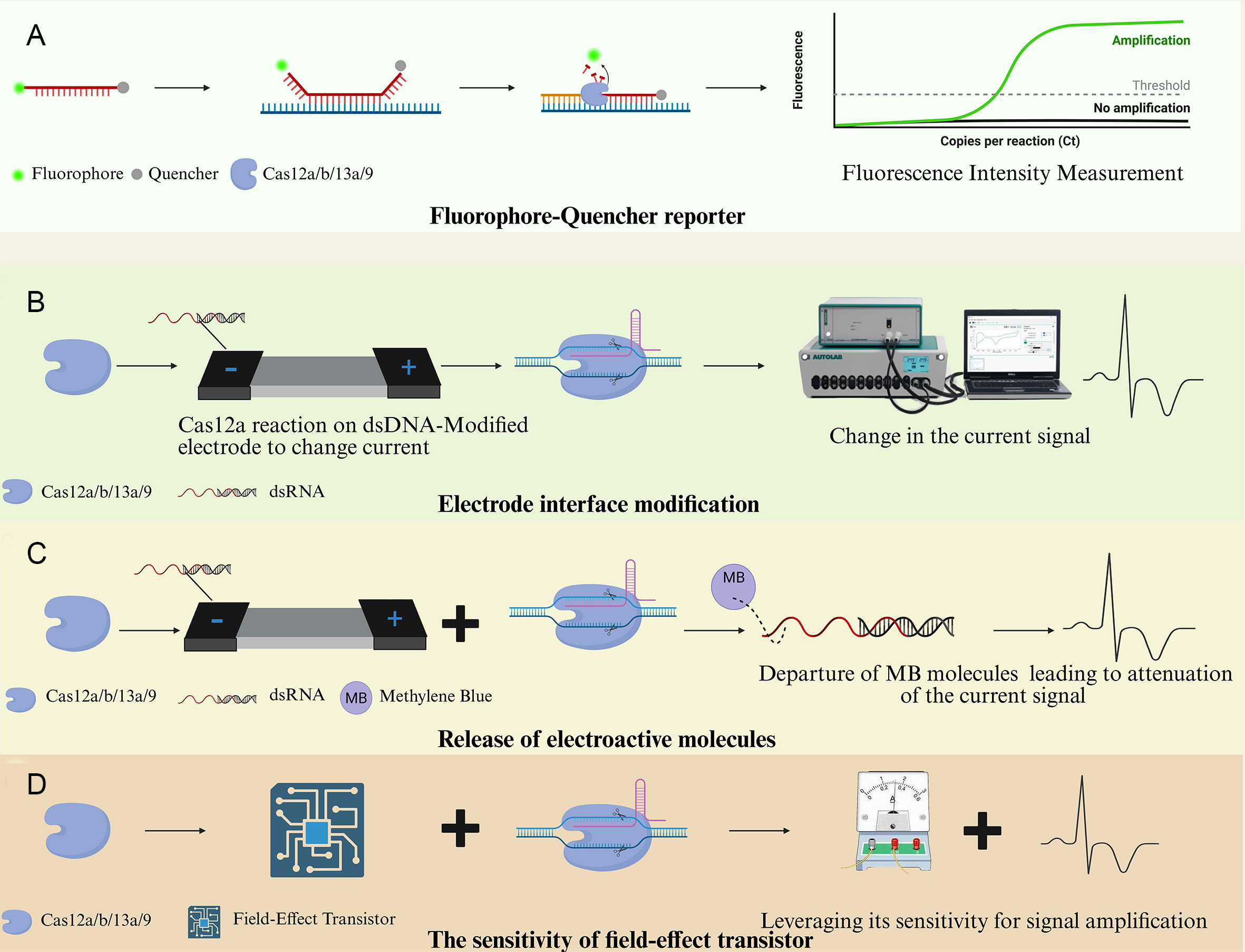
Figure 2. Mechanisms of fluorophore-quencher reporter systems. (A) Schematic of the fluorophore-quencher reporter mechanism; (B) Schematic of the signal amplification mechanism based on electrode modification; (C) Schematic of the signal amplification mechanism based on electroactive molecule release; (D) Schematic of the signal amplification mechanism based on field-effect transistor. (Created in BioRender. Xu, S. (2026) https://BioRender.com/26xqk1g).
Currently, the standard Enzyme-Linked Immunosorbent Assay (ELISA) is widely used in clinical diagnostics. Although ELISA offers high accuracy and sensitivity, the nonspecific adsorption of the Horseradish Peroxidase (HRP) label often leads to elevated background signals. Moreover, the assay typically requires several hours to complete, which limits its applicability in point-of-care testing scenarios. Lee applied the aforementioned CRISPR immuno-assay to analyze urine samples from kidney transplant patients. The results demonstrated that its detection sensitivity was approximately seven times higher than that of conventional ELISA and showed a high degree of concordance with clinical diagnostic outcomes, indicating strong potential for clinical application[55].
Electrochemical and optical signal amplification strategy
Electrochemical signals offer advantages such as ease of amplification, high degree of device miniaturization, and suitability for integration. Meanwhile, the programmable properties and cleavage activity of enzymes like Cas12a provide opportunities to construct diverse signal transduction mechanisms[56]. Therefore, integrating the CRISPR/Cas system with electrochemical analysis technology enables highly sensitive, rapid, and portable detection. The primary focus is on the following three types of signal amplification strategies:
1. Strategy based on electrode interface modification
The core of this strategy lies in utilizing activated Cas12a to cleave single-stranded DNA modified on the electrode surface, thereby altering the physicochemical properties of the electrode interface.The principle of this method is typically shown in Figure 2B. This modification affects the electron transfer process and subsequently induces significant changes in the current signal. Wu Chenshuo integrated the interface modification strategy with electrochemiluminescence (ECL). Upon activation, Cas12a cleaves a trigger chain, which then initiates the in situ assembly of a large DNA tetrahedral framework on the electrode surface. This framework efficiently enriches a high density of ECL luminescent molecules, resulting in an intense ECL signal[57]. This "cleavage-triggered DNA nanostructure assembly" strategy greatly amplifies the interface variation, enabling ultrasensitive detection. Wu Lina developed an electrochemical biosensor based on the CRISPR/Cas system for the detection of SARS-CoV-2, which was successfully applied to the analysis of pharyngeal swab and frozen food packaging samples. Among 50 clinical samples tested, the CRISPR-SPCE method yielded results that were completely consistent with those of quantitative polymerase chain reaction (qPCR) (37 positive and 13 negative cases), indicating its high reliability in real-world sample detection[58].
2. Strategy Based on Electroactive Molecule Release
As depicted in Figure 2C, this strategy typically employs a gold electrode as the substrate, where ssDNA probes labeled with electroactive molecules (such as methylene blue) at the terminal are immobilized via Au-S bonds. Under a specific voltage, methylene blue molecules located near the electrode surface undergo redox reactions, generating a distinct current signal. When Cas12a is activated and cleaves the probe, methylene blue molecules detach from the electrode surface, resulting in a decreased current signal, thereby enabling the detection of the target nucleic acid[59].
3. Strategy based on field-effect transistor (FET)
As depicted in Figure 2D, Graphene Field-Effect Transistors (GFETs), characterized by high carrier mobility, excellent electrical conductivity, and ambipolar field-effect characteristics, are highly sensitive to surface charge variation[60]. Wang Weiqi developed a Cas12a-mediated GFET sensor for the serological diagnosis of tuberculosis. In a test involving 56 clinical samples (including 30 tuberculosis patients and 26 healthy individuals), the sensor correctly identified 54 cases, demonstrating high diagnostic accuracy and strong potential for application in complex clinical samples[61].
At present, the integration of CRISPR systems with electrochemical techniques for signal amplification has been widely applied across various fields. In particular, significant progress has been made in food safety and pathogen identification. Li Xiaolong elaborated on the principles and amplification efficiencies of the three major approaches mentioned above, and proposed the development of simplified and efficient DNA circuits based on CRISPR-Cas systems[35].
Due to their high sensitivity, ease of operation, and visualizability, optical signal readout methods have emerged as one of the fastest-growing detection strategies in recent years. Optical sensing systems are capable of converting the activation state of Cas proteins into various forms of optical signals, such as fluorescence, colorimetric, or chemiluminescent outputs, thereby enabling efficient recognition and amplification of target nucleic acids. Currently, CRISPR-based optical detection primarily involves three types of mechanisms, as illustrated in Figure 3.
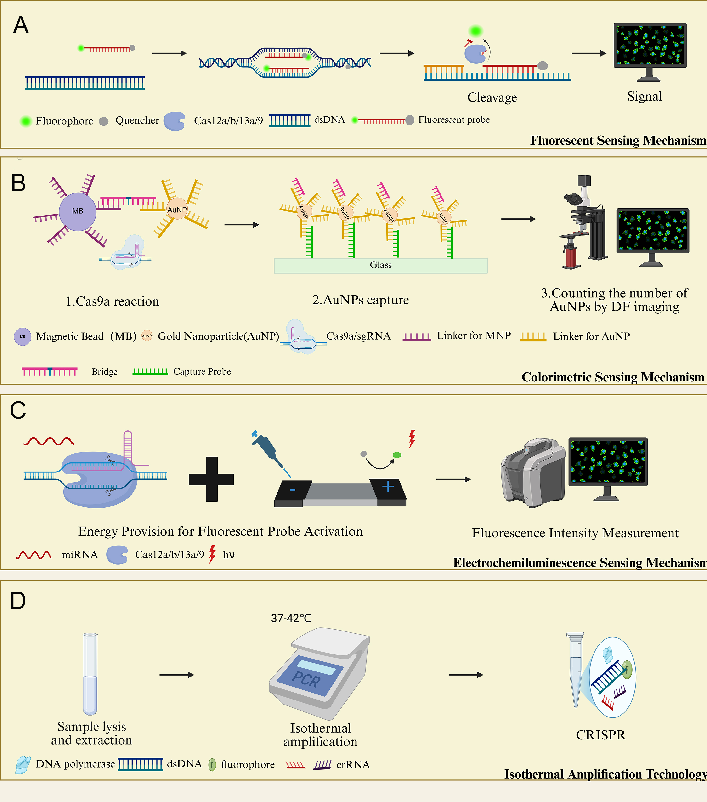
Figure 3. Optical detection strategies. (A) Fluorescent sensing mechanism diagram; (B) Colorimetric sensing mechanism diagram; (C) Chemiluminescence and electrochemiluminescence sensing mechanism diagram; (D) Isothermal amplification technology flowchart.(Created in BioRender. Xu, S. (2026) https://BioRender.com/o41cyqs).
4. Fluorescence sensing mechanism
This mechanism leverages the change in cleavage activity of Cas proteins before and after target recognition to regulate the state of the fluorescence reporting system, thereby translating the biomolecular recognition event into a fluorescent signal output. For example, Yang Rui developed a quencher-free CRISPR detection system in which, in the presence of target nucleic acids, Cas12a cleaves an amphiphilic DNA probe, releasing the hydrophobic Cy5 fluorophore. This alters the wettability of the solution and subsequently affects the fluorescence dispersion behavior, enabling visual quantification of the target. This strategy demonstrated comparable performance to conventional PCR when validated with clinical swab samples, indicating its potential as a simple, cost-effective, and sensitive approach for the diagnosis of HPV-related tumors and other infectious diseases[61].
5. Colorimetric sensing mechanism
Colorimetric sensing converts the target recognition process of the CRISPR-Cas system into a visually perceptible color change, making it suitable for on-site detection without the need for complex instrumentation. Kim Jaejoon developed a CRISPR-based method for miRNA detection and conducted cross-validation with RT-qPCR. The results showed that this method could rapidly, selectively, and accurately identify small RNAs in five breast cancer cell lines, demonstrating its potential for early tumor diagnosis[44].
6. Chemiluminescence and ECL sensing mechanisms
Chemiluminescence refers to the process in which the energy released from a chemical reaction is directly converted into light radiation, whereas ECL is a luminescent phenomenon triggered electrochemically on the surface of an electrode. Zhou Yan and colleagues developed a CRISPR/Cas13a-driven portable ECL chip (Portable ECL CRISPR, PECL-CRISPR) for ultrasensitive and highly specific analysis of miRNA. By testing four types of miRNA, including miR-17, miR-10b, miR-155, and miR-21, and validating specificity through polyacrylamide gel electrophoresis, the system demonstrated strong capability to meet the detection demands of clinical complex samples[62].
Isothermal amplification (RPA/LAMP)-based synergistic detection mechanism
Isothermal amplification techniques, such as RPA and loop-mediated isothermal amplification (LAMP), enable efficient amplification of target nucleic acids at a constant temperature. When combined with the CRISPR system, these methods can further enhance detection sensitivity and efficiency. A typical strategy involves first generating a large number of target sequences through isothermal amplification, followed by specific recognition using the CRISPR system, and finally outputting the results via fluorescence or colorimetric signals. Sun Qi and colleagues developed a photoactivated CRISPR-Cas12a strategy and integrated it with RPA into a one-pot reaction system for rapid genotyping of SARS-CoV-2 Omicron subgroups. This method demonstrated a clinical sensitivity of 97.3%, specificity of 100.0%, and a concordance rate of 98.3% with Sanger sequencing results, indicating strong potential for clinical application[45].
Technical integration, result validation, and practical application challenges
Despite the remarkable sensitivity, speed, and specificity of CRISPR-based biosensing, its translation from laboratory prototypes to routine clinical and point-of-care diagnostics remains constrained by several critical challenges related to system integration, result validation, and practical deployment[44,62].
First, workflow complexity and integration remain major barriers. Most high-performance CRISPR assays rely on a two-step strategy involving nucleic acid pre-amplification (e.g., RPA or LAMP) followed by CRISPR-based detection[45,63]. This open, multi-step workflow increases operational complexity and contamination risk, compromising result reliability. Consequently, the development of integrated “one-pot” systems has emerged as a key direction. Representative strategies employ spatiotemporal regulation mechanisms to sequentially coordinate amplification and CRISPR reactions within a single tube. For example, photoactivated CRISPR-Cas12a systems enable controlled reaction initiation, reducing enzymatic interference and contamination while maintaining high sensitivity and specificity[64]. Such approaches provide effective solutions for simplifying workflows and improving assay robustness.
Second, dependence on specialized readout equipment limits accessibility. Ultra-sensitive CRISPR sensing platforms often require sophisticated instruments, such as fluorescence readers or electrochemical workstations, restricting their use in primary healthcare and resource-limited settings. To address this, recent efforts have focused on integrating CRISPR detection with portable hardware and user-friendly readout modalities, including colorimetric outputs and smartphone-based analysis. Handheld CRISPR platforms that integrate sample processing, signal detection, and result interpretation exemplify a practical route toward instrument-free or minimally instrumented point-of-care testing. Consistent with this, Song and colleagues developed a smartphone-based CRISPR-LAMP system using colorimetric DNAzyme reaction for SARS-CoV-2 and variant detection, enabling visual readout without bulky instruments[65].
Third, the lack of standardization and quality control hampers clinical translation. Key parameters-including crRNA design, Cas enzyme activity calibration, reporter probe configuration, and buffer composition-remain poorly standardized, leading to substantial variability in assay performance across laboratories and platforms. As highlighted in recent reviews, the absence of unified protocols and validation criteria represents a major bottleneck for regulatory approval and clinical adoption[66]. Establishing standardized operating procedures, reference materials, and quality control systems is therefore essential for ensuring reproducibility, comparability, and clinical reliability.
Finally, balancing cost and multiplexing capability remains challenging. Compared with established diagnostic methods such as ELISA or lateral flow assays[67,68], CRISPR diagnostics currently rely on relatively expensive enzymes, synthetic crRNAs, and reporter molecules[27]. Reducing reagent costs through scalable enzyme production and lyophilized formulations is critical for commercialization. Meanwhile, clinical applications increasingly demand multiplex detection of multiple targets. Traditional multiplexing strategies often require multiple Cas enzymes or complex optical encoding, increasing both cost and system complexity. Emerging designs that combine single-enzyme, multi-crRNA strategies with low-cost spatial encoding-such as lateral flow-based multiplex readouts-offer promising solutions by enabling reliable multiplex detection without sophisticated instrumentation.
In summary, overcoming challenges related to workflow integration, equipment dependence, standardization, and cost is essential for advancing CRISPR biosensing toward stable, reliable, and user-friendly diagnostic applications[38]. Future research should prioritize simplification, robustness, and economic feasibility alongside performance enhancement to facilitate widespread clinical and public health adoption. This imperative for holistic development-balancing technical performance with practical deployability-is a common theme across the translational pipeline of disruptive biomedical technologies, from next-generation diagnostics like CRISPR to therapeutic modalities such as cancer gene therapy[69].
APPLICATION OF CRISPR IN PATHOGEN DETECTION
The CRISPR/Cas system, owing to its high specificity in nucleic acid recognition, programmability, and signal amplification capability, has emerged as a revolutionary technology in the field of pathogen detection[66]. Its core advantage lies in overcoming the limitations of traditional methods, such as culture-based techniques and PCR, which often require sophisticated instrumentation, involve complex procedures, and are time-consuming. The underlying mechanism involves the use of guide RNA (gRNA) to precisely target pathogen-specific nucleic acid sequences, including conserved gene regions, antibiotic resistance genes, thereby activating the cis-cleavage and collateral cleavage activities of Cas proteins (such as Cas12 and Cas13). This leads to the degradation of fluorescent reporter molecules for signal output. When combined with isothermal amplification technology, the system significantly enhances the detection sensitivity for low-abundance targets, making it well-suited for point-of-care testing and large-scale screening applications. As shown in Figure 4
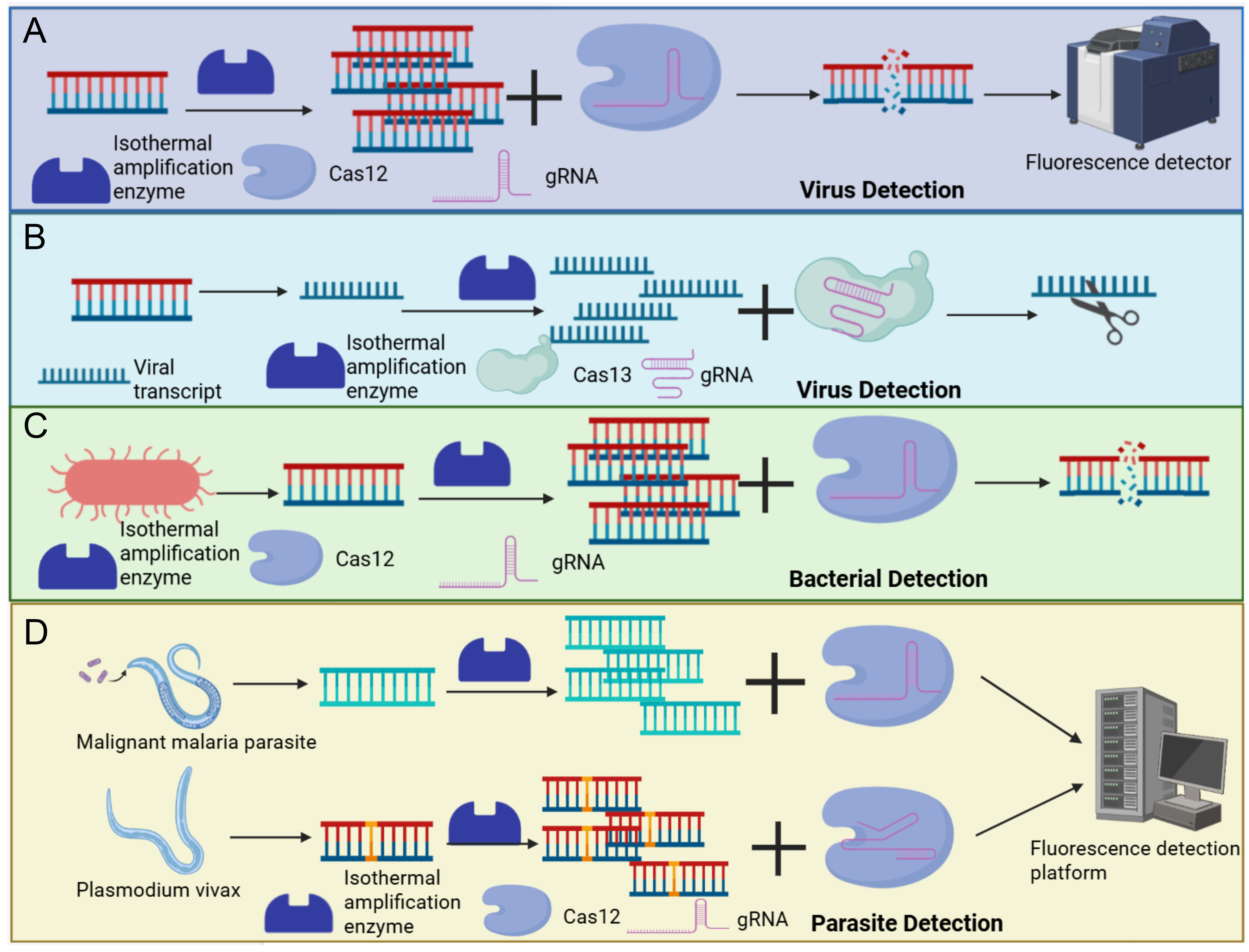
Figure 4. Mechanism of CRISPR-based pathogen detection. (A) illustrates the mechanism of the SHERLOCK detection platform based on Cas12a for specific detection of SARS-CoV-2. (B) depicts the CRISPR/Cas13 system combined with T7 RNA polymerase signal amplification technology for specific detection of HIV virus. (C) demonstrates the CRISPR/Cas12a detection system designed with specific gRNAs targeting beta-lactamase resistance genes (blaTEM and blaCTX-M) for rapid screening of antibiotic resistance in Enterobacteriaceae. (D) shows the mechanism by which the CRISPR/Cas12a detection platform distinguishes two Plasmodium species with high specificity, utilizing RPA isothermal amplification, CRISPR signal amplification, and specific gRNAs.(Created in BioRender. Gao, W. (2026) https://BioRender.com/mfu8j2d).
Virus detection
The primary objectives of virus detection are the rapid identification of low-abundance viral nucleic acids and the differentiation of closely related viral strains. The CRISPR system achieves early infection diagnosis and circulating strain monitoring by targeting viral structural protein genes or enzyme genes, thereby addressing the limitations of traditional methods in terms of sensitivity and specificity. SARS-CoV-2 spreads rapidly and has a high proportion of asymptomatic carriers, placing stringent demands on the speed and convenience of diagnostic technologies. The SHERLOCK detection platform based on Cas12a employs specifically designed gRNAs targeting the spike protein (S) gene. Through a cascade reaction involving RPA isothermal amplification and CRISPR collateral cleavage, this system ensures both rapid detection and enhanced sensitivity, delivering fluorescent readouts within 1 h with a detection limit of 10 copies/µL, and without the need for large-scale equipment such as a PCR instrument[70]. Clinical studies published in Cell have validated the specificity of this approach, demonstrating a 99.5% specificity for SARS-CoV-2 detection, with high concordance with RT-qPCR results and effective avoidance of cross-reactivity with other coronaviruses. Subsequent refinements reported in Nature Biotechnology further improved the detection limit to 1 copy/µL by optimizing the reaction buffer and gRNA design, enabling early screening of asymptomatic carriers and providing critical technological support for epidemic prevention and control[71].
Unlike the high transmission rate of SARS-CoV-2, the occult nature of Hepatitis B virus (HBV) infection-often missed by conventional methods-and its genotypic diversity pose unique challenges for clinical diagnosis. A CRISPR/Cas13 system targeting the HBV core antigen (C) gene has been developed to specifically recognize viral DNA transcripts within hepatocytes. When combined with T7 RNA polymerase signal amplification technology, this approach significantly enhances the detection capability for low viral loads[72]. Extensive studies have demonstrated that this method offers a tenfold increase in sensitivity compared to conventional PCR, effectively identifying occult HBV infections that are frequently missed by traditional ELISA, thereby reducing the risk of liver cirrhosis and hepatocellular carcinoma. Moreover, it exhibits robust cross-reactivity across HBV genotypes A through H, with no significant differences in specificity, making it suitable for detecting a wide range of globally circulating strains. This provides a unified technical solution for cross-border healthcare and epidemiological monitoring.
Bacterial detection
The primary challenges in bacterial detection lie in the prolonged turnaround time of traditional culture methods (1-3 days) and the rapid spread of drug-resistant bacteria, which renders empirical treatment ineffective. The CRISPR system enables integrated rapid bacterial identification and antibiotic resistance screening by targeting 16S rRNA (for species identification) or antibiotic resistance genes (for antibiotic resistance phenotype prediction)[73,74].
Mycobacterium tuberculosis grows slowly (requiring 2-4 weeks for culture), and extrapulmonary tuberculosis samples (such as cerebrospinal fluid and peripheral blood) often contain very low bacterial loads, resulting in low detection rates with conventional methods. A CRISPR/Cas12 system targeting the conserved region of Mycobacterium tuberculosis 16S rRNA, combined with RPA isothermal amplification technology, reduces detection time to 2 h and achieves a sensitivity of 10 CFU/mL for sputum samples[75]. Critically, the assay has been validated in cerebrospinal fluid and peripheral blood specimens, maintaining reliable performance in these clinically relevant matrices. This processing workflow overcomes the limitations of detecting non-sputum samples, enabling non-invasive testing of extrapulmonary tuberculosis specimens such as peripheral blood and cerebrospinal fluid. It offers a novel diagnostic approach for populations unsuitable for sputum sample collection, including children and the elderly.
In addition to the slow growth of certain bacterial colonies, the increasing prevalence of drug resistance presents a major challenge in bacterial detection. Enterobacteriaceae can acquire resistance to a wide range of antibiotics through multiple mechanisms, and the overuse of β-lactam antibiotics has exacerbated the spread of such resistant strains. Rapid differentiation of antibiotic resistance genotypes is essential for precise antimicrobial therapy. A CRISPR/Cas12a detection system employing specific gRNAs targeting β-lactamase resistance genes (blaTEM, blaCTX-M) enables rapid screening of resistance profiles in Enterobacteriaceae. Studies have confirmed that this method can differentiate between various types of antibiotic resistance genes within 1.5 h, achieving a concordance rate of 98.7% with whole-genome sequencing results. It eliminates the waiting time associated with traditional antimicrobial susceptibility testing (2-3 days). Additionally, it demonstrates a detection accuracy of 97.2% for antibiotic-resistant Escherichia coli in urine samples, enabling clinicians to formulate targeted antimicrobial treatment strategies before bacterial culture results are available, thereby reducing the misuse of broad-spectrum antibiotics and the risk of antibiotic-resistant bacteria transmission.
Parasitic infection detection
Parasitic infections are predominantly concentrated in resource-limited regions, where nucleic acid abundance in low-intensity infection samples is extremely low. Traditional methods, such as microscopic examination, often lack sensitivity and are labor-intensive. The CRISPR system, by targeting parasite-specific antigen genes and incorporating signal amplification techniques, enables highly sensitive and user-friendly detection. A large number of low parasite load infected individuals are present in malaria-endemic regions, and since treatment regimens differ between Plasmodium falciparum and Plasmodium vivax, accurate differentiation is essential. A CRISPR/Cas12a-based detection platform targeting the pfhrp2 gene of Plasmodium falciparum, combined with RPA isothermal amplification and CRISPR-mediated signal amplification, enables detection of parasitemia as low as 0.1 parasites/μL in blood samples. Specific gRNAs allow for precise discrimination between the two Plasmodium species[76]. Moreover, the assay has been validated against genetically diverse field isolates from endemic regions, accounting for natural sequence variation across parasite populations. This method achieved a detection rate of 95.6% in clinical samples from malaria-endemic areas, significantly outperforming the 82.1% detection rate of rapid malaria antigen diagnostic tests, thereby reducing missed diagnoses. Moreover, the assay does not require trained personnel or sophisticated equipment, offering both accuracy and ease of use in resource-limited settings, and meeting the requirements for epidemiological screening. This provides a robust technical foundation for malaria elimination programs[77].
In addition to Plasmodium, Toxoplasma gondii often establishes latent infections in immunocompromised individuals, such as pregnant women and patients with acquired immune deficiency syndrome, where it can become reactivated[78]. Therefore, screening methods with high specificity and sensitivity are required. A CRISPR/Cas13 system targeting the B1 gene of T. gondii, combined with RT-RPA amplification technology, enables precise detection of T. gondii nucleic acid in serum samples. This method has a detection limit of 5 copies per reaction and can effectively identify latent infections in immunocompromised patients, allowing for early intervention to prevent severe complications. Compared with traditional molecular diagnostics (qPCR/LAMP), this CRISPR-based assay offers lower cost, simpler operation, and better scalability for large-scale population screening, especially in low-resource settings. Moreover, it demonstrates a detection specificity of 99.1% for serum samples from pregnant women, making it suitable for early screening of congenital toxoplasmosis. This helps reduce the risk of fetal malformation and miscarriage, addressing the limitations of conventional methods in detecting infections in special populations.
APPLICATION OF CRISPR IN EARLY CANCER SCREENING (FOCUSING ON NUCLEIC ACID BIOMARKERS)
Early diagnosis of genetic rare diseases and tumors is critical for improving prognosis. However, conventional detection methods such as sequencing and immunoassays often suffer from high costs, limited sensitivity, and complex procedures. The CRISPR/Cas system, with its single-base resolution, high sensitivity, and signal amplification capability, enables precise identification of nucleic acid biomarkers, including gene mutations, methylation abnormalities, fusion genes, providing a non-invasive and efficient molecular basis for early disease screening, diagnosis, and personalized treatment, as illustrated in Figure 5.
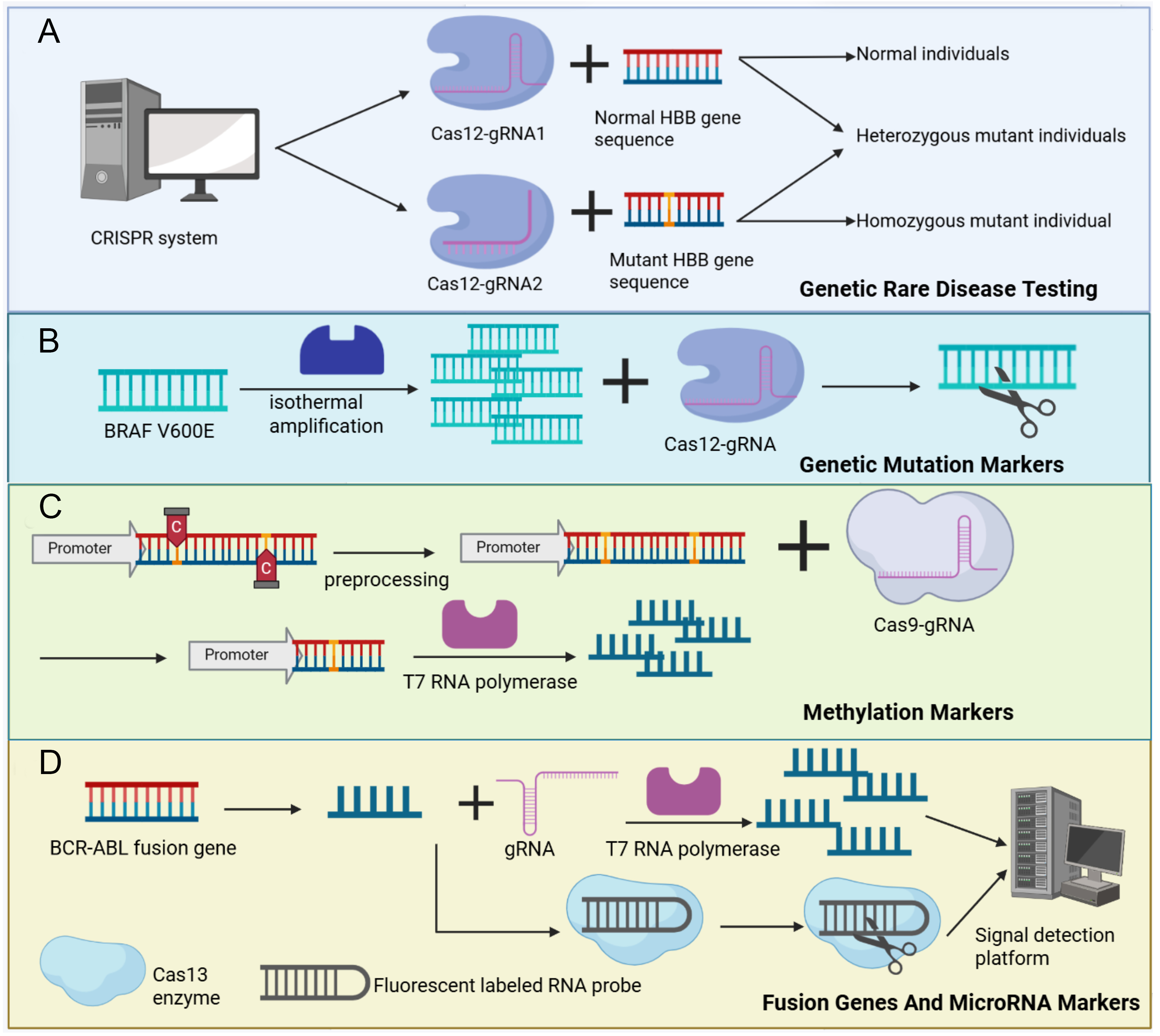
Figure 5. Mechanisms of CRISPR application in genetic rare diseases and early cancer screening. (A) Illustrates the CRISPR/Cas12 detection system incorporating a specifically designed gRNA, which rapidly distinguishes wild-type, heterozygous, and homozygous genotypes of the HBB gene for sickle cell disease screening. (B) Demonstrates the principle of the CRISPR/Cas12a detection system, which integrates RPA isothermal amplification technology and optimized reaction conditions for rapid detection of the BRAF V600E mutation in thyroid cancer. (C) Shows the mechanism by which Cas9 nickase, combined with T7 promoter amplification technology, enables methylation-dependent detection of the SEPT9 gene for early-stage colorectal cancer screening. (D) Presents the CRISPR/Cas13 system targeting the BCR-ABL fusion breakpoint, which specifically recognizes the fusion transcript and employs signal amplification technology to achieve rapid and highly sensitive detection of the BCR-ABL fusion gene for chronic myelogenous leukemia monitoring.(Created in BioRender. Gao, W. (2026) https://BioRender.com/i6qvsh1).
Genetic rare disease detection
Genetic rare diseases are often caused by single-gene pathogenic mutations, necessitating precise differentiation between wild-type and mutant sequences in clinical settings. The CRISPR/Cas system enables targeted detection of specific pathogenic mutation sites, facilitating carrier screening, newborn diagnosis, and disease subtyping. This approach effectively addresses the limitations of traditional methods, such as long turnaround times and high costs[79].
Sickle cell disease is caused by the Glu6Val mutation in the HBB (Hemoglobin Beta Gene, HBB) gene. Early diagnosis and intervention can significantly reduce mortality, highlighting the need for rapid and accurate mutation typing methods. A CRISPR/Cas12 detection system designed with mutation-specific gRNA for this site leverages single-base resolution to rapidly distinguish among wild-type, heterozygous mutant, and homozygous mutant individuals. This method achieved a detection accuracy of 99.3% for whole blood samples, demonstrating strong concordance with the results of the gold standard hemoglobin electrophoresis. Moreover, the detection time was reduced to within 1 h. Importantly, testing can be completed within 72 h after birth, providing a critical time window for early intervention strategies such as blood transfusion or gene therapy[80]. This makes the method particularly suitable for large-scale applications in newborn screening.
Early tumor screening: detection of gene mutation-based biomarkers
Driver mutations in tumor-derived ctDNA serve as key biomarkers for early diagnosis; however, the extremely low abundance of ctDNA (with variant allele frequency [VAF] often < 1%) poses significant challenges for detection using conventional methods. The CRISPR system, when combined with signal amplification technologies such as microfluidics and isothermal amplification, enables the precise capture of ultra-low abundance mutations. This provides a non-invasive solution for early tumor screening and efficacy monitoring[81].
For example, the BRAF V600E mutation is the most common driver mutation in thyroid cancer, with an incidence of approximately 40%-60%. Its detection is critical for pathological diagnosis and the selection of targeted therapies. An optimized CRISPR/Cas12a detection system targeting this mutation (1799T>A) has been developed by integrating RPA isothermal amplification technology with improved reaction conditions (14 mM magnesium acetate as a buffer additive and 150 nM specific gRNA). Direct comparisons with traditional PCR-based methods confirmed that removing the thermal cycler does not compromise analytical sensitivity or specificity. This system enhances detection specificity while simplifying the workflow, eliminating the need for a thermal cycler[82]. It is applicable not only for pathological diagnosis using preoperative fine-needle aspiration samples but also for dynamic monitoring of ctDNA changes in patients with advanced-stage disease following treatment. This enables rapid assessment of the efficacy of targeted drugs such as vemurafenib and addresses the limitations of conventional methods in detecting low-abundance mutations and conducting real-time monitoring.
In addition, the PIK3CA H1047R mutation is a key driver mutation in breast cancer and is closely associated with tumor invasion and drug resistance. The detection of this mutation in ctDNA holds significant value for early diagnosis and efficacy monitoring. The team from the Tumor Hospital of Harbin Medical University developed the innovative “TIDE-Cas14a” detection system, which integrates RPA-T7 isothermal amplification, engineered crRNA recognition, and micro-well digital microfluidic chip technology with
Early tumor screening: detection of methylation biomarkers
Gene methylation abnormality is a key early event in tumorigenesis and occurs prior to gene mutation, making it an ideal early screening biomarker. However, traditional detection methods, such as MSP (Methylation-Specific PCR, MSR), suffer from limited sensitivity and poor specificity. Following demethylation pretreatment, the CRISPR system targets sequences corresponding to methylated sites and, in combination with a fluorescence reporter system, enables precise qualitative and quantitative assessment of methylation status.
Promoter region methylation of the SEPT9 gene is a specific early biomarker for colorectal cancer, but its abundance in plasma is extremely low, necessitating highly sensitive detection methods. A CRISPR-based detection system targeting this methylation site employs Cas9 nickase in combination with T7 promoter amplification technology[84]. Through demethylation pretreatment to expose the target sequence, followed by signal amplification, this system enables the detection of low-abundance methylated fragments. This method achieved a detection rate of 87.5% for early-stage colorectal cancer (stages I-II) and 62.3% for precancerous lesions such as adenomas, significantly outperforming the traditional fecal occult blood test (detection rate < 50%) in identifying early pathological changes. It also demonstrated a specificity of 96.8% and a concordance rate of 91.2% with pathological diagnosis of colorectal cancer. Moreover, the test requires only peripheral blood samples, eliminating the need for invasive procedures and thereby improving screening compliance. This provides an efficient and non-invasive technological solution for early colorectal cancer screening[84].
Early tumor screening: detection of fusion genes and MicroRNA biomarkers
Fusion genes and tumor-specific miRNAs serve as characteristic biomarkers for certain tumors, such as leukemia and lung cancer. However, their detection requires precise identification of breakpoint sequences or low-abundance miRNAs, which traditional methods often fail to achieve[85,86]. The CRISPR system enables early diagnosis and efficacy monitoring by targeting fusion gene breakpoints or miRNA sequences in combination with signal amplification technologies[87-89].
The BCR-ABL fusion gene is a hallmark biomarker of chronic myelogenous leukemia, and its detection and quantitative monitoring are critical for diagnosis and therapeutic evaluation. A CRISPR/Cas13 system targeting the BCR-ABL fusion breakpoint achieves rapid and highly sensitive detection by specifically recognizing the fusion transcript and incorporating signal amplification techniques[90]. This method enables detection within 1 h, with a sensitivity of 10 copies per reaction for bone marrow samples, allowing effective identification of fusion genes in early-stage patients. In terms of sensitivity, specificity, and clinical utility, this CRISPR/Cas13 assay performs comparably to standardized qPCR, the current gold standard for minimal residual disease (MRD) monitoring. Studies have demonstrated that this system can accurately monitor dynamic changes in MRD following treatment, providing early warning of relapse risk. This molecular insight supports timely adjustment of tyrosine kinase inhibitor (TKI) therapy regimens, thereby significantly improving patient prognosis[89].
POINT-OF-CARE NUCLEIC ACID TESTING DEVICE BASED ON CRISPR: TECHNOLOGICAL EVOLUTION FROM CLOSED MICROFLUIDICS TO PORTABLE SYSTEM
Early CRISPR diagnostics typically employed a two-step method, involving isothermal amplification followed by open-lid transfer of the amplified products to the CRISPR reaction system. This process is prone to aerosol contamination, leading to false positive results[91-93], as illustrated in Figure 6A. For instance, the CARE device developed by Hui Ge integrates RPA with CRISPR-Cas12a Detection within a fully Closed Microfluidics Chip[94]. By precisely controlling the Liquid Flow with a Syringe Pump, the sample undergoes sequential Lysis, RPA Amplification, and CRISPR Detection within the chip, achieving Zero Lid-Opening Operation throughout the entire process. This device supports the simultaneous detection of seven Aquatic or Livestock and Poultry Pathogens, with Singleplex Detection Sensitivity reaching 1 copy/μL and Multiplex Detection Limit ranging from 10 to 100 copies/μL. The entire procedure is completed within 1 h, and the detection results are highly consistent with qPCR, significantly enhancing the Bio-Safety and Throughput of on-site detection. To address this bottleneck, researchers have designed various closed systems that feature physical isolation but automatic connection[95].
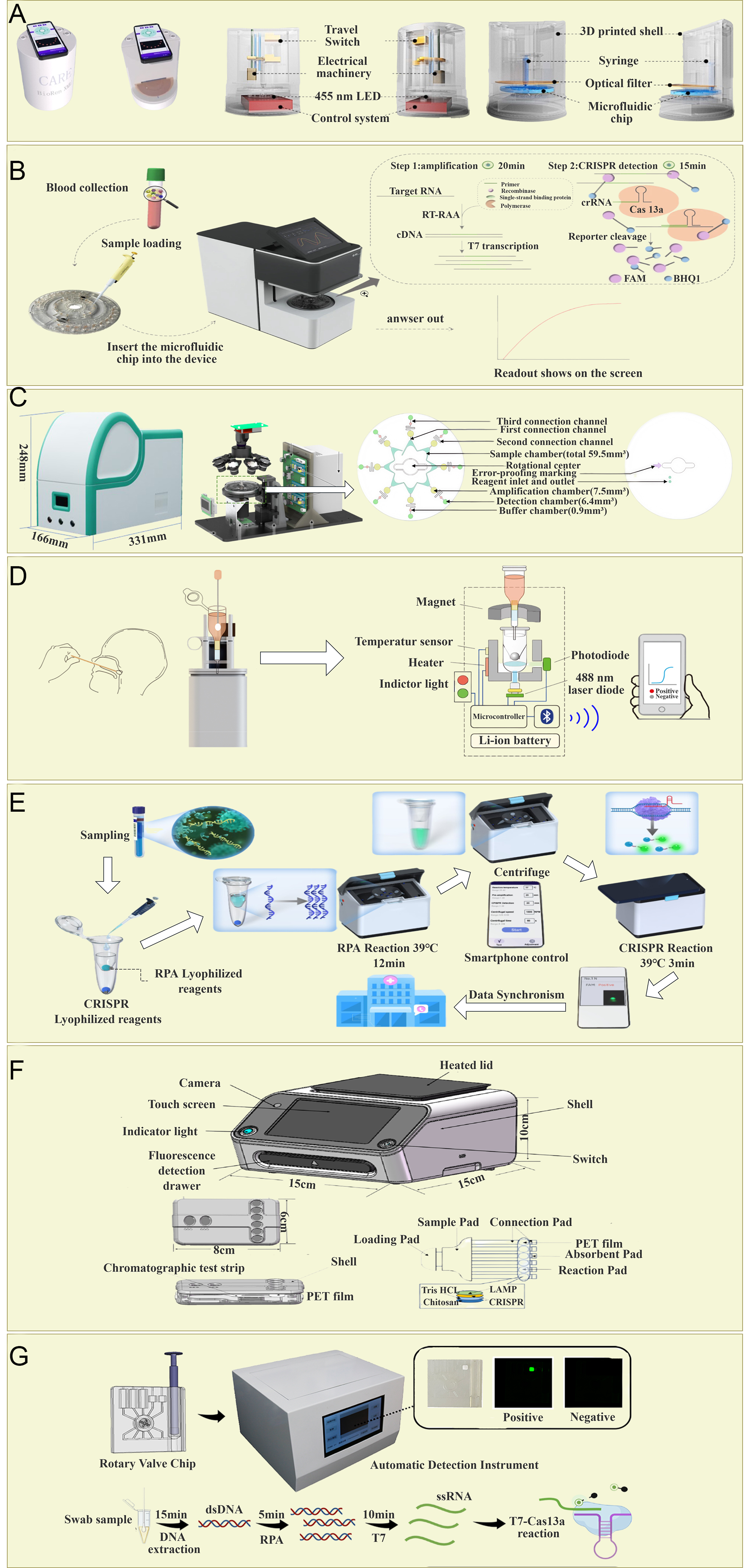
Figure 6. Representative CRISPR-Cas integrated detection platforms. Schematics and/or photographs illustrate the key working principles and designs of the various integrated systems discussed. (A) CARE device[94]: A fully sealed, pressure-driven Microfluidic Chip integrating RPA and CRISPR-Cas12a Detection for multiplex pathogen detection. (B) Portable integrated centrifugal system[99]: A CD-sized disk that automates Nucleic Acid Extraction, RAA, and CRISPR-Cas13a Detection. (C) Lift-CM Platform[101]: A centrifugal microfluidics system employing a spatially encoded disk and ascending heating to transfer amplicons to the CRISPR Detection chamber, compatible with various Isothermal Amplification methods. (D) CRAFT Platform[102]: A palm-sized device utilizing a mobile magnetic head valve within a single tube to compartmentalize RPA and CRISPR reactions. (E) Smartphone-controlled portable device[103]: A handheld device integrating heating, centrifugation, and Fluorescence Detection for extraction-free detection. (F) Integrated paper-based platform[107]: A Test Strip integrating bacterial Lysis, Chitosan Nucleic Acid purification, LAMP, and CRISPR-Cas12a Detection, driven by Capillary Action. (G) Automated microfluidics platform[109]: A Rotary Valve and plunger-assisted chip for fully automated sample pretreatment, Nucleic Acid Extraction, and one-pot RPA-T7-Cas13a reaction.
Centrifugal microfluidics technology, which utilizes centrifugal force generated by rotation as the primary driving force, offers a simplified solution for fluid manipulation without the need for external pump valves[96-98]. This characteristic makes it an ideal platform for integrating multistep Biochemical Reactions, especially for realizing Sample-In-Result-Out Fully Automated Molecular Diagnostics. To achieve true Sample-In-Result-Out diagnostics, integrating the time-consuming Nucleic Acid Extraction step upfront into the device is a crucial step toward full automation. The Portable Fully Integrated Centrifugal Microfluidic System developed by Ya Zhang has achieved significant breakthroughs in this regard[99], as shown in Figure 6B. This system automates the entire process of Magnetic Bead-Based Nucleic Acid Extraction, RAA Amplification, and CRISPR-Cas13a Detection on a CD-sized Centrifugal Chip through precise Addressable Thermally-Triggered Paraffin Valves and Magnetic Switch Structures. The user only needs to add 200μL of plasma sample, and the device can automatically complete the simultaneous detection of 10 High-Risk Infectious Disease Viruses (such as Ebola Virus and Zika Virus) within 45 min. The Limit of Detection for plasmid and Simulated Plasma Sample is as low as 1 copy/reaction and 5 copies/reaction, respectively, which is highly consistent with Laboratory RT-PCR results, highlighting its great potential for Rapid Screening at the Epidemic Site[100]. Similarly, the Lift-CM Platform reported by Taowei Shu adopts an innovative Lifting Heating Centrifugal Microfluidic System[101], as shown in Figure 6C. Its core component is a Spatially Encoded Centrifugal Disc, which employs two program-controlled centrifugation steps to seamlessly transfer amplification products from the Amplification Chamber to the Detection Chamber preloaded with CRISPR Reagents. The platform's unique Dual-Temperature Zone Heating Module enables simultaneous compatibility with Isothermal Amplification technologies requiring different temperatures, such as RPA (approximately 39 °C) and LAMP (approximately 63 °C), demonstrating excellent Platform Compatibility and achieving Precise Subtyping Detection of various Influenza Virus Subtypes within 30 min.
Significant progress has also been made in device miniaturization, cost reduction, and user-friendliness. The CRAFT Platform developed by Jiajun Li is a truly Handheld CRISPR Sensing Device[102], as shown in
Paper-Based Microfluidic Systems offer another attractive technical approach for on-site detection due to their low cost, environmental friendliness, and excellent Capillary Action Fluid Driving Capability[104-106]. The Integrated Paper-Based Microfluidic Platform developed by Siqi Cui[107]. innovatively integrates LAMP-CRISPR/Cas12a Detection with a chitosan-based Nucleic Acid Purification Technology on a single Test Strip, as shown in Figure 6F. Driven by capillary action-driven fluid, this platform automates sample processing and is equipped with portable devices, enabling high-sensitivity (1 copy/μL) and multiplex simultaneous detection of five common pathogenic bacteria, providing a robust solution for on-site screening in network-free environments[108].
Furthermore, the automated microfluidic nucleic acid detection platform developed by Zheng Li represents another significant direction in highly integrated and automated systems[109], as shown in Figure 6G. This platform utilizes a rotary valve and piston-assisted microfluidic chip to seamlessly integrate sample pretreatment, nucleic acid extraction, and RPA-T7-Cas13a Single Chamber Reaction. Its innovative PDMS in-situ Formed Sealing Ring design effectively addresses the common Leakage Problem in Microfluidic Chip. This platform completed the detection of Group B Streptococcus within 30 min, achieving a sensitivity of up to 8 copies/reaction, which is superior to traditional qPCR, and demonstrated 100% accuracy in 40 Clinical Samples. Through Lyophilized Reagent Pre-loading and Fully Automated Fluid Control, the platform truly achieves Unmanned Operation with a "Sample-In-Result-Out" process, showcasing the significant potential of microfluidic technologies in enhancing detection automation, reproducibility, and throughput[110].
CONCLUSIONS AND PERSPECTIVES
The CRISPR-Cas system, distinguished by its exceptional specificity and programmability, has rapidly evolved from a groundbreaking genome-editing technology into a powerful paradigm for molecular biosensing. Its applications have expanded beyond rapid pathogen detection to encompass the more challenging domain of early tumor screening. This evolution has been propelled in parallel by significant advances in device engineering, transitioning from conventional in-tube assays to integrated, closed microfluidic chips, and ultimately to portable platforms that combine precise temperature control, optical signal readout, and smartphone-based data processing. Collectively, these innovations have effectively miniaturized the “molecular laboratory” into a handheld format, greatly enhancing accessibility and usability[111-114].
Looking forward, the realization of fully automated and intelligent “sample-in, result-out” diagnostic systems remains a central objective. Achieving this goal will require the development of highly efficient sample preprocessing modules and multiplexed microfluidic architectures capable of sensitively detecting low-abundance targets and multidimensional biomarker signatures within complex biological matrices, as encountered in early-stage cancer screening[115-117]. In parallel, the integration of nanostructured materials with nucleic acid recognition elements offers promising solutions to long-standing challenges related to instrumentation cost, assay affordability, and signal stability[118-120]. Moreover, synergistic incorporation of artificial intelligence-both for optimizing sensing interfaces and for deciphering complex, high-dimensional signals-has the potential to substantially improve sensitivity, robustness, and multiplexing capacity[121].
Despite these advances, several critical challenges must be addressed to enable widespread clinical implementation and point-of-care deployment. One of the foremost concerns is the risk of off-target activity, whereby CRISPR effectors may inadvertently recognize and cleave non-target sequences, leading to false-positive diagnostic results. Mitigation strategies include rigorous guide RNA design, engineering of high-fidelity Cas variants, and the application of computational tools for off-target prediction and optimization[121]. In addition, integration barriers remain; although portability has improved, many current platforms still rely on specialized instrumentation or multi-step workflows, limiting their applicability in resource-constrained settings. Ongoing efforts to simplify operation through lyophilized reagents and user-friendly interfaces must also ensure reproducibility, long-term stability, and cost-effectiveness. Furthermore, comprehensive clinical validation and standardization are indispensable. While promising performance is frequently demonstrated under controlled laboratory conditions, robust evaluation using diverse, real-world clinical samples is essential to establish reliability and clinical relevance. Concurrently, evolving regulatory frameworks will necessitate stringent analytical and clinical validation to ensure safety and efficacy[122].
Accordingly, future research should focus on several strategic directions. First, continuous optimization of Cas proteins and guide RNA architectures is essential to further enhance specificity and minimize off-target effects. Second, deeper integration of CRISPR-based assays with microfluidic technologies and consumer electronics is required to streamline workflows and enable truly decentralized diagnostics. Third, the establishment of standardized protocols, reference materials, and performance benchmarks will be crucial for accelerating regulatory approval and clinical translation. Finally, the exploration of novel Cas enzymes and advanced signal amplification strategies may broaden detection modalities and further improve sensitivity and multiplexing performance[123].
In summary, CRISPR-based molecular biosensing stands at a pivotal crossroads, offering unprecedented opportunities to redefine disease diagnosis and health monitoring. By maintaining a balanced perspective that recognizes both its transformative potential and its technical and translational challenges, and by fostering sustained interdisciplinary collaboration among molecular biologists, engineers, clinicians, and regulatory scientists, this technology is well positioned to transition from bench to bedside, ushering in a new era of earlier, more accurate, and more accessible diagnostics.
DECLARATIONS
Acknowledgments
The graphical abstract was created with BioRender.com (Created in BioRender. Tang, Z. (2026) https://BioRender.com/o2yk3mo).
Authors’ contributions
Conceptualization, methodology, supervision, project administration, funding acquisition: Chen Z
Validation: Liu J
Formal analysis: Gao W, Mao X
Investigation, Resources: Tang Z, Liu J
Data curation: Xu S, Yan S
Writing - original draft, writing - review & editing: Tang Z, Xu S, Yan S, Gao W, Mao X
Visualization: Xu S, Yan S, Gao W, Mao X
All authors have read and agreed to the published version of the manuscript.
Availability of data and materials
The original contributions presented in this study are included in the article. Further inquiries can be directed to the corresponding author.
AI and AI-assisted tools statement
Not applicable.
Financial support and sponsorship
This study was funded by the Natural Science Foundation of Hunan Province (No. 2026JJ50050), the Science and Technology Innovation Program of Hunan Province (No.2025RC3190), and the College Students’ Innovative Entrepreneurial Training Plan Program (No. 202510555071).
Conflict of interests
Chen Z is the Section Editor of the Journal of Translational Genetics and Genomics. Chen Z was not involved in any steps of the editorial process, notably including reviewer selection, manuscript handling, or decision-making, while the other authors have declared that they have no conflicts of interest.
Ethical approval and consent to participate
Not applicable.
Consent for publication
Not applicable.
Copyright
© The Author(s) 2026.
REFERENCES
1. Silva S, Ayoub HH, Johnston C, Atun R, Abu-Raddad LJ. Estimated economic burden of genital herpes and HIV attributable to herpes simplex virus type 2 infections in 90 low- and middle-income countries: a modeling study. PLoS Med. 2022;19:e1003938.
2. Bray F, Laversanne M, Sung H, et al. Global cancer statistics 2022: GLOBOCAN estimates of incidence and mortality worldwide for 36 cancers in 185 countries. CA Cancer J Clin. 2024;74:229-63.
3. World Health Organization. World health statistics 2020: monitoring health for the SDGs, sustainable development goals. Available from: https://www.who.int/publications/i/item/9789240005105 [Last accessed on 27 May 2026].
4. Sung H, Ferlay J, Siegel RL, et al. Global cancer statistics 2020: GLOBOCAN estimates of incidence and mortality worldwide for 36 cancers in 185 countries. CA Cancer J Clin. 2021;71:209-49.
5. Wu Z, Mcgoogan JM. Characteristics of and important lessons from the coronavirus disease 2019 (COVID-19) outbreak in China: summary of a report of 72 314 cases from the Chinese center for disease control and prevention. JAMA. 2020;323:1239.
6. Wang H, Paulson KR, Pease SA, et al. Estimating excess mortality due to the COVID-19 pandemic: a systematic analysis of COVID-19-related mortality, 2020-21. Lancet. 2022;399:1513-36.
7. Rolando JC, Melkonian AV, Walt DR. The present and future landscapes of molecular diagnostics. Annu Rev Anal Chem. 2024;17:459-74.
8. Barrangou R, Marraffini LA. CRISPR-Cas systems: prokaryotes upgrade to adaptive immunity. Mol Cell. 2014;54:234-44.
10. Wang KC, Zheng T, Hubbard BP. CRISPR/Cas technologies for cancer drug discovery and treatment. Trends Pharmacol Sci. 2025;46:437-52.
11. Wei L, Wang Z, She Y, Fu H. CRISPR/Cas multiplexed biosensing: advances, challenges, and perspectives. Anal Chem. 2025;97:11943-58.
12. Zhou X, Zhou SJ, Liu J, Wang YX. CRISPR/Cas system targeting RNA and its derivative technology. Yi Chuan. 2025;47:842-860.
13. Cao L, Wang Z, Lei C, Nie Z. Engineered CRISPR/Cas ribonucleoproteins for enhanced biosensing and bioimaging. Anal Chem. 2025;97:5866-79.
14. Navarro C, Díaz MP, Duran P, et al. CRISPR-Cas systems: a functional perspective and innovations. Int J Mol Sci. 2025;26:3645.
15. Zhou J, Li Z, Seun Olajide J, Wang G. CRISPR/Cas-based nucleic acid detection strategies: trends and challenges. Heliyon. 2024;10:e26179.
16. Hryhorowicz M, Lipiński D, Zeyland J. Evolution of CRISPR/Cas systems for precise genome editing. Int J Mol Sci. 2023;24:14233.
17. Wang C, Liu M, Wang Z, Li S, Deng Y, He N. Point-of-care diagnostics for infectious diseases: from methods to devices. Nano Today. 2021;37:101092.
18. Liu R, Han H, Liu F, et al. Positive rate of RT-PCR detection of SARS-CoV-2 infection in 4880 cases from one hospital in Wuhan, China, from Jan to Feb 2020. Clin Chim Acta. 2020;505:172-5.
19. Laleh S, Ibarlucea B, Stadtmüller M, Cuniberti G, Medina-Sánchez M. Portable microfluidic impedance biosensor for SARS-CoV-2 detection. Biosens Bioelectron. 2023;236:115362.
20. Dai Y, Wu Y, Liu G, Gooding JJ. CRISPR mediated biosensing toward understanding cellular biology and point-of-care diagnosis. Angew Chem Int Ed. 2020;59:20754-66.
21. Chhipa AS, Radadiya E, Patel S. CRISPR-Cas based diagnostic tools: Bringing diagnosis out of labs. Diagn Microbiol Infect Dis. 2024;109:116252.
22. Liang Y, Xie S, Lv Y, et al. A novel single-tube LAMP-CRISPR/Cas12b method for rapid and visual detection of zoonotic Toxoplasma gondii in the environment. Infect Dis Poverty. 2024;13:94.
23. Belo Dos Santos S, Van Tricht C, Lammertyn J, Spasic D. Zoonotic disease detection at the point-of-care: the best of RPA and CRISPR-Cas. Biosens Bioelectron. 2026;293:118139.
24. Mallozzi A, Fusco V, Ragazzini F, Di Bernardo D. A biomolecular circuit for automatic gene regulation in mammalian cells with CRISPR technology. ACS Synth Biol. 2024;13:3917-25.
25. Liu Y, Liu H, Yu G, et al. One-tube RPA-CRISPR Cas12a/Cas13a rapid detection of methicillin-resistant Staphylococcus aureus. Anal Chim Acta. 2023;1278:341757.
26. Manghwar H, Lindsey K, Zhang X, Jin S. CRISPR/Cas system: recent advances and future prospects for genome editing. Trends Plant Sci. 2019;24:1102-25.
27. Kostyusheva A, Brezgin S, Babin Y, et al. CRISPR-Cas systems for diagnosing infectious diseases. Methods. 2022;203:431-46.
28. Hillary VE, Ceasar SA. A review on the mechanism and applications of CRISPR/Cas9/Cas12/Cas13/Cas14 proteins utilized for genome engineering. Mol Biotechnol. 2022;65:311-25.
30. Smith QM, Whittle S, Aramayo RJ, et al. Structural basis of supercoiling-induced CRISPR-Cas9 off-target activity. Nature. 2026;653:627-35.
31. Pardee K, Green AA, Takahashi MK, et al. Rapid, low-cost detection of zika virus using programmable biomolecular components. Cell. 2016;165:1255-66.
32. Tyumentseva M, Tyumentsev A, Akimkin V. CRISPR/Cas9 landscape: current state and future perspectives. Int J Mol Sci. 2023;24:16077.
33. Yan F, Wang W, Zhang J. CRISPR-Cas12 and Cas13: the lesser known siblings of CRISPR-Cas9. Cell Biol Toxicol. 2019;35:489-92.
34. Singh M, Bindal G, Misra CS, Rath D. The era of Cas12 and Cas13 CRISPR-based disease diagnosis. Crit Rev Microbiol. 2022;48:714-29.
35. Li X, Li C, Shi C, et al. CRISPR-Cas systems in DNA functional circuits: Strategies, challenges, prospects. Chin Chem Lett. 2025;36:110507.
36. Badon IW, Oh Y, Kim H, Lee SH. Recent application of CRISPR-Cas12 and OMEGA system for genome editing. Mol Ther. 2024;32:32-43.
37. Yu H, Feng M, Liu C, et al. CRISPR-Cas12a2-based rapid and sensitive detection system for target nucleic acid. Int J Biol Macromol. 2025;290:138996.
38. Chen JS, Ma E, Harrington LB, et al. CRISPR-Cas12a target binding unleashes indiscriminate single-stranded DNase activity. Science. 2018;360:436-9.
39. Zhuang S, Hu T, Zhou H, et al. CRISPR-HOLMES-based NAD+ detection. Front Bioeng Biotechnol. 2024;12:1355640.
40. Li S, Cheng Q, Wang J, et al. CRISPR-Cas12a-assisted nucleic acid detection. Cell Discov. 2018;4:20.
41. Li L, Li S, Wu N, et al. HOLMESv2: a CRISPR-Cas12b-assisted platform for nucleic acid detection and DNA methylation quantitation. ACS Synth Biol. 2019;8:2228-37.
42. Scholz P, Thompson J, Crosby KT, et al. RNA-triggered cell killing with CRISPR-Cas12a2. Nature. 2026:10466.
43. Yang H, Patel DJ. Structures, mechanisms and applications of RNA-centric CRISPR-Cas13. Nat Chem Biol. 2024;20:673-88.
44. Gootenberg JS, Abudayyeh OO, Lee JW, et al. Nucleic acid detection with CRISPR-Cas13a/C2c2. Science. 2017;356:438-42.
45. Kellner MJ, Koob JG, Gootenberg JS, Abudayyeh OO, Zhang F. SHERLOCK: nucleic acid detection with CRISPR nucleases. Nat Protoc. 2019;14:2986-3012.
46. Gootenberg JS, Abudayyeh OO, Kellner MJ, Joung J, Collins JJ, Zhang F. Multiplexed and portable nucleic acid detection platform with Cas13, Cas12a, and Csm6. Science. 2018;360:439-44.
47. Harrington LB, Burstein D, Chen JS, et al. Programmed DNA destruction by miniature CRISPR-Cas14 enzymes. Science. 2018;362:839-42.
48. Aquino-Jarquin G. CRISPR-Cas14 is now part of the artillery for gene editing and molecular diagnostic. Nanomed Nanotechnol Biol Med. 2019;18:428-31.
49. Zhuang S, Bai B, Liu Y. CRISPR-based SNP detection technologies advance from classical methods to cutting-edge innovations. Chem Commun. 2025;61:13345-58.
50. Khmeleva S, Ptitsyn K, Kurbatov L, et al. Biosensing platforms for DNA diagnostics based on CRISPR/Cas nucleases: towards the detection of nucleic acids at the level of single molecules in non-laboratory settings. Biomed Khim. 2024;70:287-303.
51. Sardaru M, Marangoci N, Palumbo R, Roviello GN, Rotaru A. Nucleic acid probes in bio-imaging and diagnostics: recent advances in ODN-based fluorescent and surface-enhanced raman scattering nanoparticle and nanostructured systems. Molecules. 2023;28:3561.
52. Min Y, Hong Y, Kim C, Lee K, Shin Y, Byun J. Split probe-induced protein translational amplification for nucleic acid detection. ACS Appl Bio Mater. 2024;7:8389-97.
53. Chen Z, Luo G, Ren J, et al. Recent advances in and application of fluorescent microspheres for multiple nucleic acid detection. Biosensors. 2024;14:265.
54. Li P, Zhang H, Yang Z, et al. Rapid fluorescent nucleic acid sensing with ultra-thin gold nanosheets. Anal Chim Acta. 2024;1317:342872.
55. Lee I, Kwon S, Sorci M, Heeger PS, Dordick JS. Highly sensitive immuno-CRISPR assay for CXCL9 detection. Anal Chem. 2021;93:16528-34.
56. Shi N, Jia H, Zhang J, et al. Accurate expression of neck motion signal by piezoelectric sensor data analysis. Chin Chem Lett. 2024;35:109302.
57. Wu C, Chen Z, Li C, et al. CRISPR-Cas12a-empowered electrochemical biosensor for rapid and ultrasensitive detection of SARS-CoV-2 delta variant. Nano Micro Lett. 2022;14:159.
58. Wu L, Wang X, Wu C, et al. Ultrasensitive SARS-CoV-2 diagnosis by CRISPR-based screen-printed carbon electrode. Anal Chim Acta. 2022;1221:340120.
59. Dong J, Ma W, Zhou S, et al. Tri-mode CRISPR-based biosensor for miRNA detection: enhancing clinical diagnostics with cross-validation. Anal Chem. 2025;97:10938-46.
60. Wang W, Du H, Dai C, et al. Amplification-free detection of Mycobacterium tuberculosis using CRISPR-Cas12a and graphene field-effect transistors. Nanoscale. 2025;17:4603-9.
61. Yang R, Guan X, Zhang J, et al. Quencher-free CRISPR-based molecular detection using an amphiphilic DNA fluorescence probe. Biosens Bioelectron. 2025;271:117054.
62. Kim J, Hong J, Kim H, et al. CRISPR/Cas13a-assisted amplification-free miRNA biosensor via dark-field imaging and magnetic gold nanoparticles. Sens Diagn. 2024;3:1310-8.
63. Tian T, Qiu Z, Jiang Y, Zhu D, Zhou X. Exploiting the orthogonal CRISPR-Cas12a/Cas13a trans-cleavage for dual-gene virus detection using a handheld device. Biosens Bioelectron. 2022;196:113701.
64. Liu H, Yin H, Xiu L, et al. One-pot isothermal nucleic acid amplification assisted CRISPR/Cas detection technology: challenges, strategies, and perspectives. Adv Sci. 2025;12:e06716.
65. Song J, Cha B, Moon J, et al. Smartphone-based SARS-CoV-2 and variants detection system using colorimetric DNAzyme reaction triggered by loop-mediated isothermal amplification (LAMP) with clustered regularly interspaced short palindromic repeats (CRISPR). ACS Nano. 2022;16:11300-14.
66. Park S, Lee GE, Cho SM, Choi E. Recent applications, future perspectives, and limitations of the CRISPR-Cas system. Mol Ther Nucl Acids. 2025;36:102634.
67. Broughton JP, Deng X, Yu G, et al. CRISPR-Cas12-based detection of SARS-CoV-2. Nat Biotechnol. 2020;38:870-4.
68. Joung J, Ladha A, Saito M, et al. Detection of SARS-CoV-2 with SHERLOCK one-pot testing. N Engl J Med. 2020;383:1492-4.
69. Ibor O, Bassey E, Agu CM, Mirinn KE. Review on gene and cell therapies in prostate cancer treatment: prospects and challenges. J Transl Genet Genom. 2025;9:62-75.
70. Ganbaatar U, Liu C. CRISPR-based COVID-19 testing: toward next-generation point-of-care diagnostics. Front Cell Infect Microbiol. 2021;11:663949.
71. Yin W, Li L, Yang Y, et al. Ultra-sensitive detection of the SARS-CoV-2 nucleocapsid protein via a clustered regularly interspaced short palindromic repeat/Cas12a-mediated immunoassay. ACS Sens. 2024;9:3150-7.
72. Tian Y, Fan Z, Xu L, et al. CRISPR/Cas13a-assisted rapid and portable HBV DNA detection for low-level viremia patients. Emerg Microbes Infect. 2023;12:e2177088.
73. Sam IK, Chen Y, Ma J, et al. TB-QUICK: CRISPR-Cas12b-assisted rapid and sensitive detection of Mycobacterium tuberculosis. J Infect. 2021;83:54-60.
74. Shen Y, Li B, Hao G, et al. A CRISPR/Cas12a-based direct transverse relaxation time biosensor via hydrogel sol-gel transition for Salmonella detection. Food Chem. 2025;470:142693.
75. Patel AK, Singh N, Sachan N, Chandra P. Advancing antibacterial strategies: CRISPR-phage-mediated gene therapy targeting bacterial resistance genes. Curr Gene Ther. 2025:25.
76. Wei H, Li J, Liu Y, et al. Rapid and ultrasensitive detection of Plasmodium spp. parasites via the RPA-CRISPR/Cas12a platform. ACS Infect Dis. 2023;9:1534-45.
77. Zhu H, Zhu D, Li Y, et al. Rapid detection of mutations in the suspected piperaquine resistance gene E415G-exo in Plasmodium falciparum exonuclease via AS-PCR and RAA with CRISPR/Cas12a. Int J Parasitol Drugs Drug Resist. 2024;26:100568.
78. Sidik SM, Huet D, Ganesan SM, et al. A genome-wide CRISPR screen in toxoplasma identifies essential apicomplexan genes. Cell. 2016;166:1423-35.e12.
79. Musunuru K, Grandinette SA, Wang X, et al. Patient-specific in vivo gene editing to treat a rare genetic disease. N Engl J Med. 2025;392:2235-43.
80. Park SH, Bao G. CRISPR/Cas9 gene editing for curing sickle cell disease. Transfus Apher Sci. 2021;60:103060.
81. Bai L, Pang Y, Wang T, et al. SPEAR: CRISPR-mediated ultrasensitive, specific and rapid one-pot detection strategy for cancer-related SNPs. Theranostics. 2025;15:3275-88.
82. Etemadzadeh A, Salehipour P, Motlagh FM, et al. An optimized CRISPR/Cas12a assay to facilitate the BRAF V600E mutation detection. J Clin Lab Anal. 2024;38:e25101.
83. Yu Y, Jin M, Yuan W, et al. Engineered crRNA drives RPA-T7-CRISPR/Cas14a cascade for ultrasensitive detection of ctDNA PIK3CA H1047R. Adv Sci. 2025;12:e07126.
84. Sun X, Li X, Jiang H, Shan Y, Zhou S, Wang Z. Accurate diagnosis of colorectal cancer using a combination of lectin-induced recombinase polymerase amplification and CRISPR/Cas12a assay on a point-of-care testing platform with deep learning assistant. Anal Chem. 2025;97:13086-94.
85. Jin L, Zhang J, Nie L, Deng Y, Khana GJ, He N. Chitosan nanoparticles act as promising carriers of microRNAs to brain cells in neurodegenerative diseases. Chin Chem Lett. 2025;36:110774.
86. Wei W, Lin H, Hao T, et al. DNA walker-mediated biosensor for target-triggered triple-mode detection of Vibrio parahaemolyticus. Biosens Bioelectron. 2021;186:113305.
87. Tian Y, Fan Z, Zhang X, et al. CRISPR/Cas13a-assisted accurate and portable hepatitis D virus RNA detection. Emerg Microbes Infect. 2023;12:2276337.
88. Xu J, Liu Y, Zhang J, et al. Single-droplet dual-target quantification of circRNA biomarkers for colorectal cancer screening. Adv Sci. 2025;12:e06159.
89. Fu Y, Zhang P, Chen F, et al. CRISPR detection of cardiac tumor-associated microRNAs. Mol Biol Rep. 2025;52:114.
90. Singh A, Bhatia P. Effective downregulation of BCR-ABL tumorigenicity by RNA targeted CRISPR-Cas13a. Curr Gene Ther. 2021;21:270-7.
91. Bao M, Chen Q, Xu Z, et al. Challenges and opportunities for clustered regularly interspaced short palindromic repeats based molecular biosensing. ACS Sens. 2021;6:2497-522.
92. Bhatia S, Pooja, Yadav SK. CRISPR-Cas for genome editing: Classification, mechanism, designing and applications. Int J Biol Macromol. 2023;238:124054.
93. Kaur R, Gupta S, Chauhan A, Mishra V, Sharma MK, Singh J. Harnessing the power of clustered regularly interspaced short palindromic repeats (CRISPR) based microfluidics for next-generation molecular diagnostics. Mol Biol Rep. 2024;51:896.
94. Ge H, Feng J, Huang L, et al. Development of a highly sensitive, high-throughput and automated CRISPR-based device for the contamination-free pathogen detection. Biosens Bioelectron. 2025;278:117323.
95. Zhang X, Chen H, Fan T, Zhu X, Tan Y, Gao D. A microfluidic biosensor based on multiple signal amplification for rapid and sensitive detection of E. coli O157:H7 and Staphylococcus aureus. Chin Chem Lett. 2026;37:111705.
96. Woolf M, Dignan L, Karas S, et al. Characterization of a centrifugal microfluidic orthogonal flow platform. Micromachines. 2022;13:487.
97. Dignan LM, Karas SM, Mighell IK, Treene WR, Landers JP, Woolf MS. A novel method for inward fluid displacement in centrifugal microdevices for highly integrated nucleic acid processing with long-term reagent storage. Anal Chim Acta. 2022;1221:340063.
98. Chen B, Li Y, Xu F, Yang X. Powerful CRISPR-based biosensing techniques and their integration with microfluidic platforms. Front Bioeng Biotechnol. 2022;10:851712.
99. Zhang Y, Guo Y, Liu G, et al. Portable all-in-one microfluidic system for CRISPR-Cas13a-based fully integrated multiplexed nucleic acid detection. Lab Chip. 2024;24:3367-76.
100. Hu H, Ye Z, Zhang L, et al. A PAM-free and universal CRISPR-Cas12a activation model for ultra-sensitive DNA methylation detection. Chin Chem Lett. 2026;37:111184.
101. Shu T, Yin X, Xiong Q, et al. Lift-CM: an integrated lift-heater centrifugal microfluidic platform for point-of-care pathogen nucleic acid detection using isothermal amplification and CRISPR/Cas12a. Biosens Bioelectron. 2025;274:117178.
102. Li J, Huang H, Song Z, et al. Palm-sized CRISPR sensing platform for on-site mycoplasma pneumoniae detection. Biosens Bioelectron. 2025;281:117458.
103. Hu F, Zhang Y, Yang Y, et al. A rapid and ultrasensitive RPA-assisted CRISPR-Cas12a/Cas13a nucleic acid diagnostic platform with a smartphone-based portable device. Biosens Bioelectron. 2025;280:117428.
104. Avaro AS, Santiago JG. A critical review of microfluidic systems for CRISPR assays. Lab Chip. 2023;23:938-63.
105. Nafian F, Esfahani KS, Hobabi Aghmiuni M, et al. Emerging microfluidic technologies for CRISPR-based diagnostics: an overview. Anal Methods. 2025;17:1962-76.
106. Peleg-Chen D, Shuvali G, Brio L, et al. Microfluidic tool for rapid functional characterization of CRISPR complexes. New Biotechnol. 2022;68:1-8.
107. Cui S, Wang K, Yang Y, Lv X, Li X. An integrated and paper-based microfluidic system employing LAMP-CRISPR and equipped with a portable device for simultaneous detection of pathogens. Anal Bioanal Chem. 2024;417:785-97.
108. Zhao M, Liu X, Ye H, et al. Digital microfluidics actuated multiplexed heterogeneous immunoassay with enhanced automation and efficiency. Chin Chem Lett. 2026;37:111788.
109. Li Z, Hua L, Xie L, Wang D, Jiang X. Automated microfluidic nucleic acid detection platform-integrated RPA-T7-Cas13a for pathogen diagnosis. Anal Chem. 2023;95:6940-7.
110. Wang Y, Long W, He B, Xiong M, Li J, Gao Y. A dual-signal amplification strategy combining DNA-functionalized covalent organic frameworks and CRISPR-Cas12a for early detection of ovarian cancer biomarkers. Chin Chem Lett. 2026;37:112061.
111. Li Y, Zhang J, Chen J, et al. Detection of SARS-CoV-2 based on artificial intelligence-assisted smartphone: A review. Chin Chem Lett. 2024;35:109220.
112. Oboma YI, Ekpenyong B, Charles M. A, Kenneth E.M. Review on gene and cell therapies in prostate cancer treatment: prospects and challenges. J Transl Genet Genom. 2025;9:62-75.
113. Li Z, Ding X, Yin K, Avery L, Ballesteros E, Liu C. Instrument-free, CRISPR-based diagnostics of SARS-CoV-2 using self-contained microfluidic system. Biosens Bioelectron. 2022;199:113865.
114. Tan LL, Mohamad NS. Electrochemical aptamer-based biosensors for sepsis diagnosis: recent advances, challenges, and future perspectives (2020-2025). Biosensors. 2025;15:402.
115. Chen F, Chen L. CRISPR/Cas-mediated macromolecular DNA methylation editing: Precision targeting of DNA methyltransferases in cancer therapy. Int J Biol Macromol. 2025;308:142401.
116. Tian W, Liu T, Niu X, et al. CRISPR/Cas12a-mediated DNAzyme/split-aptamer cascade for label-free detection of site-specific DNA methylation. Biosens Bioelectron. 2025;287:117720.
117. Broto M, Kaminski MM, Adrianus C, et al. Nanozyme-catalysed CRISPR assay for preamplification-free detection of non-coding RNAs. Nat Nanotechnol. 2022;17:1120-6.
118. Liu Y, Zhang S, Jiang K, et al. Near-infrared light responsive upconversion-DNA nanocapsules for remote-controlled CRISPR-Cas9 genome editing. Chin Chem Lett. 2025;36:110282.
119. Choi MY, Li C, Choi J, Choi J. Intracellular biosensors by functional nanomaterial-integrated CRISPR technologies for real-time molecular sensing. Chem Commun. 2026;62:58-70.
120. Zhang Y, Wang T, Zhang J, et al. Formation mechanism for stable system of nanoparticle/protein corona and phospholipid membrane. Chin Chem Lett. 2024;35:108619.
121. Escalona-Noguero C, López-Valls M, Sot B. CRISPR/Cas technology as a promising weapon to combat viral infections. Bioessays. 2021;43:2000315.
122. Xie S, Ji Z, Suo T, Li B, Zhang X. Advancing sensing technology with CRISPR: from the detection of nucleic acids to a broad range of analytes - a review. Anal Chim Acta. 2021;1185:338848.
Cite This Article
How to Cite
Download Citation
Export Citation File:
Type of Import
Tips on Downloading Citation
Citation Manager File Format
Type of Import
Direct Import: When the Direct Import option is selected (the default state), a dialogue box will give you the option to Save or Open the downloaded citation data. Choosing Open will either launch your citation manager or give you a choice of applications with which to use the metadata. The Save option saves the file locally for later use.
Indirect Import: When the Indirect Import option is selected, the metadata is displayed and may be copied and pasted as needed.
About This Article
Special Topic
Copyright

Data & Comments
Data
0


Comments
Comments must be written in English. Spam, offensive content, impersonation, and private information will not be permitted. If any comment is reported and identified as inappropriate content by OAE staff, the comment will be removed without notice. If you have any queries or need any help, please contact us at [email protected].