

Multimodal artificial intelligence for vascular aging: toward proactive detection and management
 0
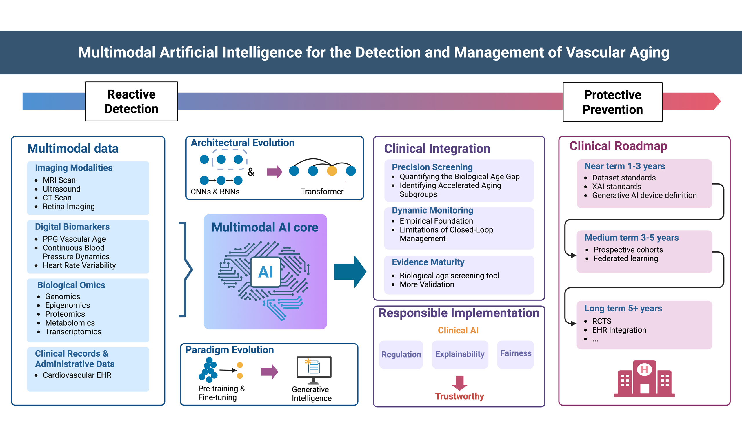
Graphical Abstract
Abstract
Vascular aging is a fundamental driver of cardiovascular diseases (CVD), yet identifying individuals undergoing accelerated vascular aging - and therefore at elevated cardiovascular risk - remains a significant clinical challenge. The traditional “one-size-fits-all” health risk assessments are often insensitive to early, subclinical vascular changes. The recent explosion of medical big data, coupled with the advent of Artificial Intelligence (AI) and Multimodal Foundation Models (MFMs), offer a transformative opportunity to shift vascular aging management from reactive detection toward proactive, predictive protection. This review systematically explores the integration of multimodal artificial intelligence (MMAI) and large-scale foundation models in the identification, stratification, and personalized management of vascular aging. We first delineate the multidimensional data ecosystem of vascular aging, including electronic health records (EHRs), imaging, omics, and continuous physiological signals from wearables, and analyze how deep learning architectures enable the detection of vascular aging by capturing subclinical structural and functional alterations. We then focus on the emerging role of MFMs, which transcend task-specific AI by integrating heterogeneous data types to provide holistic phenomapping. Furthermore, we discuss the utility of Large Language Models (LLMs) in clinical workflows, enhancing patient adherence through natural language interaction, and accelerating research through knowledge synthesis. Despite the potential of foundation models, their clinical translation faces technical and institutional barriers. We analyze four systemic challenges: algorithmic fairness, regulatory uncertainty, explainable AI (XAI) and the lack of real-world validation. We propose a strategic roadmap aligned with international frameworks that progresses from standardized performance reporting for elderly subgroups in the near term to cost-effective, individualized interventions that intercept the health-to-disease transition in the long term. The convergence of big data and foundation models represents a shift from reactive cardiovascular care to proactive, precision-based longevity management, positioning early, AI-enabled identification of high-risk vascular aging phenotypes as a primary target for the next generation of preventive medicine.
Keywords


 Download PDF
INTRODUCTION
Vascular aging
Vascular aging is a physiological phenomenon in which large arteries undergo structural and functional changes with advancing age[1]. Early vascular aging refers to, in populations with increased cardiovascular risk, the acceleration of these age-related changes[2,3]. Vascular aging is the result of multidimensional interactions between intrinsic and extrinsic factors across the life course. These interactions are associated with both structural and functional changes in the arterial wall[4]. Structural changes include vascular smooth muscle cell proliferation, irreversible elastin fragmentation, progressive collagen deposition, and advanced glycation end-product-mediated collagen cross-linking[5-7]. Functional changes include Vascular Smooth Muscle Cell (VSMC) phenotype switching, endothelial dysfunction, and perivascular adipose tissue dysfunction[8,9]. Large artery vascular aging leads to a range of adverse hemodynamic consequences: loss of arterial compliance increases pulsatility, which is transmitted to high-flow, low-resistance end organs, triggering vascular remodeling, perfusion imbalance, and organ-specific injury cascades in the heart, brain, and kidney that further accelerate disease progression[10-12]. Collectively, these processes represent a continuum from risk exposure to molecular and cellular dysfunction, culminating in clinical manifestations and measurable changes in diagnostic biomarkers [Figure 1]. Vascular aging has become one of the most fundamental drivers of cardiovascular disease, which remains the leading cause of death globally, making accurate assessment of vascular health essential for effective management and prevention strategies[13,14]. Traditional cardiovascular aging assessment relies on static, single-modality clinical indicators: structural changes are measured primarily by ultrasound assessment of arterial wall thickness and luminal diameter[15]; functional changes are assessed through flow-mediated dilation to measure endothelium-dependent vasodilation[16], and through arterial stiffness indices including the widely applied carotid-femoral pulse wave velocity and augmentation index[17]; serological markers can also reflect the degree of vascular aging to some extent[18,19]. However, vascular aging unfolds as a continuous and gradual process - a form of suboptimal health, or reversible, pre-pathological state preceding overt cardiovascular disease - rather than a discrete clinical event[20]. Because of this dynamic trajectory, these static clinical indicators are inherently limited in their ability to capture this progression, underscoring the rationale for early, artificial intelligence (AI)-enabled detection before structural damage becomes irreversible.
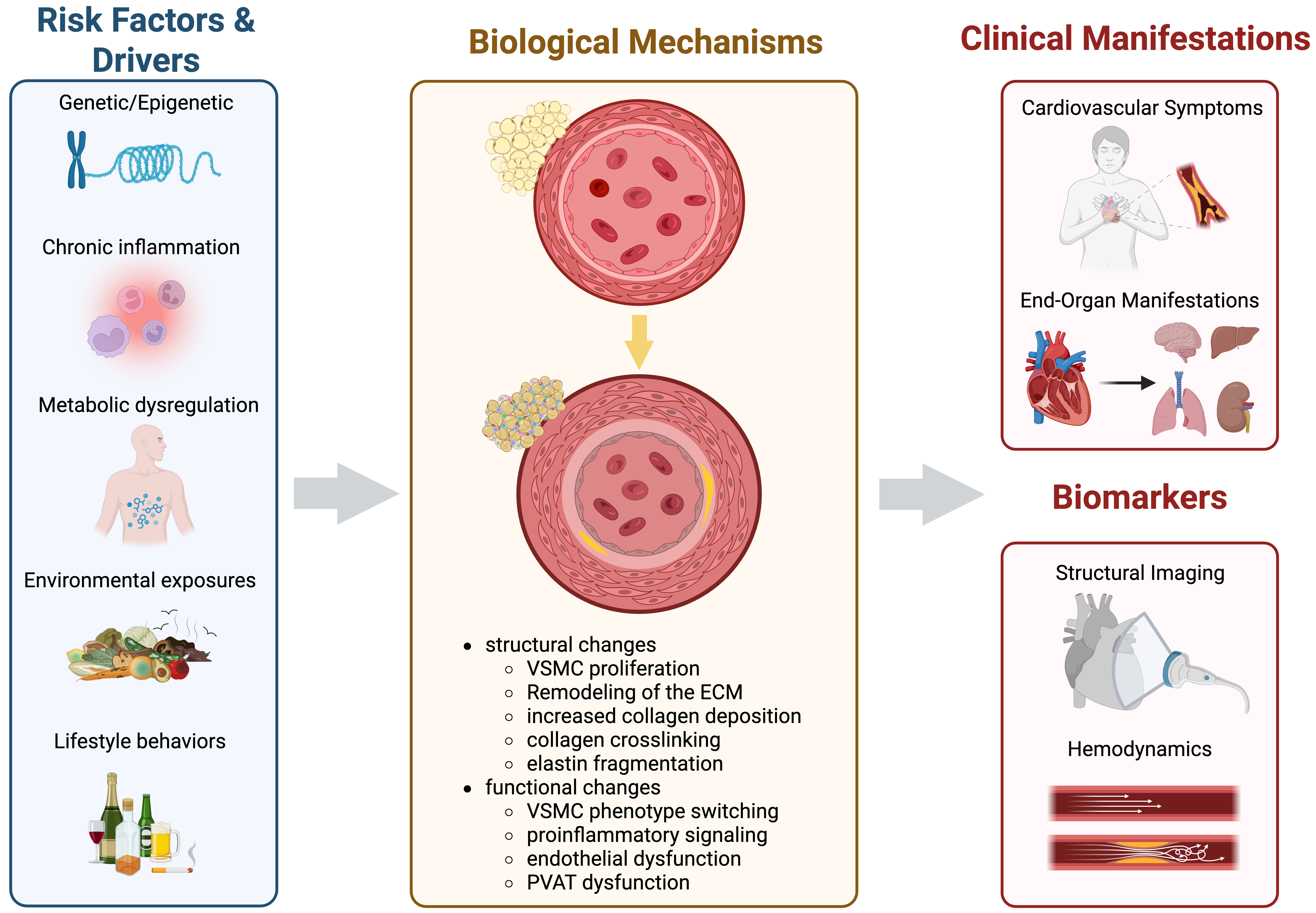
Figure 1. Schematic flow chart of the pathophysiological processes in vascular aging. The schematic illustrates the transition from risk factors (e.g., chronic inflammation, lifestyle behaviors, and metabolic dysregulation) to the molecular and cellular mechanisms. These processes ultimately culminate in clinical manifestations and alterations in diagnostic biomarkers. Created in BioRender. Wang Z (2026) https://BioRender.com/77p8ijz. VSMC: Vascular smooth muscle cell; ECM: extracellular matrix; PVAT: perivascular adipose tissue.
The digital revolution
Over the past decade, the rapid development of AI has profoundly transformed the landscape of biomedical research and clinical practice. Multimodal AI is reshaping the framework of next-generation healthcare by integrating large-scale data from diverse sources, including electronic health records (EHRs), medical imaging, wearable devices, and genomic data[21]. AI can reveal complex patterns and relationships that traditional methods struggle to detect[22], enabling precise quantification and prediction of cardiovascular aging[23,24]. Vascular aging is shaped by the interaction of multiple factors across biological scales, including genetic, metabolic, inflammatory, environmental, and behavioral dimensions[18,25], making assessment through the single clinical indicators typically used in practice prone to bias, yet well suited to a multimodal AI data-driven analytical paradigm[24]. Deep learning and large-scale foundation models are capable of processing these heterogeneous, high-dimensional data streams and extracting latent features and non-linear relationships that are imperceptible to human observers[26,27]. This capacity is progressively repositioning health risk assessment from a static, one-time classification exercise into a continuous, predictive process capable of anticipating the health-to-disease transition before clinical symptoms emerge.
Literature search strategy
A literature search was conducted in PubMed (MEDLINE) through April 2026, using combinations of the following search terms: “vascular aging”, “arterial stiffness”, “early vascular aging”, “biological age”, “epigenetic clock”, “proteomic aging”, “artificial intelligence” combined with “cardiovascular”, “electrocardiographic age”, “photoplethysmography age”, “wearable sensors”, “heart rate variability”, “multimodal artificial intelligence”, “large language models”, “explainable artificial intelligence”, and “sex differences” combined with “cardiovascular aging”. Google Scholar was used as a supplementary source to identify additional relevant articles, particularly recent preprints and conference proceedings not yet indexed in PubMed. No language restriction was applied beyond a preference for English-language publications. While the search focused primarily on literature published in the past decade (2015-2026) to capture rapid advances in AI-based vascular aging assessment, seminal earlier studies establishing foundational concepts and measurement techniques (e.g., carotid intima-media thickness ultrasound methodology) were also included regardless of publication date. Reference lists of key reviews were additionally screened to identify further relevant studies.
BIG DATA FOUNDATIONS FOR EARLY VASCULAR AGING
Vascular aging arises from systemic physiological perturbations spanning genetic, metabolic, inflammatory, and behavioral domains, rather than isolated organ-level dysfunction - underscoring the value of a holistic, multi-source assessment paradigm[28]. Currently, the medical field employs various methods to assess vascular aging, encompassing imaging modalities, omics, digital biomarkers, and clinical text data [Figure 2]. However, in clinical practice, these data dimensions have long been fragmented, making it difficult to form a comprehensive assessment of the degree of vascular aging. Multimodal AI can integrate these diverse and heterogeneous data, identifying cross-modal correlations that are difficult for humans to perceive, thereby enabling the prediction of early vascular aging. This diverse and massive amount of data also constitutes an indispensable data foundation for training multimodal AI models.
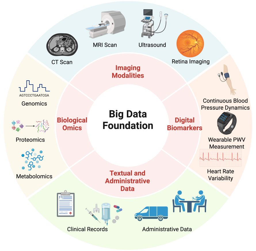
Figure 2. Multi-source data foundation in training multimodal medical AI. The diagram illustrates the integration of multi-source clinical data for AI training. Created in BioRender. Wang Z (2026) https://BioRender.com/w2g3l16. AI: Artificial intelligence; MRI: magnetic resonance imaging; CT: computed tomography.
Imaging modalities
Different imaging modalities capture the structural and functional features of vascular aging from complementary dimensions, providing indirect but clinically validated measures of age-related vascular changes.
Traditional clinical structural markers
Computed Tomography (CT) quantifies coronary artery calcification (CAC) using the Agatston method, stratifying individuals into four cardiovascular risk tiers and guiding preventive therapy decisions in asymptomatic populations[29]. As a clinical risk stratification tool, CAC scoring is well-validated and widely deployed. However, in the context of vascular aging assessment, calcification represents a downstream structural consequence of age-related and pathological vascular change rather than a direct measure of the aging process, limiting its specificity as a vascular aging biomarker for AI-based vascular evaluation. Multi-vendor variability further compounds this limitation, with studies demonstrating that differing CT scanner hardware can lead to risk reclassification in up to 6.5% of intermediate-risk individuals[30].
Ultrasound provides non-invasive structural assessment of vascular aging through measurement of carotid intima-media thickness (cIMT). cIMT has been shown to be a strong predictor of future vascular events[31]. However, an individual patient data meta-analysis demonstrated that mean carotid IMT is independently associated with the risk of cardiovascular composite [Hazard Ratio (HR) = 1.16], while the annual rate of cIMT progression showed a relatively weaker prognostic association[32]. These findings suggest that the association between cIMT progression and cardiovascular risk in the general population remains unproven, limiting its potential for precisely tracking the progression of vascular aging. Furthermore, ultrasound is operator-dependent, and standardization of measurements across different operators and instruments remains a significant challenge[33].
Functional and microvascular aging biomarkers
Magnetic resonance imaging (MRI) non-invasively quantifies aortic pulse wave velocity (PWV) and aortic strain using phase-contrast sequences. It provides precise measurement of local arterial stiffness with validated methodological frameworks supporting its clinical and research application[34]. A population-based 4D flow MRI study demonstrated a significant age-related increase in aortic PWV, from 4.93 ± 0.54 m/s in the 20-30 age group to 8.06 ± 1.03 m/s in the 70-80 age group, independent of blood pressure and cardiovascular risk factors[35]. These findings support the utility of MRI-derived PWV as a sensitive functional marker of age-related arterial stiffening, consistent with its role as a biomarker of vascular aging. However, the high cost and operational complexity of MRI result in relatively limited vascular aging MRI dataset sizes, constraining the training and generalization capacity of deep learning models.
Retinal imaging is currently the most mature vascular aging imaging modality in combination with artificial intelligence. The retinal microvasculature serves as a non-invasive window into systemic microvascular aging, and deep learning algorithms applied to fundus photographs have demonstrated the capacity to predict cardiovascular risk factors and outcomes well beyond conventional clinical assessment[36]. The deep learning-derived RetiPhenoAge marker showed significant independent associations with all-cause mortality (HR = 1.92), cardiovascular mortality (HR = 1.97), and cardiovascular events (HR = 1.70) across the UK Biobank and two external validation cohorts[37]. The primary challenge is interpretability: current AI retinal aging models largely operate as black-box systems. Establishing mechanistic links between extracted microvascular features and systemic vascular aging biology remains an open research frontier.
Biological omics
Biological omics data offer intrinsic molecular-level signatures of aging processes, capable of providing mechanistic insights into vascular and systemic senescence that routine imaging may not fully capture.
Genomics and epigenomics
Genomics and epigenomics currently represent the most mature omics pathway for quantifying biological age. Epigenetic clocks, including landmark tools such as the Horvath clock, PhenoAge, and GrimAge, estimate biological age from DNA methylation patterns and have demonstrated capacity to predict age-related disease risk and mortality beyond chronological age[38-40]. However, the predictive accuracy of epigenetic clocks is influenced by genetic background, lifestyle, environmental exposures, and technical variation[41]. Furthermore, most validated epigenetic clocks are derived from peripheral blood DNA[42], which may not accurately reflect the local epigenetic aging status of specific vascular tissues such as the aortic wall or coronary arteries. Epigenomics faces the challenge of optimizing predictive accuracy and improving the generalizability of these clocks across diverse populations and tissue types.
Proteomics
Proteomics captures dynamic changes in the vascular microenvironment by monitoring abundance changes in circulating plasma proteins. A proteomic clock based on 45,441 UK Biobank participants and 2,897 plasma proteins identified 204 proteins that accurately predicted chronological age in the held-out test set [n = 13,633; Pearson r = 0.94, Mean Absolute Error (MAE) = 2.24 years], with proteomic aging independently associated with the incidence of 18 major chronic diseases, including cardiac, hepatic, renal, and pulmonary conditions as well as multimorbidity and all-cause mortality risk[43]. Building on this, organ-specific proteomic clock research further revealed substantial heterogeneity in aging trajectories across organs at the proteomic level. Proteomic profiles across 13 human tissues spanning five decades revealed that blood vessels represent one of the earliest and most susceptible tissues to aging, with an inflection point emerging around age 50[44]. However, clinical application remains constrained by the cost barrier of high-throughput protein detection, the tissue specificity limitation that may incompletely reflect local arterial aging, and cross-platform technical variability[45].
Metabolomics and transcriptomics
Metabolomics constructs metabolic aging clocks through analysis of lipid metabolites, amino acid profiles, and glycerophospholipid metabolic changes. A targeted metabolomics study comparing plasma metabolic profiles between healthy young adults (aged 21-40 years, n = 75) and older individuals (aged 65 and above, n = 76) demonstrated age-associated impairment in fatty acid oxidation and amino acid metabolism[46]. These changes may point to potential biomarkers and metabolic pathways that reflect the physiological degradation associated with aging. However, challenges remain in optimizing standardization protocols and addressing population heterogeneity[47]. Transcriptomic approaches, including RNA sequencing and single-cell transcriptomics, have identified age-associated gene expression signatures in vascular smooth muscle cells and endothelial cells[48]; however, the clinical translation of transcriptomic biomarkers remains at an early stage.
Omics data provide high molecular resolution, but their clinical adoption still faces three core challenges: the high cost of sequencing and complex bioinformatic analysis pipelines; tissue specificity issues, whereby circulating biomarkers may not fully reflect the local aging status of specific large arteries; and difficulties in data standardization arising from inconsistent sample processing protocols across laboratories.
Looking further ahead, emerging biosensing paradigms - such as mitochondria-based physiological monitoring, proposed as a holistic indicator of systemic health status - may eventually complement imaging-based and omics-based vascular aging assessment, offering a pathway toward more cost-effective, individualized protection against the health-to-disease transition[28,49]
Digital biomarkers
Digital biomarkers derived from wearable devices and mobile health terminals offer the core advantage of capturing the dynamic fluctuation characteristics of the vasculature under the load of daily life, thereby describing a continuous vascular aging trajectory.
Photoplethysmography vascular age
Photoplethysmography (PPG) signals contain rich information on vascular elasticity and aging, making them one of the most scalable digital vascular aging biomarkers currently available. A deep learning model developed from 212,231 UK Biobank PPG recordings defines the difference between AI-PPG age and actual age (AI-PPG age gap) as a novel cardiovascular digital biomarker. Individuals with an age gap exceeding nine years show a significantly elevated risk of major adverse cardiovascular and cerebrovascular events (MACCE) (HR = 2.37, P = 8.46 × 10-80), with significant gradient associations across coronary heart disease, myocardial infarction, stroke, and all-cause mortality, and external validation cohorts confirmed its independent association with in-hospital mortality[50]. These findings were further presented at the European Society of Cardiology Congress, reinforcing the translational relevance of AI-PPG vascular age as a scalable cardiovascular screening tool[51].
Continuous blood pressure dynamics
Continuous blood pressure monitoring, enabled by ambulatory and wearable cuffless systems, captures hemodynamic fluctuations. In particular, it can unmask hypertensive phenotypes invisible to conventional clinic measurements, including masked hypertension and early morning hypertension, both of which carry independent risk for target organ damage[52-54]. Beyond mean pressure levels, real-time blood pressure dynamics captured by AI-enhanced wearable platforms provide novel insights into vascular aging trajectories, enabling individualized risk profiling that static measurements cannot offer[55,56].
Autonomic nervous aging and heart rate variability
Deterioration of the autonomic nervous system is an important driver of vascular aging and can be non-invasively quantified through heart rate variability (HRV). Decreases in time-domain indices Root Mean Square of Successive Differences (RMSSD) and Standard Deviation of Normal-to-Normal intervals (SDNN) reflect reduced vagal tone[57]. In frequency-domain analysis, the high-frequency component (HF,
Wearable arterial stiffness indices
Arterial stiffness, an independent predictor of cardiovascular risk[61], can be captured by wearable devices through multiple complementary indices. The comparative validity of noninvasive vascular aging indices has been systematically evaluated in a pioneering in silico study of 3,837 virtual subjects (aged 25-75 yr), benchmarking 19 pulse wave indices against aortic Young’s modulus (EAo) under simulated measurement noise. Carotid-femoral PWV, Cardio-Ankle Vascular Index (CAVI), and carotid distensibility demonstrated superior EAo correlations (rp > 0.95; rs < -0.99) and noise resilience, establishing them as preferred clinical markers of arterial stiffness. PPG-based indices, while most susceptible to noise and showing weaker EAo correlations (|rs| < 0.84), retain potential for continuous daily-life monitoring when signal quality is optimized[62]. Systematic review evidence confirms the considerable promise of consumer-grade wearable devices for cardiovascular screening, risk prediction, and continuous monitoring; however, a definitive assessment of their measurement accuracy is currently impeded by pervasive heterogeneity in research methodologies and outcome definitions[63]. Consumer-grade wearable devices provide a feasible data acquisition framework for large-scale population-level dynamic monitoring of vascular aging[64], but the establishment of standardized validation protocols and collaborative industry partnerships are necessary to enhance the reliability and practical applicability of wearable-based evaluations[63].
Clinical records and administrative data
Clinical medical record text and administrative data contain patients’ long-term health narratives and medical behavior trajectories, capturing subjective symptom descriptions, treatment adherence, and social determinant information that structured imaging and omics data cannot readily quantify. Compared with structured data, the unstructured components of cardiovascular disease-related EHRs - such as progress notes, discharge summaries, and imaging reports - typically contain richer contextualized clinical information, making this data source particularly well suited to natural language processing (NLP) applications in cardiology[65].
NLP and large language models in cardiovascular EHR
AI-based NLP technology can rapidly and automatically process the unstructured portions of EHRs - including physician notes, radiology reports, and discharge summaries - and has been applied across a range of cardiology tasks, including cardiovascular disease cohort identification, sign and symptom extraction, risk factor recognition, comorbidity identification, and clinical reasoning reconstruction, substantially reducing reliance on manual annotation[65,66]. At the level of multimodal data integration, Large Language Models (LLMs) have demonstrated capacity to jointly process clinical text alongside structured and semi-structured data sources, supporting risk stratification and clinical decision-making in cardiovascular care[67,68]. A multimodal deep learning study integrating admission notes with clinical tabular data from MIMIC-III (Medical Information Mart for Intensive Care III), MIMIC-IV (Medical Information Mart for Intensive Care IV), and the eICU Collaborative Research Database further confirmed that joint modeling of text and structured data significantly improved the prediction of in-hospital mortality in heart failure, with the multimodal model achieving an Area Under the Curve (AUC) of 0.838-0.849 across internal and prospective validation sets, outperforming unimodal approaches[69]. At the system level, LLMs have also shown potential for automated phenotyping, registry construction, and quality surveillance, with system-facing applications capable of processing large volumes of clinical narrative text to detect complex disease states and risk phenotypes, though safe deployment remains constrained by hallucinations, temporal obsolescence, and data privacy concerns[68].
FROM TASK-SPECIFIC AI TO MULTIMODAL FOUNDATION MODELS
This evolution can be understood along two intertwined but conceptually distinct axes: architectural evolution - from discriminative networks to the transformer, and training-paradigm evolution - from task-specific supervision to large-scale pretraining and generative synthesis, both of which converge in current Multimodal Foundation Models (MFMs). Table 1 provides a representative overview of AI architectures applied to vascular aging research across these developmental stages, summarizing study design, input modalities, cohort characteristics, and validation approaches.
Overview of AI architectures in vascular aging research
| Ref. | Author, year | Journal | Study design | Architecture | Training paradigm | Input modality | Cohort (n) | Key result | Validation |
| [37] | Nusinovici et al., 2024 | Lancet Healthy Longev. | Retrospective cohort development(from prospective biobank)& validation | Deep CNN (RetiPhenoAge) | End-to-end/from scratch | Retinal fundus photograph; PhenoAge composite | Train: 34,061; val1: 9,429; val2: 3,986; total: 47,476 | HR = 1.92 all-cause mortality; HR = 1.97 CV death; retinal biological age predicts outcomes | External; multi-cohort (UK Biobank + SEED + AREDS) |
| [55] | Guimarães et al., 2025 | Hypertension | Retrospective cohort | DNN + GBDT + Logistic Regression | End-to-end/from scratch | Office + ambulatory BP | n = 59,124 | AI-derived BP risk score outperforms conventional risk calculators for CV events | Internal (cross-validation) |
| [71] | Kim et al., 2025 | Eur Radiol. | Retrospective cohort | Fully automated DL | Pre-trained (commercial system) | ECG-gated CT + Non-gated chest CT | n = 652 paired CT (SNUH) + 425 ECG-gated + 199 non-gated (Stanford); multivendor | DL CAC scoring comparable to manual scoring in both CT types | External; multi-vendor |
| [72] | Attia et al., 2019 | Circ Arrhythm Electrophysiol. | Retrospective cohort | CNN | End-to-end/from scratch | 12-lead ECG | Train: n = 499,727; test: n = 275,056; longitudinal sub-cohort: n = 100 | AI-estimated ECG biological age correlates with chronological age; sex estimation feasible | Internal; single-center |
| [73] | Rawlani et al., 2025 | npj Digit Med. | Retrospective cohort | DL (echo clock; multi-view video CNN) | End-to-end/from scratch | Multi-view echocardiography | Train/val/test: 90,738 patients, 166,508 studies, 2,610,266 videos (CSMC); external: 5,528 (SHC) + 96,380 (KP) + 4,463 (CGMH) + 129 (KU) studies | AI echocardiographic biological age independently associated with cardiovascular risk | External; multi-national |
| [74] | Lima et al., 2021 | Nat Commun. | Retrospective cohort | ResNet (DNN) | End-to-end/from scratch | 12-lead ECG | Train/val/test: n = 1,558,415; external: n = 14,236 (ELSA-Brasil) + n = 1,631 (SaMi-Trop) | HR = 1.79; ECG age gap > 8yr → all-cause mortality | External; multi-cohort |
| [75] | Linardos et al., 2022 | Sci Rep. | Federated learning simulation | Federated learning + CNN | Pre-trained + Federated transfer learning | Cardiac cine MRI | n = 180 (4 simulated federated centers: vall d’Hebron n = 46, Sagrada Familia n = 70, SantPau n = 24, ACDC n = 40) | Federated learning achieves performance close to centralized training across sites | Internal simulation only (public datasets: M&M + ACDC; 4-center federated simulation) |
| [70] | Sarmun et al., 2024 | Eng Appl Artif Intell. | Retrospective cohort | Multi-stage feature fusion DL | End-to-end/from scratch | Carotid ultrasound (cIMT) | CUBS (public); n = 1,088 | Novel DL framework improves cIMT segmentation accuracy over single-stage methods | External; single-dataset |
| [103] | Hannun et al., 2019 | Nat Med. | Retrospective cohort | End-to-end deep neural network | End-to-end/from scratch | Single-lead ambulatory ECG | n = 53,549 (patients) | Cardiologist-level arrhythmia detection and classification from single-lead ECG | Internal |
| [36] | Poplin et al., 2018 | Nat Biomed Eng. | Retrospective cohort | Deep CNN (Inception-v3) | Pre-trained (ImageNet) + fine-tuned | Retinal fundus photograph | Training: n = 284,335; validation: n = 12,026 (dataset 1) + n = 999 (dataset 2); total: n = 297,360 | CV risk factors (age, sex, BP, smoking) predicted from retinal images via DL | Internal; temporal; dual-source |
| [50] | Nie et al., 2025 | Commun Med. | Retrospective cohort (from prospective biobank) + external validation | DL-based biological age clock | End-to-end/from scratch | Raw PPG signal (wearable device) | UK Biobank: n = 212,231; external: MIMIC-III-derived cohort n = 2,343; total: n = 214,574 | AI-PPG vascular age gap independently predicts MACCE and mortality | External; multi-cohort |
| [69] | Gao et al., 2024 | J Med Internet Res. | Retrospective cohort; prospective cohort | Multimodal DNN | End-to-end/from scratch | Admission notes; clinical tabular data | Dev: n = 9,989; internal val: n = 2,497; prospective val: n = 1,896; external val: n = 7,432; total: n = 21,814; | Multimodal DL outperforms unimodal models in heart failure outcome prediction | Internal; prospective; external |
| [78] | Rasmy et al., 2021 | npj Digit Med. | Retrospective cohort | Med-BERT (Transformer) | Pre-trained + fine-tuned | Structured EHR | Pretrain: 28,490,650; finetune: 2 independent clinical databases | Standard finetune: AUC + 1.21%-6.14% vs. DL without Med-BERT; small dataset scenario: AUC + > 20% or equivalent to model trained on 10× larger dataset | Internal (Cerner); external (truven) |
| [81] | Soenksen et al., 2022 | npj Digit Med. | Retrospective benchmark cohort; | HAIM modular fusion | Pre-trained + frozen probe | 4 modalities (tabular, time-series, text, images); 11 sources | n = 34,537 samples (6,485 patients; 7,279 hospitalizations) | +6%-33% over single-source across 12 clinical tasks | Internal; multi-task |
| [43] | Argentieri et al., 2024 | Nat Med. | Retrospective cohort | Gradient boosting | End-to-end/from scratch | Plasma proteomics | Train: n = 45,441 (UK Biobank); external val: n = 3,977 (CKB) + n = 1,990 (FinnGen); total: n = 51,408 | Pearson r = 0.94 (UKB); r = 0.92 (CKB); r = 0.94 (FinnGen); ProtAgeGap associated with heart disease, hypertension, diabetes and all-cause mortality | External; multi-cohort |
Architectural foundations I: discriminative AI
The evolution of AI in cardiovascular medicine marks a paradigm shift from “specialized tools” toward “general intelligence”. Early research relied primarily on discriminative AI, in which convolutional neural networks (CNNs) dominated medical image feature extraction. Deep learning algorithms have since achieved automated segmentation and quantification of carotid intima-media complex from ultrasound[70] and fully automated CAC scoring from both ECG-gated and non-gated low-dose chest CT, providing quantitative vascular aging information without dedicated scanning protocols[71]. Concurrently, recurrent neural networks (RNNs) and their long short-term memory (LSTM) variants were widely applied to time-series signals such as the ECG to estimate individual “cardiac age” - a biological age surrogate derived from the divergence between AI-estimated and chronological age[72]. This approach was further extended to echocardiography-derived cardiac age estimation as an independent cardiovascular risk marker[73]. Lima et al. demonstrated in 1,558,415 individuals that an ECG age gap exceeding eight years independently predicted all-cause mortality (HR = 1.79), establishing a large-scale proof that discriminative AI could extract clinically actionable biological aging signals from a single physiological signal[74].
However, these models were designed for single tasks and struggled to integrate heterogeneous patient data across modalities. They confronted a fundamental “data silo” problem: institutions could not pool training data without compromising patient privacy and no individual site possessed sufficient data diversity to achieve population-level generalization. Federated learning emerged as a partial solution. Simulated multi-center studies in cardiovascular imaging demonstrated that federatively trained models achieved substantially more consistent performance across repeated initializations than centrally pooled models, reflecting increased robustness in output stability; however, under out-of-domain (leave-center-out) evaluation, federated models proved more sensitive to domain shift, with performance declining once shape and intensity augmentations were introduced[75]. Yet the core constraint remained: discriminative AI optimized for one task could not transfer its learned representations to another, limiting its utility in the face of the true complexity of cardiovascular aging phenotypes.
Architectural foundations II: the transformer revolution
The advent of Transformer architecture resolved the cross-modal representation problem that constrained discriminative AI. Unlike CNNs and RNNs, Transformers employ self-attention mechanisms that capture long-range dependencies by computing pairwise relationships between all sequence elements simultaneously, enabling context-sensitive representations irrespective of the distance between positions[76]. It is a property particularly advantageous for genomic[77] and EHR data[78] where a feature's importance is inseparable from its context[26]. For vascular aging research, cross-modal learning enables imaging, omics and text data to be projected into a unified latent space, where non-linear associations that are invisible within any single modality become detectable[79].
The quantitative evidence for this architectural advance is now consolidated. A scoping review of 432 deep learning-based multimodal AI papers published between 2018 and 2024 found that multimodal AI models consistently outperformed unimodal counterparts, with an average AUC improvement of 6.2 percentage points[80]. This gain reflects genuine information complementarity across modalities rather than model complexity alone. Soenksen et al. benchmarked 14,324 independent models derived from HAIM-MIMIC-MM - a multimodal clinical database comprising 34,537 samples across 7,279 unique hospitalizations and 6,485 patients - spanning 4 data modalities (tabular, time-series, text, and images), 11 data sources, and 12 clinical prediction tasks; multimodal inputs improved performance by 6%-33% over single-source approaches[81], with Shapley value attribution confirming that different modalities contributed differentially across tasks. Together, these findings established the empirical foundation for multimodal AI as a necessary, not merely aspirational, architectural upgrade.
Training paradigm shift I: pre-training and fine-tuning
Building on the Transformer architecture introduced above, the power of MFMs further derives from the pre-training and fine-tuning paradigm. General representations acquired from large-scale corpora are subsequently refined on targeted cardiovascular datasets, enabling detection of subtle vascular aging signals that would be unattainable through task-specific training alone[26]. The canonical biomedical instantiation is BiomedCLIP, a multimodal foundation model pre-trained on PMC-15M - 15 million biomedical image-text pairs collected from 4.4 million PubMed Central articles - via contrastive learning with domain-specific adaptations tailored to biomedical vision-language processing[82]. Benchmarked across retrieval, classification, and visual question-answering tasks, BiomedCLIP achieved state-of-the-art results across a wide range of standard biomedical datasets, substantially outperforming prior approaches[82]. This substantially alleviates the annotation bottleneck that previously limited discriminative AI: rather than requiring large-scale labeled vascular aging cohorts, a fine-tuned foundation model can identify clinically relevant biological signals from a substantially smaller number of well-characterized examples.
The clinical impact of this paradigm is demonstrated by foundation models pre-trained on large-scale imaging repositories and adapted to novel downstream tasks. Nielsen et al. applied RETFound - a vision transformer pre-trained via masked autoencoding on large-scale retinal fundus images - to retinal age prediction by fine-tuning only a lightweight regression head, achieving performance on par with fully centralized training even under federated and distributed learning settings[83]. Together, these studies confirm that the pre-training and fine-tuning paradigm transfers effectively to vascular biological age estimation, producing accurate and scalable biomarkers without requiring full model retraining.
Training paradigm shift II: generative intelligence
Within this same Transformer-based paradigm, generative intelligence further endows MFMs with the capacity to synthesize information, moving beyond the discriminative outputs described in Section Architectural foundations I: discriminative AI. Rather than returning a single “vascular age” value, a generative model can integrate findings across imaging, laboratory, and wearable data with relevant medical literature to produce structured diagnostic reports, transforming AI from a passive classifier into an active clinical reasoning tool[84,85].
The LLM represents the primary vehicle for this transition. Singhal et al. demonstrated that Med-PaLM encoded sufficient clinical knowledge to achieve performance comparable to physicians on medical licensing benchmarks, establishing that LLMs could serve as the integrative reasoning layer in a multimodal clinical system[86]. When augmented with Retrieval-Augmented Generation (RAG), LLMs can dynamically retrieve patient-specific evidence from unstructured clinical records, converting heterogeneous EHR data[87] into a structured vascular health timeline - a transformation that would require prohibitive manual annotation using discriminative approaches alone.
Characteristics and limitations of current clinical applications
Existing MFMs can achieve high detection accuracy under controlled conditions and consistently outperform traditional risk scores in multi-center validation[80]. However, the current clinical landscape is characterized by a clear “laboratory ahead of clinic” pattern, driven by three unresolved institutional barriers.
First, the “black-box” interpretability problem: clinicians are frequently unable to trace the reasoning pathway by which an MFM reaches an early vascular aging (EVA) conclusion. Explainability methods such as SHapley Additive exPlanations (SHAP) and Gradient-weighted Class Activation Mapping (Grad-CAM) exist but lack standardized evaluation frameworks, and their impact on clinical trust and decision quality has rarely been assessed in real care environments[88].
Second, the hallucination risk of generative models: when fabricated clinical details are embedded in prompts, LLMs elaborate on the false information in 50%-83% of cases across leading models, with mitigation prompts halving but not eliminating the error rate[89]. In a structured clinical summarization framework, hallucination and omission rates of 1.47% and 3.45%, respectively, were observed across 12,999 clinician-annotated sentences[90] - rates that, while low in absolute terms, carry serious patient safety implications when deployed at scale in cardiovascular risk management.
Third, and most critically, the randomized controlled trials (RCTs) evidence gap: a systematic review of cardiovascular AI trials identified only 11 RCTs meeting inclusion criteria, conducted between 2021 and 2024. Among these, 6 studies (54.5%) reported improvements in diagnostic accuracy and early detection, 5 (45.5%) demonstrated improvements in clinical events, and 3 (27.3%) showed enhanced resource utilization[91]. No prospective RCT has yet demonstrated that MFM-guided vascular aging interventions improve long-term cardiovascular outcomes, leaving a critical evidence gap specific to MFM-guided EVA interventions.
CLINICAL INTEGRATION
Multimodal AI systems are progressively being integrated across the full spectrum of clinical decision-making. The diverse data types that clinicians rely upon, including patient history, physical signs, imaging, and laboratory data, constitute the natural domain of multimodal artificial intelligence (MMAI). However, data heterogeneity and integration complexity remain the principal barriers to clinical implementation[84]. In the field of early vascular aging, the clinical integration of MMAI can be understood along three logical processes: screening, monitoring, and intervention, though it must be noted that the current maturity of evidence differs substantially across these three domains[80].
Precision screening: quantifying the biological age gap and identifying accelerated aging subgroups
The core challenge of cardiovascular aging screening is to capture the discrepancy between biological age and chronological age in the asymptomatic stage and convert it into actionable clinical risk signals.
The first pathway is evidence from single-modality biological age clocks. Deep learning models based on 12-lead ECG have been confirmed in large-scale cohorts to produce an AI-estimated ECG age that is an independent predictor of mortality in cardiovascular and acute care patients, outperforming traditional clinical scores[74]. A systematic review and meta-analysis of 17 studies further confirmed that an elevated heart delta age is independently associated with all-cause mortality (HR = 1.62, 95%CI: 1.49-1.77) and cardiovascular mortality (HR = 2.12, 95%CI: 1.71-2.63)[92]. Individuals with an AI-PPG age gap exceeding nine years show significantly elevated MACCE risk (HR = 2.37) in the UK Biobank cohort[50]. Both signal types are acquirable through standard clinical devices or consumer-grade wearable devices, making the screening potentially low-cost[93]. At the retinal microvascular level, the AI-derived retinal biological age marker RetiPhenoAge has been independently validated across three cohorts for its association with all-cause mortality (HR = 1.92) and cardiovascular mortality (HR = 1.97), with predictive performance exceeding traditional aging markers such as grip strength, telomere length, and physical activity[37].
The second pathway is multimodal feature fusion for risk stratification. MMAI approaches integrating large biobanks, EHRs, medical imaging, wearable sensors, and genomic data can capture the inherent complexity of human health and disease, demonstrating systematic advantages over single biomarkers in personalized medicine and risk stratification[81,84]. Validation of an early vascular aging construct model using external risk indicators confirmed that multidimensional integration of clinical and biochemical biomarkers improves comprehensive cardiovascular risk assessment beyond traditional single-marker approaches[94]. However, the performance advantage of multimodal models, an average AUC improvement of 6.2 percentage points, remains infrequently confirmed in external validation studies, with most existing research relying on internal validation[80], indicating that the generalization capacity of current screening models awaits systematic evaluation.
Dynamic monitoring and early intervention: empirical foundation and limitations of closed-loop management
MMAI holds potential in preventive medicine by integrating demographic characteristics, social determinants, immune status, omics, imaging, and pharmacogenomic data to identify high-risk individuals who can benefit from early intervention, enabling continuous health trajectory tracking based on real-time data streams.
In the area of dynamic monitoring combining wearable devices with AI, the evidence chain is forming but remains incomplete. AI-driven real-time monitoring systems have been demonstrated to provide early warning hours before events such as atrial fibrillation or ischemia occur, with a scoping review of 47 studies confirming that AI-based wearable monitoring significantly shortens the time from anomaly detection to clinical intervention across multiple cardiovascular conditions[95]. New-generation flexible bioelectronic devices can now attach stably to the skin for extended periods, acquiring high-fidelity cardiovascular signals without compromising patient quality of life and providing high-quality input data for AI algorithms[96]. In heart failure management, a systematic review confirmed that remote monitoring combined with AI analysis effectively reduces rehospitalization rates by identifying early subtle changes in fluid retention and hemodynamics[97]. However, this evidence derives predominantly from single-disease cohorts, and long-term closed-loop management data specifically targeting the continuous process of early vascular aging remain absent.
At the level of therapeutic decision support, AI-based tools have shown auxiliary value in personalizing blood pressure targets[98] and continuously monitoring treatment adherence and response[99] and in management of complex dyslipidemia, where AI-assisted systems demonstrate more rigorous adherence to current guidelines and recommendation of more aggressive combination lipid-lowering regimens compared with human experts[100]. In cardiovascular pharmacotherapy more broadly, AI has shown potential in predicting drug interactions, assessing adverse reaction risks, and optimizing dosing regimens in complex antithrombotic and lipid-lowering therapies[101]. However, as of November 2024, only 11 RCTs meeting inclusion criteria have been identified in cardiovascular AI, of which 54.5% showed enhanced diagnostic accuracy and early detection and 45.5% reported improvements in clinical events, mostly concentrated on single-modality scenarios such as ECG analysis and CT assessment[91]. High-quality RCT evidence that multimodal AI-driven intervention strategies improve long-term vascular aging outcomes remains entirely absent.
Integrated assessment of evidence maturity
Foundation model-driven MMAI systems such as LLaVA-Med and BiomedCLIP provide flexible and potentially generalizable reasoning capabilities, but their clinical translation still requires overcoming four challenges: computational resource demands, domain adaptation, interpretability, and rigorous real-world validation[82,102].
Synthesizing the available evidence, the maturity of MMAI in clinical integration for early vascular aging can be summarized as a gradient: biological age quantification and risk stratification - evidence relatively robust, with consistent conclusions supporting multimodal superiority over unimodal approaches, as supported by previous reviews[22,84] - progressing to dynamic monitoring and early warning - evidence accumulating, single-disease data relatively sufficient but vascular aging-specific data limited[72,95,103] - and finally to personalized therapeutic decision support - proof-of-concept stage, lacking prospective RCTs specifically targeting early vascular aging[21,104]. This gradient suggests that the highest-value clinical translation entry point at present is to position MMAI as a biological age screening tool for asymptomatic high-risk populations rather than as a direct therapeutic decision system. Table 2 summarizes the clinical evidence supporting AI-driven vascular aging assessment across major clinical domains, including study design, cohort characteristics, validation status, and an overall assessment of evidence maturity for each application area.
Clinical evidence of AI-driven vascular aging assessment by clinical domain
| Domain | Ref. | Author, year | Journal | Study design | Input modality | Cohort (n) | Key result | Validation | Evidence maturity |
| I · Vascular age estimation | [74] | Lima et al., 2021 | Nat Commun. | Retrospective cohort | 12-lead ECG | Train/val/test: n = 1,558,415; external: n = 14,236 (ELSA-Brasil) + n = 1,631 (SaMi-trop) | HR = 1.79; ECG age gap > 8yr → all-cause mortality | External; multi-cohort | Strong |
| I · Vascular age estimation | [92] | Mossavarali et al., 2025 | npj Digit Med. | Systematic review & meta-analysis | 12-lead ECG | 17 studies | AI-ECG age gap consistently predicts CV events and all-cause mortality across cohorts | Systematic | Strong |
| I · Vascular age estimation | [72] | Attia et al., 2019 | Circ Arrhythm Electrophysiol. | Retrospective cohort | 12-lead ECG | Train: n = 499,727; test: n = 275,056; longitudinal sub-cohort: n = 100 | AI-estimated ECG biological age correlates with chronological age; sex estimation feasible | Internal; single-center | Emerging |
| I · Vascular age estimation | [73] | Rawlani et al., 2025 | npj Digit Med. | Retrospective cohort | Multi-view echocardiography | Train/val/test: n = 90,738 patients, 166,508 studies, 2,610,266 videos (CSMC); external: 5,528 (SHC) + 96,380 (KP) + 4,463 (CGMH) + 129 (KU) studies | AI echocardiographic biological age independently associated with cardiovascular risk | External; multi-national | Strong |
| I · Vascular age estimation | [50] | Nie et al., 2025 | Commun Med. | Retrospective cohort (from prospective biobank) + external validation | PPG (wearable) | UK Biobank: n = 212,231; external: MIMIC-III-derived cohort n = 2,343; total: n = 214,574 | AI-PPG vascular age gap independently predicts MACCE and all-cause mortality | External; multi-cohort | Strong |
| I · Vascular age estimation | [37] | Nusinovici et al., 2024 | Lancet Healthy Longev. | Retrospective cohort development (from prospective biobank) & validation | Retinal fundus photograph | Train: n = 34,061; val1: n = 9,429; val2: n = 3,986; total: n = 47,476 | HR = 1.92 all-cause mortality; HR = 1.97 CV death; retinal biological age predicts outcomes | External; multi-cohort (UK Biobank + SEED + AREDS) | Strong |
| I · Vascular age estimation | [43] | Argentieri et al., 2024 | Nat Med. | Retrospective cohort | Plasma proteomics | Train: n = 45,441 (UK Biobank); external val: n = 3,977 (CKB) + n = 1,990 (FinnGen); total: n = 51,408 | Proteomic age clock predicts mortality and 18 age-related diseases across diverse populations | External; multi-cohort | Strong |
| II · Structural vascular imaging | [71] | Kim et al., 2025 | Eur Radiol. | Retrospective cohort | ECG-gated CT + non-gated chest CT | n = 652 paired CT (SNUH) + 425 ECG-gated + 199 non-gated (Stanford) | DL CAC scoring comparable to manual scoring in both CT types | External; multi-vendor | Moderate |
| II · Structural vascular imaging | [36] | Poplin et al., 2018 | Nat Biomed Eng. | Retrospective cohort | Retinal fundus photograph | Train: n = 284,335 (UK Biobank + EyePACS 2007-2015); val: n = 12,026 (UK Biobank) + n = 999 (EyePACS May 2015-October 2015); total: n = 297,360 | CV risk factors (age, sex, BP, smoking) predicted from retinal fundus images via DL | Internal; temporal; dual-source | Emerging |
| III · Functional cardiovascular monitoring | [103] | Hannun et al., 2019 | Nat Med. | Retrospective cohort | Ambulatory ECG | n = 53,549 (patients) | Cardiologist-level arrhythmia detection and classification from single-lead ECG | Internal | Emerging |
| III · Functional cardiovascular monitoring | [55] | Guimarães et al., 2025 | Hypertension | Retrospective cohort | Office + ambulatory BP | n = 59,124 | AI-derived BP risk score outperforms conventional risk calculators for cardiovascular mortality risk | Internal (cross-validation) | Emerging |
| III · Functional cardiovascular monitoring | [75] | Linardos et al., 2022 | Sci Rep. | Federated learning simulation | Cardiac cine MRI | n = 180 (4 simulated federated centers: vall d’Hebron n = 46, Sagrada Familia n = 70, SantPau n = 24, ACDC n = 40) | Federated learning achieves performance comparable to centralized training across sites | Internal simulation only (public datasets: M&M + ACDC; 4-center federated simulation) | Emerging |
| IV · Multimodal data fusion | [81] | Soenksen et al., 2022 | npj Digit Med. | Retrospective benchmark cohort | 4 modalities (tabular, time-series, text, images); 11 sources | n = 34,537 samples (6,485 patients; 7,279 hospitalizations) | +6%-33% performance over single-source models across 12 clinical tasks | Multi-task; internal | Emerging |
| IV · Multimodal data fusion | [69] | Gao et al., 2024 | J Med Internet Res. | Retrospective cohort; prospective cohort | Admission notes + clinical tabular data | Dev: n = 9,989; internal val: n = 2,497; prospective val: n = 1,896; external val: n = 7,432; total: n = 21,814 | Multimodal DL outperforms unimodal models in heart failure outcome prediction | Internal; prospective; external | Moderate |
| IV · Multimodal data fusion | [80] | Schouten et al., 2025 | Med Image Anal. | Scoping review | Multimodal (all modalities) | 432 studies | AUC +6.2% multimodal vs. unimodal across clinical tasks including CV | Scoping | Strong |
| V · Foundation models & LLMs | [78] | Rasmy et al., 2021 | npj Digit Med. | Pretraining + retrospective evaluation | Structured EHR | Pretrain: 28,490,650 (cerner); finetune: DHF-cerner n = 672,647 + PaCa-cerner n = 29,405 + PaCa-truven n = 42,721 (external) | Standard finetune: AUC +1.21%-6.14% vs. DL without Med-BERT; small dataset scenario: AUC + > 20% OR equivalent to model trained on 10× larger dataset | Internal (cerner); external (truven) | Strong |
| V · Foundation models & LLMs | [26] | Moor et al., 2023 | Nature | Perspective/framework | Multimodal medical data | - | Framework defining foundation model roles and requirements in generalist medical AI | Landmark paper | Foundational |
| VI · Clinical outcome & RCT evidence | [91] | Hadida Barzilai et al., 2025 | JACC Adv. | Systematic review of RCTs | Mixed CV AI interventions | 11 RCTs | 45.5% RCTs improved clinical events; 54.5% improved diagnostic accuracy & early detection; 27.3% improved resource utilization; overall AI shows promise but high-quality RCT evidence remains critically limited | Systematic; limited RCTs; no meta-analysis | Moderate |
CHALLENGES, ETHICS, AND THE ROADMAP TO CLINIC
The clinical translation of MMAI in cardiovascular aging faces barriers that are not only technical but institutional and ethical. Translating models from the laboratory to clinical practice requires simultaneously addressing four intertwined systemic challenges - algorithmic fairness, regulatory frameworks, interpretability, and real-world validation - which collectively constitute the multifaceted barriers impeding MMAI clinical adoption[105].
Algorithmic fairness and digital ageism
MMAI in vascular aging assessment faces a unique internal paradox: its core target population - elderly individuals - is precisely the group most likely to be systematically underrepresented in training data. Digital ageism can occur throughout the full lifecycle of medical AI: historical bias arises from training data originating from an era of low digital participation among older adults; representational bias arises from the longstanding systematic exclusion of elderly individuals from clinical trials, resulting in models trained on younger populations being applied to older ones[106,107].
At the empirical level, earlier research found that a commercial risk prediction algorithm assigned similar risk scores to Black patients with more severe disease and White patients with less severe disease, resulting in the systematic under-referral of the former to complex care programs[108]. This bias carries particular harm in the vascular aging context: if models systematically misestimate biological age in elderly populations, the consequence is failure of precision screening in the highest-risk group, not merely reduced generalization performance. The STANDING Together international consensus recommendations - developed with input from over 350 representatives from 58 countries - explicitly identify the core pathway for addressing dataset bias as mandating transparent reporting of training dataset limitations and prospective evaluation of performance across population subgroups[109], a framework directly applicable to data governance for vascular aging AI.
Beyond age and race, sex remains an underexamined axis of bias in vascular aging AI. Cardiovascular aging is sex-dimorphic: women present atypically before menopause but converge toward male-typical presentation afterward, and their age-specific mortality rises steadily across the lifespan even as men’s declines after midlife[110]. Guidelines have been slow to reflect this - a 2002-2024 audit of 34 European Society of Cardiology (ESC) guidelines found menopause largely overlooked despite its cardiovascular significance, and blood pressure targets remain sex-neutral[111].
AI offers a practical path to close this gap. Natural language processing and federated learning can enable sex-stratified reanalysis of existing study data, while multimodal machine-learning models can integrate imaging, hemodynamic, and clinical variables into sex-specific risk scores. A UK Biobank study exemplifies this approach: by combining cardiac MRI, pulse wave analysis, ECG, and carotid ultrasound data, the resulting model outperformed the traditional Framingham Risk Score and quantified the extent of cardiovascular disease underdiagnosis in women[112]. Yet most vascular age AI models still treat sex only as an adjustment covariate rather than a stratification variable[113]. Sex-specific modeling can reveal genuinely distinct risk targets: an AI model of epicardial adipose tissue found density-based features drove risk more in women and volume-based features more in men, consistent with women’s earlier, inflammatory-stage disease[114]; the HUNT Study trained separate sex-specific models motivated by female-specific risk factors like premature menopause[115]; and the ongoing CARAMEL protocol targets AI-based risk tools specifically for menopausal women[116]. These efforts show sex-specific AI modeling is feasible and informative but still far from standard - a gap that MMAI development in vascular aging should close by reporting sex-stratified performance and modeling inflection points like menopause explicitly.
Regulatory framework: institutional challenges for foundation models
As of December 20, 2024, the Food and Drug Administration (FDA) had granted 1,016 AI/Machine Learning (ML) medical device authorizations encompassing 736 unique devices. Among signal-based devices, cardiovascular medicine represents the largest specialty, accounting for 64.5% of this category[117]. As of April 3, 2026, the FDA’s AI/ML-Enabled Medical Devices list included 1,431 authorized devices, of which 1,377 (96.2%) were cleared via the 510(k) substantial equivalence pathway. The remaining 54 devices (3.8%) received authorization through more rigorous regulatory routes, including De Novo classification (n = 37, 2.6%) and Premarket Approval (PMA) (n = 17, 1.2%)[118]. This reveals the core tension in the current regulatory landscape: the threshold for AI medical device market entry is relatively low, while the potential clinical impact may be far-reaching.
The FDA’s Total Product Life Cycle (TPLC) regulatory framework requires manufacturers to submit Predetermined Change Control Plans (PCCPs) that pre-specify the boundaries of permissible algorithm modifications in pre-market applications, representing a shift toward proactive lifecycle management of AI-enabled medical devices. Transparency in the premarket development process is equally critical; sponsors must be transparent about AI use and regulators must develop proficiency in evaluating AI-based submissions. However, the emergence of generative AI and LLMs fundamentally challenges existing frameworks, necessitating special evaluation mechanisms that current regulatory structures were not designed to accommodate[119]. Compounding these challenges, deep learning models are widely characterized as “black box” systems that lack transparency and interpretability, creating a gap between algorithmic outputs and the understanding required by clinicians and regulators[120]. These limitations underscore the need for explainable AI (XAI) approaches that render model outputs both transparent and understandable to end users[88]. At the international level, regulatory frameworks across countries differ substantially, posing institutional barriers to cross-national data sharing and international market access[121].
Explainability: from technical tool to clinical institutional need
The value of XAI in MMAI vascular aging applications lies not only in improving physician understanding, but in satisfying three institutional requirements simultaneously: regulatory compliance, patient right to information, and clinical judgment[122,123]. However, current XAI practice falls substantially short of these requirements[88]. A systematic review of XAI methods in medical image analysis found that while explainability approaches such as SHAP and Grad-CAM are increasingly adopted to address transparency and trust requirements in clinical AI, standardized evaluation frameworks remain absent, and the medicolegal and clinical implications of explanation quality are rarely systematically assessed[124,125]. To address this standardization gap, the first clinician-informed XAI evaluation checklist with metrics (CLIX-M) has been proposed, comprising 14 items across four categories - purpose, clinical attributes, decision attributes, and model attributes[126]. By aiding authors and reviewers in the transparent reporting and evaluation of XAI methods in AI-powered clinical decision support systems, CLIX-M represents a critical step in the evolution of XAI from a technical tool to an institutional standard.
In the specific context of vascular aging, XAI encounters challenges that extend beyond those faced in conventional diagnostic AI. Biological age is a continuous, multidimensional construct shaped by the interplay of hemodynamic, structural, and metabolic factors[127]. Although XAI can identify the individual biomarker signatures that most strongly influence a vascular age prediction for a given patient, a single feature importance ranking remains insufficient for clinical interpretation. Vascular aging reflects systemic interdependencies - across hemodynamic, metabolic, and inflammatory pathways - that require integrated, multi-biomarker explanations rather than isolated feature rankings, a challenge that current XAI methods have yet to fully resolve[128].
Clinical translation roadmap: from evidence to institutional embedding
In light of the above challenges, the clinical translation roadmap for MMAI in early vascular aging should encompass the following institutional milestones.
Near term (1-3 years): Standardization and Regulatory Foundations. Establish standardized reporting norms for MMAI vascular aging assessment datasets drawing on the STANDING Together framework[109], mandating stratified performance reporting for elderly subgroups; drive FDA/European Medicines Agency (EMA) clarification of device definitions for generative AI-assisted vascular risk reporting[119]; and promote adoption of the CLIX-M checklist as a minimum XAI reporting standard for cardiovascular AI submissions[126].
Medium term (3-5 years): Prospective Evidence and Federated Infrastructure. Conduct multi-center prospective cohort studies[74]; establish cross-institutional federated learning networks to accumulate training data from diverse elderly populations under a privacy-protective framework[129,75].
Long-term (beyond 5 years): Clinical Integration and Global Harmonization. Design RCTs with long-term cardiovascular event improvement as the endpoint to verify the net clinical benefit of MMAI-driven personalized intervention strategies[91]; embed AI vascular aging scores into electronic health record systems for seamless integration with routine clinical workflows[78]; and establish cross-national regulatory mutual recognition mechanisms to advance global market access coordination for MMAI products[122].
The three horizons are deliberately sequential yet overlapping: near-term standardization creates the conditions for medium-term evidence generation, which in turn provides the outcome data necessary to justify long-term systemic integration. Progress across all three domains is interdependent, and delays in regulatory clarification or federated infrastructure development will propagate forward, compressing the timeline available for the high-quality RCT evidence that long-term clinical embedding ultimately requires.
Beyond these procedural dependencies, the roadmap represents a broader clinical ambition: to reorient cardiovascular preventive medicine from reactive disease management toward proactive, biology-informed risk stratification, in which vascular aging trajectories are identified and modulated years before irreversible damage accumulates [Figure 3]. At the population level, scalable MMAI vascular aging screening could translate individual-level precision into measurable reductions in cardiovascular disease burden.
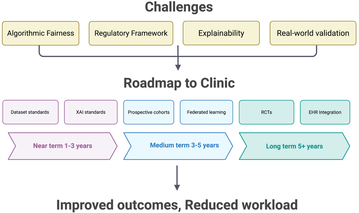
Figure 3. Roadmap for the clinical integration of multimodal AI. The schematic identifies primary obstacles to clinical implementation, including algorithmic fairness, regulatory hurdles, model explainability and real-world validation. A tiered roadmap is proposed over a horizon extending beyond 5 years, transitioning from the establishment of dataset and Explainable AI (XAI) standards in the near term to large-scale randomized controlled trials (RCTs) and seamless EHR integration in the long term. The ultimate objective of this pipeline is to enhance clinical outcomes while simultaneously reducing provider workload. Created in BioRender. Wang Z (2026) https://BioRender.com/o1iqjcw. AI: Artificial intelligence; EHR: electronic health record.
CONCLUSION
This review demonstrates that MMAI, integrating imaging, omics, digital biomarkers, and clinical data, may overcome the limitations of traditional static, single-modality assessment. Biological age clocks derived from PPG, ECG, and retinal imaging have achieved externally validated prognostic performance, with multimodal models improving AUC by an average of 6.2 percentage points over unimodal approaches.
Despite this promise, key barriers persist: underrepresentation of elderly and minority populations in training data, immature regulatory and explainability frameworks, and the absence of prospective RCTs demonstrating improved outcomes under MMAI guidance. Addressing these barriers will require progress along the roadmap outlined above - establishing standardization and regulatory foundations, building prospective evidence and federated infrastructure, and ultimately achieving clinical integration and global harmonization. Technical progress must be matched by algorithmic fairness, regulatory transparency, and rigorous clinical validation. With the convergence of big data and foundation models, vascular aging is positioned as a potential target for next-generation preventive medicine. Realizing this potential now depends on building the necessary institutional infrastructure.
DECLARATIONS
Acknowledgments
The Graphical Abstract was created in BioRender. Wang Z (2026) https://BioRender.com/dgfzcz5.
Authors’ contributions
Made substantial contributions to the conception and design of the study and performed data analysis and interpretation: Wang Z, Dai Y, Li C, Hu Y
Performed data acquisition and provided administrative, technical, and material support: Ding Z, Huang Z, Chen T
Availability of data and materials
Not applicable.
AI and AI-assisted tools statement
During the preparation of this manuscript, the AI tool Claude (version Claude Sonnet 5, Anthropic, released 2026-06-30) was used solely for language editing. The tool did not influence the study design, data collection, analysis, interpretation, or the scientific content of the work. All authors take full responsibility for the accuracy, integrity, and final content of the manuscript.
Financial support and sponsorship
This work was supported by one grant from the National Key Research and Development Program of China (No. 2023YFC3606500); a grants from National Natural Science Foundation of China (No. 82470428); one grant from the Natural Science Foundation of Zhejiang Province (No. LZ26H020001).
Conflicts of interest
All authors declared that there are no conflicts of interest.
Ethical approval and consent to participate
Not applicable.
Consent for publication
Not applicable.
Copyright
© The Author(s) 2026.
REFERENCES
2. Nilsson PM, Boutouyrie P, Laurent S. Vascular aging: a tale of EVA and ADAM in cardiovascular risk assessment and prevention. Hypertension. 2009;54:3-10.
3. Bruno RM, Nilsson PM, Engström G, et al. Early and supernormal vascular aging: clinical characteristics and association with incident cardiovascular events. Hypertension. 2020;76:1616-24.
4. Herzog MJ, Müller P, Lechner K, et al. Arterial stiffness and vascular aging: mechanisms, prevention, and therapy. Signal Transduct Target Ther. 2025;10:282.
5. van der Linden J, Stefens SJM, Heredia-Genestar JM, et al. Ercc1 DNA repair deficiency results in vascular aging characterized by VSMC phenotype switching, ECM remodeling, and an increased stress response. Aging Cell. 2024;23:e14126.
6. Beeche C, Zhao B, Tavolinejad H, et al. Early vascular aging determined by 3-dimensional aortic geometry: genetic determinants and clinical consequences. Circulation. 2025;152:748-61.
7. Lakatta EG. Arterial and cardiac aging: major shareholders in cardiovascular disease enterprises: Part III: cellular and molecular clues to heart and arterial aging. Circulation. 2003;107:490-7.
8. Donato AJ, Machin DR, Lesniewski LA. Mechanisms of dysfunction in the aging vasculature and role in age-related disease. Circ Res. 2018;123:825-48.
9. Fleenor BS, Carlini NA, Ouyang A, Du B, Harber MP. Greater aortic perivascular adipose tissue density is associated with aging, aortic stiffness, and central blood pressure in humans. J Appl Physiol. 2023;134:703-9.
10. Safar ME, Nilsson PM, Blacher J, Mimran A. Pulse pressure, arterial stiffness, and end-organ damage. Curr Hypertens Rep. 2012;14:339-44.
11. Safar ME, London GM, Plante GE. Arterial stiffness and kidney function. Hypertension. 2004;43:163-8.
12. van Sloten TT, Protogerou AD, Henry RM, Schram MT, Launer LJ, Stehouwer CD. Association between arterial stiffness, cerebral small vessel disease and cognitive impairment: a systematic review and meta-analysis. Neurosci Biobehav Rev. 2015;53:121-30.
13. 2023 Causes of Death Collaborators. Global burden of 292 causes of death in 204 countries and territories and 660 subnational locations, 1990-2023: a systematic analysis for the global burden of disease study 2023. Lancet. 2025;406:1811-72.
14. Lakatta EG, Levy D. Arterial and cardiac aging: major shareholders in cardiovascular disease enterprises: part I: aging arteries: a “set up” for vascular disease. Circulation. 2003;107:139-46.
15. Pignoli P, Tremoli E, Poli A, Oreste P, Paoletti R. Intimal plus medial thickness of the arterial wall: a direct measurement with ultrasound imaging. Circulation. 1986;74:1399-406.
16. Celermajer DS, Sorensen KE, Gooch VM, et al. Non-invasive detection of endothelial dysfunction in children and adults at risk of atherosclerosis. Lancet. 1992;340:1111-5.
17. Van Bortel LM, Laurent S, Boutouyrie P, et al. Expert consensus document on the measurement of aortic stiffness in daily practice using carotid-femoral pulse wave velocity. J Hypertens. 2012;30:445-8.
18. Ferrucci L, Fabbri E. Inflammageing: chronic inflammation in ageing, cardiovascular disease, and frailty. Nat Rev Cardiol. 2018;15:505-22.
19. Salvioli S, Basile MS, Bencivenga L, et al. Biomarkers of aging in frailty and age-associated disorders: state of the art and future perspective. Ageing Res Rev. 2023;91:102044.
20. Wang W, Yan Y, Guo Z, et al.; Suboptimal Health Study Consortium and European Association for Predictive. All around suboptimal health - a joint position paper of the Suboptimal Health Study Consortium and European Association for Predictive, Preventive and Personalised Medicine. EPMA J. 2021;12:403-33.
22. Topol EJ. High-performance medicine: the convergence of human and artificial intelligence. Nat Med. 2019;25:44-56.
23. Johnson KW, Torres Soto J, Glicksberg BS, et al. Artificial intelligence in cardiology. J Am Coll Cardiol. 2018;71:2668-79.
24. Yang XY, Li YM, Wang JY, Jia YH, Yi Z, Chen M. Utilizing multimodal artificial intelligence to advance cardiovascular diseases. Precis Clin Med. 2025;8:pbaf016.
25. North BJ, Sinclair DA. The intersection between aging and cardiovascular disease. Circ Res. 2012;110:1097-108.
26. Moor M, Banerjee O, Abad ZSH, et al. Foundation models for generalist medical artificial intelligence. Nature. 2023;616:259-65.
27. Bommasani R, Hudson DA, Adeli E, et al. On the opportunities and risks of foundation models. arXiv. 2022;arXiv:2108.07258.
28. Shao Q, Ndzie Noah ML, Golubnitschaja O, Zhan X. Mitochondrial medicine: “from bench to bedside” 3PM-guided concept. EPMA J. 2025;16:239-64.
29. Greenland P, Blaha MJ, Budoff MJ, Erbel R, Watson KE. Coronary calcium score and cardiovascular risk. J Am Coll Cardiol. 2018;72:434-47.
30. Willemink MJ, Vliegenthart R, Takx RA, et al. Coronary artery calcification scoring with state-of-the-art CT scanners from different vendors has substantial effect on risk classification. Radiology. 2014;273:695-702.
31. Lorenz MW, Markus HS, Bots ML, Rosvall M, Sitzer M. Prediction of clinical cardiovascular events with carotid intima-media thickness: a systematic review and meta-analysis. Circulation. 2007;115:459-67.
32. Lorenz MW, Polak JF, Kavousi M, et al.; PROG-IMT Study Group. Carotid intima-media thickness progression to predict cardiovascular events in the general population (the PROG-IMT collaborative project): a meta-analysis of individual participant data. Lancet. 2012;379:2053-62.
33. Bianchini E, Guala A, Golemati S, et al. The ultrasound window into vascular ageing: a technology review by the VascAgeNet COST action. J Ultrasound Med. 2023;42:2183-213.
34. Spronck B, Terentes-Printzios D, Avolio AP, et al. 2024 recommendations for validation of noninvasive arterial pulse wave velocity measurement devices. Hypertension. 2024;81:183-92.
35. Harloff A, Mirzaee H, Lodemann T, et al. Determination of aortic stiffness using 4D flow cardiovascular magnetic resonance - a population-based study. J Cardiovasc Magn Reson. 2018;20:43.
36. Poplin R, Varadarajan AV, Blumer K, et al. Prediction of cardiovascular risk factors from retinal fundus photographs via deep learning. Nat Biomed Eng. 2018;2:158-64.
37. Nusinovici S, Rim TH, Li H, et al. Application of a deep-learning marker for morbidity and mortality prediction derived from retinal photographs: a cohort development and validation study. Lancet Healthy Longev. 2024;5:100593.
39. Lu AT, Quach A, Wilson JG, et al. DNA methylation GrimAge strongly predicts lifespan and healthspan. Aging. 2019;11:303-27.
40. Levine ME, Lu AT, Quach A, et al. An epigenetic biomarker of aging for lifespan and healthspan. Aging. 2018;10:573-91.
41. Yousefi PD, Suderman M, Langdon R, Whitehurst O, Davey Smith G, Relton CL. DNA methylation-based predictors of health: applications and statistical considerations. Nat Rev Genet. 2022;23:369-83.
42. Liang R, Tang Q, Chen J, Zhu L. Epigenetic clocks: beyond biological age, using the past to predict the present and future. Aging Dis. 2024;16:3520-45.
43. Argentieri MA, Xiao S, Bennett D, et al. Proteomic aging clock predicts mortality and risk of common age-related diseases in diverse populations. Nat Med. 2024;30:2450-60.
44. Ding Y, Zuo Y, Zhang B, et al. Comprehensive human proteome profiles across a 50-year lifespan reveal aging trajectories and signatures. Cell. 2025;188:5763-5784.e26.
45. Raffield LM, Dang H, Pratte KA, et al.; NHLBI Trans-Omics for Precision Medicine (TOPMed) Consortium. Comparison of proteomic assessment methods in multiple cohort studies. Proteomics. 2020;20:e1900278.
46. Jasbi P, Nikolich-Žugich J, Patterson J, et al. Targeted metabolomics reveals plasma biomarkers and metabolic alterations of the aging process in healthy young and older adults. Geroscience. 2023;45:3131-46.
47. Kočar E, Šket R, Vasle AH, et al. Measuring biological age: insights from omics studies. Ageing Res Rev. 2026;114:102988.
48. Emechebe U, Nelson JW, Alkayed NJ, Kaul S, Adey AC, Barnes AP. Age-dependent transcriptional alterations in cardiac endothelial cells. Physiol Genomics. 2021;53:295-308.
49. Golubnitschaja O. How to use an extensive Flammer syndrome phenotyping for a holistic protection against health-to-disease transition - facts and practical recommendations. EPMA J. 2025;16:535-9.
50. Nie G, Zhao Q, Tang G, Li Y, Hong S. Artificial intelligence-derived photoplethysmography age as a digital biomarker for cardiovascular health. Commun Med. 2025;5:481.
51. Zhao Q, Nie G, Nie W, Tang G, Hong S. Artificial intelligence-derived vascular age from photoplethysmography: a novel digital biomarker for cardiovascular health. Eur Heart J. 2025;46:ehaf784.4449.
52. Sheikh AB, Sobotka PA, Garg I, et al. Blood pressure variability in clinical practice: past, present and the future. J Am Heart Assoc. 2023;12:e029297.
53. Kario K. Morning surge in blood pressure and cardiovascular risk: evidence and perspectives. Hypertension. 2010;56:765-73.
54. Drawz PE, Pajewski NM, Bates JT, et al. Effect of intensive versus standard clinic-based hypertension management on ambulatory blood pressure: results from the SPRINT (systolic blood pressure intervention trial) ambulatory blood pressure study. Hypertension. 2017;69:42-50.
55. Guimarães P, Keller A, Böhm M, et al. Artificial intelligence-derived risk prediction: a novel risk calculator using office and ambulatory blood pressure. Hypertension. 2025;82:46-56.
56. Xie H, Yang L, Jiang B, Huang Z, Lin Y. State-of-the-art wearable sensors for cardiovascular health: a review. NPJ Cardiovasc Health. 2025;2:53.
57. Task Force of the European Society of Cardiology the North American Society of Pacing Electrophysiology. Heart rate variability: standards of measurement, physiological interpretation, and clinical use. Circulation. 1996;93:1043-65.
58. Goldstein DS, Bentho O, Park MY, Sharabi Y. Low-frequency power of heart rate variability is not a measure of cardiac sympathetic tone but may be a measure of modulation of cardiac autonomic outflows by baroreflexes. Exp Physiol. 2011;96:1255-61.
59. Hillebrand S, Gast KB, de Mutsert R, et al. Heart rate variability and first cardiovascular event in populations without known cardiovascular disease: meta-analysis and dose-response meta-regression. Europace. 2013;15:742-9.
60. Theurl F, Schreinlechner M, Sappler N, et al. Smartwatch-derived heart rate variability: a head-to-head comparison with the gold standard in cardiovascular disease. Eur Heart J Digit Health. 2023;4:155-64.
61. Vlachopoulos C, Aznaouridis K, Stefanadis C. Prediction of cardiovascular events and all-cause mortality with arterial stiffness: a systematic review and meta-analysis. J Am Coll Cardiol. 2010;55:1318-27.
62. Hong J, Nandi M, Charlton PH, Alastruey J. Noninvasive hemodynamic indices of vascular aging: an in silico assessment. Am J Physiol Heart Circ Physiol. 2023;325:H1290-303.
63. Doherty C, Baldwin M, Keogh A, Caulfield B, Argent R. Keeping pace with wearables: a living umbrella review of systematic reviews evaluating the accuracy of consumer wearable technologies in health measurement. Sports Med. 2024;54:2907-26.
64. Jamieson A, Chico TJA, Jones S, Chaturvedi N, Hughes AD, Orini M. A guide to consumer-grade wearables in cardiovascular clinical care and population health for non-experts. NPJ Cardiovasc Health. 2025;2:44.
65. Boonstra MJ, Weissenbacher D, Moore JH, Gonzalez-Hernandez G, Asselbergs FW. Artificial intelligence: revolutionizing cardiology with large language models. Eur Heart J. 2024;45:332-45.
66. Pons E, Braun LM, Hunink MG, Kors JA. Natural language processing in radiology: a systematic review. Radiology. 2016;279:329-43.
67. Ferreira Santos J, Ladeiras-Lopes R, Leite F, Dores H. Applications of large language models in cardiovascular disease: a systematic review. Eur Heart J Digit Health. 2025;6:540-53.
68. Ferreira Santos J, Dores H. Large language models in cardiovascular prevention: a narrative review and governance framework. Diagnostics. 2026;16:390.
69. Gao Z, Liu X, Kang Y, et al. Improving the prognostic evaluation precision of hospital outcomes for heart failure using admission notes and clinical tabular data: multimodal deep learning model. J Med Internet Res. 2024;26:e54363.
70. Sarmun R, Kabir S, Prithula J, et al. Enhancing intima-media complex segmentation with a multi-stage feature fusion-based novel deep learning framework. Eng Appl Artif Intell. 2024;133:108050.
71. Kim S, Park EA, Ahn C, et al. Performance of fully automated deep-learning-based coronary artery calcium scoring in ECG-gated calcium CT and non-gated low-dose chest CT. Eur Radiol. 2025;35:7084-95.
72. Attia ZI, Friedman PA, Noseworthy PA, et al. Age and sex estimation using artificial intelligence from standard 12-lead ECGs. Circ Arrhythm Electrophysiol. 2019;12:e007284.
73. Rawlani M, Ieki H, Binder C, et al. Artificial intelligence prediction of age from echocardiography as a marker for cardiovascular disease. npj Digit Med. 2025;8:688.
74. Lima EM, Ribeiro AH, Paixão GMM, et al. Deep neural network-estimated electrocardiographic age as a mortality predictor. Nat Commun. 2021;12:5117.
75. Linardos A, Kushibar K, Walsh S, Gkontra P, Lekadir K. Federated learning for multi-center imaging diagnostics: a simulation study in cardiovascular disease. Sci Rep. 2022;12:3551.
77. Avsec Ž, Agarwal V, Visentin D, et al. Effective gene expression prediction from sequence by integrating long-range interactions. Nat Methods. 2021;18:1196-203.
78. Rasmy L, Xiang Y, Xie Z, Tao C, Zhi D. Med-BERT: pretrained contextualized embeddings on large-scale structured electronic health records for disease prediction. npj Digit Med. 2021;4:86.
79. Stahlschmidt SR, Ulfenborg B, Synnergren J. Multimodal deep learning for biomedical data fusion: a review. Brief Bioinform. 2022;23:bbab569.
80. Schouten D, Nicoletti G, Dille B, et al. Navigating the landscape of multimodal AI in medicine: a scoping review on technical challenges and clinical applications. Med Image Anal. 2025;105:103621.
81. Soenksen LR, Ma Y, Zeng C, et al. Integrated multimodal artificial intelligence framework for healthcare applications. npj Digit Med. 2022;5:149.
82. Zhang S, Xu Y, Usuyama N, et al. A multimodal biomedical foundation model trained from fifteen million image-text pairs. NEJM AI. 2025;2:AIoa2400640.
83. Nielsen C, Souza R, Wilms M, Forkert ND. Foundation model-driven distributed learning for enhanced retinal age prediction. J Am Med Inform Assoc. 2024;31:2550-9.
84. Acosta JN, Falcone GJ, Rajpurkar P, Topol EJ. Multimodal biomedical AI. Nat Med. 2022;28:1773-84.
85. Zhang K, Zhou R, Adhikarla E, et al. A generalist vision-language foundation model for diverse biomedical tasks. Nat Med. 2024;30:3129-41.
86. Singhal K, Azizi S, Tu T, et al. Large language models encode clinical knowledge. Nature. 2023;620:172-80.
87. Zakka C, Shad R, Chaurasia A, et al. Almanac - retrieval-augmented language models for clinical medicine. NEJM AI. 2024;1:AIoa2300068.
88. Salih A, Boscolo Galazzo I, Gkontra P, et al. Explainable artificial intelligence and cardiac imaging: toward more interpretable models. Circ Cardiovasc Imaging. 2023;16:e014519.
89. Omar M, Sorin V, Collins JD, et al. Multi-model assurance analysis showing large language models are highly vulnerable to adversarial hallucination attacks during clinical decision support. Commun Med. 2025;5:330.
90. Asgari E, Montaña-Brown N, Dubois M, et al. A framework to assess clinical safety and hallucination rates of LLMs for medical text summarisation. npj Digit Med. 2025;8:274.
91. Hadida Barzilai D, Sudri K, Goshen G, et al. Randomized controlled trials evaluating artificial intelligence in cardiovascular care: a systematic review. JACC Adv. 2025;4:102152.
92. Mossavarali S, Vaezi A, Gholami Z, et al. Determinants of artificial intelligence electrocardiogram-derived age and its association with cardiovascular events and mortality: a systematic review and meta-analysis. npj Digit Med. 2025;8:322.
93. Pedroso AF, Khera R. Leveraging AI-enhanced digital health with consumer devices for scalable cardiovascular screening, prediction, and monitoring. NPJ Cardiovasc Health. 2025;2:34.
94. Cavero-Redondo I, Saz-Lara A, Martínez-García I, Otero-Luis I, Martínez-Rodrigo A. Validation of an early vascular aging construct model for comprehensive cardiovascular risk assessment using external risk indicators for improved clinical utility: data from the EVasCu study. Cardiovasc Diabetol. 2024;23:33.
95. Abedi A, Verma A, Jain D, et al. AI-driven real-time monitoring of cardiovascular conditions with wearable devices: scoping review. JMIR Mhealth Uhealth. 2025;13:e73846.
96. Huang G, Chen X, Liao C. AI-driven wearable bioelectronics in digital healthcare. Biosensors. 2025;15:410.
97. Noci F, Capodici A, Nuti S, Passino C, Emdin M, Giannoni A. Wearable technologies to predict and prevent and heart failure hospitalizations: a systematic review. Eur Heart J Digit Health. 2025;6:868-77.
98. Oikonomou EK, Spatz ES, Suchard MA, Khera R. Individualising intensive systolic blood pressure reduction in hypertension using computational trial phenomaps and machine learning: a post-hoc analysis of randomised clinical trials. Lancet Digit Health. 2022;4:e796-805.
99. Wang TW, Syu JY, Chu HW, et al. Intelligent bio-impedance system for personalized continuous blood pressure measurement. Biosensors. 2022;12:150.
100. Ucdal M, Yurtsever K, Yildiz P, Akalin A, Mert KU, Guven GS. Comparison of artificial intelligence models and human experts in managing dyslipidemia: assessment of adherence to clinical guidelines. Cureus. 2025;17:e91363.
101. Costa F, Gomez Doblas JJ, Díaz Expósito A, et al. Artificial intelligence in cardiovascular pharmacotherapy: applications and perspectives. Eur Heart J. 2025;46:3616-27.
102. Li C, Wong C, Zhang S, et al. LLaVA-Med: training a large language-and-vision assistant for biomedicine in one day. arXiv 2023;arXiv:2306.00890.
103. Hannun AY, Rajpurkar P, Haghpanahi M, et al. Cardiologist-level arrhythmia detection and classification in ambulatory electrocardiograms using a deep neural network. Nat Med. 2019;25:65-9.
104. Liu X, Cruz Rivera S, Moher D, Calvert MJ, Denniston AK; SPIRIT-AI and CONSORT-AI Working Group. Reporting guidelines for clinical trial reports for interventions involving artificial intelligence: the CONSORT-AI extension. Lancet Digit Health. 2020;2:e537-48.
105. Veneziano FA, Cocco N, Veneziano R. Artificial intelligence in interventional cardiology: current applications and future clinical integration. Vessel Plus. 2025;9:3.
106. van Kolfschooten H. The AI cycle of health inequity and digital ageism: mitigating biases through the EU regulatory framework on medical devices. J Law Biosci. 2023;10:lsad031.
107. Chen RJ, Wang JJ, Williamson DFK, et al. Algorithmic fairness in artificial intelligence for medicine and healthcare. Nat Biomed Eng. 2023;7:719-42.
108. Obermeyer Z, Powers B, Vogeli C, Mullainathan S. Dissecting racial bias in an algorithm used to manage the health of populations. Science. 2019;366:447-53.
109. Alderman JE, Palmer J, Laws E, et al. Tackling algorithmic bias and promoting transparency in health datasets: the STANDING Together consensus recommendations. Lancet Digit Health. 2025;7:e64-88.
111. Lashkarinia SS, Lee AWC, Baptiste TMG, et al. Representation of women in cardiovascular disease management: a systematic analysis of ESC guidelines. Open Heart. 2025;12:e003320.
112. St Pierre SR, Kaczmarski B, Peirlinck M, Kuhl E. Sex-specific cardiovascular risk factors in the UK Biobank. Front Physiol. 2024;15:1339866.
113. Mitchell GF, Rong J, Larson MG, et al. Vascular age assessed from an uncalibrated, noninvasive pressure waveform by using a deep learning approach: the AI-vascularage model. Hypertension. 2024;81:193-201.
114. Singh P, Hu T, Hoori A, Al-Kindi S, Wilson DL, Rajagopalan S. Sex-specific cardiovascular risk prediction using AI-derived epicardial adipose tissue measurements on CT calcium scoring exams. Am J Prev Cardiol. 2026;25:101367.
115. De Martin Topranin V, Wiig-Fisketjøn A, Botten E, Dalen H, Langaas M, Bye A. Sex-specific cardiovascular disease risk prediction using statistical learning and explainable artificial intelligence: the HUNT study. Eur J Prev Cardiol. 2026;33:2400-11.
116. Romero LGL. CARAMEL: retrospective study for personalized risk assessment of cardiovascular disease in menopausal and perimenopausal women using real world data (CARAMEL RS). Available from: https://clinicaltrials.gov/study/NCT06999317 [accessed 12 October 2026].
117. Singh R, Bapna M, Diab AR, Ruiz ES, Lotter W. How AI is used in FDA-authorized medical devices: a taxonomy across 1,016 authorizations. npj Digit Med. 2025;8:388.
118. U.S. Food and Drug Administration. List of artificial intelligence-enabled medical devices. Available from: https://www.fda.gov/medical-devices/software-medical-device-samd/artificial-intelligence-enabled-medical-devices [accessed 12 October 2026].
119. Warraich H, Tazbaz T, Califf RM. Regulation of artificial intelligence in health care and biomedicine-reply. JAMA. 2025;333:1003-4.
120. Muehlematter UJ, Daniore P, Vokinger KN. Approval of artificial intelligence and machine learning-based medical devices in the USA and Europe (2015-20): a comparative analysis. Lancet Digit Health. 2021;3:e195-203.
121. Gerke S, Babic B, Evgeniou T, Cohen IG. The need for a system view to regulate artificial intelligence/machine learning-based software as medical device. npj Digit Med. 2020;3:53.
123. Doshi-Velez F, Kim B. Towards a rigorous science of interpretable machine learning. arXiv. 2017;arXiv:1702.08608.
124. Muhammad D, Bendechache M. Unveiling the black box: a systematic review of explainable artificial intelligence in medical image analysis. Comput Struct Biotechnol J. 2024;24:542-60.
125. Loh HW, Ooi CP, Seoni S, Barua PD, Molinari F, Acharya UR. Application of explainable artificial intelligence for healthcare: a systematic review of the last decade (2011-2022). Comput Methods Programs Biomed. 2022;226:107161.
126. Brankovic A, Cook D, Rahman J, et al. Clinician-informed XAI evaluation checklist with metrics (CLIX-M) for AI-powered clinical decision support systems. npj Digit Med. 2025;8:364.
127. López-Otín C, Blasco MA, Partridge L, Serrano M, Kroemer G. Hallmarks of aging: an expanding universe. Cell. 2023;186:243-78.
128. Kalyakulina A, Yusipov I, Moskalev A, Franceschi C, Ivanchenko M. eXplainable artificial intelligence (XAI) in aging clock models. Ageing Res Rev. 2024;93:102144.
129. Strangio A, Leo I, Sabatino J, et al. Is artificial intelligence the new kid on the block? Sustainable applications in cardiology. Vessel Plus. 2024;8:12.
Cite This Article
 Open Access
How to Cite
Wang Z, Dai Y, Li C, Hu Y, Ding Z, Huang Z, Chen T. Multimodal artificial intelligence for vascular aging: toward proactive detection and management. J Cardiovasc Aging. 2026;6:51. https://dx.doi.org/10.20517/jca.2026.35
Download Citation
If you have the appropriate software installed, you can download article citation data to the citation manager of your choice. Simply select your manager software from the list below and click on download.
Export Citation File
Type of Import
Tips on Downloading Citation
Citation Manager File Format
Type of Import
Direct Import: When the Direct Import option is selected (the default state), a dialogue box will give you the option to Save or Open the downloaded citation data. Choosing Open will either launch your citation manager or give you a choice of applications with which to use the metadata. The Save option saves the file locally for later use.
Indirect Import: When the Indirect Import option is selected, the metadata is displayed and may be copied and pasted as needed.
About This Article
Copyright

Data & Comments
Data
0










Comments
Comments must be written in English. Spam, offensive content, impersonation, and private information will not be permitted. If any comment is reported and identified as inappropriate content by OAE staff, the comment will be removed without notice. If you have any queries or need any help, please contact us at [email protected].