

Bacterial extracellular vesicles in priority pathogens: Klebsiella pneumoniae, Staphylococcus aureus, and Streptococcus pneumoniae
 0

Graphical Abstract
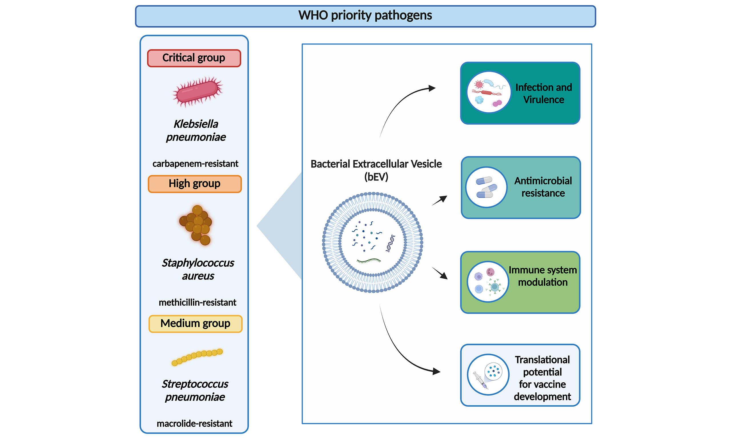
Abstract
Antimicrobial resistance (AMR) is a major global health threat that requires innovative approaches to infection prevention and treatment. In 2024, the World Health Organization (WHO) updated its Bacterial Priority Pathogens List (BPPL) to guide research toward the pathogens of highest clinical relevance. Among these, carbapenem-resistant Klebsiella pneumoniae, methicillin-resistant Staphylococcus aureus, and macrolide-resistant Streptococcus pneumoniae remain major contributors to infectious disease burden and AMR-associated mortality worldwide. Recent evidence identifies bacterial extracellular vesicles (bEVs) as important mediators of microbial adaptation, host interaction, and antimicrobial resistance. Released under physiological and stress conditions, bEVs carry proteins, lipids, and nucleic acids that can influence bacterial survival and infection outcomes. In this mini-review, we summarize current knowledge on bEVs produced by these WHO priority pathogens and discuss their emerging roles in infection biology. We highlight how bEVs contribute to antimicrobial resistance through the transport of resistance-associated proteins, the horizontal transfer of resistance genes and antibiotic-degrading activity. We further examine the involvement of bEVs in inflammation, host tissue damage and immune modulation, including their capacity to carry virulence factors or sequester antimicrobial agents. Finally, we discuss recent advances in the use of bEVs as vaccine platforms. We prioritize studies on extracellular vesicles (EVs) from antibiotic-resistant strains of the three WHO priority pathogens. When these were unavailable, we also included studies using antibiotic-susceptible strains, particularly for S. pneumoniae, for which data on macrolide-resistant strains are lacking. Overall, bEVs are increasingly recognized as active components of bacterial physiology and promising targets for developing new strategies against antimicrobial-resistant infections.
Keywords


 Download PDF
INTRODUCTION
Antimicrobial resistance (AMR) is a major threat to public health worldwide and was associated with approximately 5 million deaths in 2019[1]. By 2050, AMR-associated deaths are estimated to increase by 70%[2] and the global AMR cost to be around USD 100 trillion[3]. Despite this burden, only a small fraction of the antibiotics approved over the past 40 years belong to new compound classes[4].
AMR develops naturally when microorganisms adapt to overcome inhibitory agents, allowing them to survive and grow despite the presence of drugs[5]. Bacteria have developed diverse resistance mechanisms, such as reduced membrane permeability, efflux pumps, antibiotic degradation or target modification, which compromise the efficacy of most antibiotics[6,7]. Global Burden of Disease (GBD) estimates identify 33 bacterial pathogens (both resistant and susceptible to antimicrobials) responsible for 11 major infectious syndromes. Three of them, Klebsiella pneumoniae, Staphylococcus aureus and Streptococcus pneumoniae, are among the most significant contributors to deaths[8].
Accordingly, the 2024 World Health Organization (WHO) Bacterial Priority Pathogens List (BPPL) classifies carbapenem- and third-generation cephalosporin-resistant K. pneumoniae, methicillin-resistant S. aureus and macrolide-resistant S. pneumoniae as critical-, high- and medium-priority pathogens, respectively[1].
For these pathogens, treatment options are limited, or access to therapies is restricted in some populations or geographical areas[1]. Despite active research, no licensed vaccines are available for K. pneumoniae and
Innovative strategies are needed to limit infections caused by these pathogens, particularly antibiotic-resistant strains. Recently, extracellular vesicles (EVs) have emerged as important mediators of cell-to-cell communication[12,13] and environmental adaptation[14-16], and many studies have highlighted their contribution to microbial resistance[7].
Bacterial extracellular vesicles (bEVs) are typically 40-400 nm in diameter and carry proteins, lipids and nucleic acids, including ribonucleic acid (RNA) and deoxyribonucleic acid (DNA). In Gram-negative bacteria, EVs form by membrane blebbing, generating outer membrane vesicles (OMVs) and outer-inner membrane vesicles (OIMVs), or by explosive cell lysis, producing explosive outer membrane vesicles (EOMVs) and explosive outer-inner membrane vesicles (EOIMVs). In Gram-positive bacteria, EV biogenesis is less well understood and may involve membrane blebbing or bubbling cell death, producing cytoplasmic membrane vesicles (CMVs). Different vesiculation processes give rise to distinct EV types whose structure and content ultimately determine their function[17]. Bacterial EVs also contribute to antibiotic resistance by acting as decoys for membrane-targeting antibiotics, degrading antibiotics enzymatically or transferring resistance genes[7]. Moreover, bEVs have diagnostic potential as biomarkers of disease state and treatment efficacy, and they are being explored for vaccine development and targeted drug delivery[18].
In this review, we focus on EVs produced by K. pneumoniae, S. aureus and S. pneumoniae, summarizing recent findings that describe them as drivers of virulence, immune modulation, and antimicrobial resistance. Given the substantial global health burden posed by these pathogens, understanding the role of EVs in establishing infection, as well as their potential for vaccine development, is of particular interest. We searched PubMed for original research and review articles combining the terms “bacterial extracellular vesicles”, “outer membrane vesicles”, “Klebsiella pneumoniae”, “Staphylococcus aureus”, “Streptococcus pneumoniae”, “antimicrobial resistance” and “vaccine”. The reference lists of the retrieved articles were also manually checked for additional relevant studies. We prioritized recent, peer-reviewed studies on EVs from antibiotic-resistant strains of these WHO priority pathogens and, when these were unavailable, also considered studies on other strains. A recent book chapter covered bEVs from two Gram-negative, WHO critical-priority pathogens, Acinetobacter baumannii and Klebsiella pneumoniae, focusing on antimicrobial resistance, pathogenicity and OMV-based vaccine strategies[19]. Our mini-review differs in scope: by covering pathogens from three WHO priority tiers (critical, high and medium), including both Gram-negative and Gram-positive organisms, it allows a comparison of EV-mediated resistance and immune modulation across the priority spectrum rather than within a single tier.
EVs FROM KLEBSIELLA PNEUMONIAE
K. pneumoniae is a commensal Gram-negative bacterium that colonizes the mucosal surfaces of the nose, throat, skin and gastrointestinal tract in healthy individuals[20]. It is an opportunistic pathogen that can cause pneumonia, urinary tract, bloodstream, soft tissue and surgical wound infections, particularly in immunocompromised individuals[20,21]. Its multidrug resistance, high virulence and high transmissibility pose a significant threat to public health[20].
K. pneumoniae infections have caused approximately 800,000 deaths worldwide, 80% of them associated with AMR[22]. Its clinical impact is largely attributable to the global spread of carbapenem-resistant K. pneumoniae (CRKP), which is classified in the critical priority group of the WHO 2024 BPPL[1,9,20,21].
K. pneumoniae produces EVs, classified as OMVs, under both physiological and stress conditions. Environmental conditions, including antibiotic treatment, influence the composition and function of Klebsiella pneumoniae-derived OMVs (KP OMVs)[21]. Using proteomics, Fan et al. identified thousands of proteins in OMVs released by meropenem-treated CRKP and reported an enrichment of proteins and enzymes associated with virulence, pathogenicity, drug resistance, stress, bacterial survival and cellular metabolism[23]. Resistance-related proteins included penicillin-binding protein 1B, EmrA, Na+/antidrug transporter, ArnT, ArnA, ArnC, blaKPC, blaSHV, SHV-112, MacB, outer membrane channel protein, and outer membrane protein OmpK36[23]. The multidrug resistance-associated protein MdtQ was also detected in OMVs isolated from CRKP[24].
OMVs have also been recognized as mediators of horizontal gene transfer[7,17]. Li et al. demonstrated that OMVs of carbapenem-resistant hypervirulent K. pneumoniae (hvKP) contained genetic material, particularly carbapenemases and virulence genes, which could be transferred to recipient bacteria, enhancing their resistance-associated features[25]. CRKP OMVs were shown to mediate the transfer of the blaNDM-1 gene (encoding New Delhi metallo-β-lactamase 1) to other K. pneumoniae strains, increasing the carbapenem minimum inhibitory concentration (MIC) of the transformants[26].
In addition to their role in antimicrobial resistance, OMVs are involved in host-pathogen interactions, inducing proinflammatory responses and modulating the host immune system[27,28]. KP OMVs, derived from hvKP and clinical K. pneumoniae strains, were shown to stimulate the release of proinflammatory interleukin-8 (IL-8) from human bronchial epithelial cells, through nuclear factor kappa B (NF-κB) activation[29]. Ye et al. showed that treatment with imipenem, a widely used carbapenem antibiotic, enhanced the release of chaperonin GroEL-enriched OMVs from clinical multidrug-resistant K. pneumoniae. These OMVs were readily taken up by macrophages, inducing pyroptosis and promoting the release of proinflammatory cytokines [interleukin-1β (IL-1β), interleukin-18 (IL-18), and tumor necrosis factor-α (TNF-α)] in a mouse model of pneumonia[30]. Outer membrane proteins such as outer membrane protein A (OmpA) and outer membrane protein C (OmpC) are also components of KP OMVs (from K. pneumoniae ATCC 10031) that can induce an inflammatory response in host cells[31] and contribute to their immunogenicity[32].
OMVs are also being investigated as promising vaccine platforms[9,21,32]. Li et al. used high-pressure homogenization to induce CRKP to form stable, bacterial biomimetic vesicles (BBVs) with high yield and reduced nucleic acid content. These BBVs effectively stimulated dendritic cell maturation, elicited both humoral and cellular immune responses, and provided protection in mice against multidrug-resistant
Overall, current evidence indicates that KP OMVs contribute to antimicrobial resistance, host interaction and immune modulation, and are potential candidates for innovative vaccines. However, these studies relied on a limited number of CRKP isolates, mostly using in vitro assays or short-term mouse models, and vesicle isolation protocols differed between them, which limits the comparison and generalization of the findings.
EVs FROM STAPHYLOCOCCUS AUREUS
S. aureus is a Gram-positive bacterium that asymptomatically colonizes the human host, primarily on mucosal and epithelial surfaces[35]. It can also cause a wide range of infections and life-threatening conditions, such as bacteremia, endocarditis, osteomyelitis and pneumonia[35,36]. S. aureus was associated with more than 1 million deaths in 2019[8], and its increasing antibiotic resistance complicates clinical management[35,36]. In particular, methicillin-resistant S. aureus (MRSA) strains are resistant to β-lactam antibiotics, including penicillin derivatives, which comprise many commonly used antibiotics[37]. Consequently, MRSA is listed in the high-priority group of the 2024 WHO BPPL[1].
The role of EVs in the pathogenesis of S. aureus has received increasing attention, particularly their biogenesis and involvement in the spread of antibiotic resistance[37]. EV production is strongly influenced by environmental conditions, and studies have shown that treating bacteria, including S. aureus, with β-lactam antibiotics increases EV production[37,38]. Moreover, EVs derived from ampicillin-treated MRSA were shown to possess β-lactamase activity and to degrade antibiotics such as amoxicillin and ampicillin[38]. Kim et al. reported that proteins related to β-lactam degradation, including penicillin-binding proteins (PBP1, -2, -3), β-lactamase (BlaZ) and the UPF0173 metal-dependent hydrolase (SA1529), were abundant in EVs released from stressed bacteria[38]. In addition, MRSA-derived EVs have been investigated for their ability to transfer resistance-related factors across bacterial species[39]. Lee et al. demonstrated that these EVs could deliver β-lactamase activity to susceptible Escherichia coli, increasing its resistance to β-lactam antibiotics. EVs from resistant E. coli strains contained higher levels of β-lactamase and other antimicrobial resistance-related proteins (OmpC, OmpA, OmpT, MipA, LpoA and LpoB)[39].
Staphylococcus aureus-derived EVs (SA EVs) are also involved in other resistance mechanisms, such as the sequestration of host-derived antimicrobial fatty acids (AFAs), which normally inhibit bacterial growth on skin and mucosal surfaces[40]. Kengmo Tchoupa et al. demonstrated that both methicillin-resistant and -susceptible S. aureus strains release EVs that act as decoys, reducing the binding of AFAs to bacterial cells[40].
To investigate the role of MRSA-derived EVs in modulating host immune responses, Asano et al. purified EVs from a clinical MRSA strain and found that they stimulate proinflammatory cytokine release [TNF-α and interleukin-6 (IL-6)] via Toll-like receptors (TLRs) 2, 4 and 9, and induce cell death in mouse macrophages[41]. SA EVs also contribute to skin infections[42]. Chen et al. showed that EVs from clinical MRSA strains inhibit proliferation and induce apoptosis in human adult low-calcium high-temperature keratinocyte (HaCaT) cells, potentially via upregulation of apoptosis-related genes such as B-cell lymphoma 2 (BCL2) antagonist/killer 1 (BAK1)[43].
SA EVs have also emerged as a promising vaccine platform, because they carry multiple immunogenic components, potentially overcoming the limitations of single-antigen vaccines[36]. Wang et al. genetically engineered S. aureus (JE2 strain) to produce detoxified EVs expressing non-toxic antigens, generating a vaccine candidate that elicited neutralizing antibodies and protected mice in a sepsis model[44]. Similarly, Chen et al. developed a multi-antigen vaccine by encapsulating SA EVs (from both drug-resistant and -susceptible strains) with magnetic mesoporous silica nanoparticles (MSNs) loaded with indocyanine green (ICG). Upon laser irradiation, this nanovaccine promoted dendritic cell maturation and enhanced cluster of differentiation 8-positive (CD8+) T cell responses. In vivo, the nanovaccine was efficiently transported to lymph nodes, prevented superficial infections and reduced bacterial dissemination in mouse models[45].
Collectively, these findings show that S. aureus-derived EVs play multifaceted roles in antimicrobial resistance and host-pathogen interactions and are promising candidates for novel vaccines and therapeutic strategies. However, most mechanistic data come from a small number of clinical MRSA isolates studied
EVs FROM STREPTOCOCCUS PNEUMONIAE
S. pneumoniae is a Gram-positive bacterium that asymptomatically colonizes the nasopharynx of healthy individuals. As an opportunistic pathogen, it can cause many diseases, including community-acquired pneumonia, meningitis, otitis media and bacteremia, particularly in the elderly, children and immunocompromised people[46,47]. Among children under 5 years old, S. pneumoniae was one of the pathogens responsible for the most AMR-attributable deaths in 2021[2]. The spread of antibiotic-resistant S. pneumoniae and non-vaccine serotypes is a major global concern, reflected in the classification of macrolide-resistant S. pneumoniae in the medium-priority group of the 2024 WHO BPPL[1,46,47].
Streptococcus pneumoniae-derived EVs (SP EVs) have been studied for their role in host-pathogen interactions, as carriers of virulence factors and as promising platforms for novel vaccines[47]. SP EV production is influenced by environmental cues, including temperature, pH, host-derived factors and nutrient availability[15,48]. The protein cargo of SP EVs (from S. pneumoniae D39) includes carbon metabolism-related proteins [e.g., enolase (Eno), glyceraldehyde-3-phosphate dehydrogenase A (GapA), non-phosphorylating glyceraldehyde-3-phosphate dehydrogenase (GapN), phosphoglycerate mutase A (GpmA), phosphofructokinase A (PfkA), pyruvate kinase (PykF), and triosephosphate isomerase (Tpi)] and virulence factors, such as pneumolysin (Ply), lysozyme (LytC) and pneumococcal surface protein A (PspA)[15]. SP EVs are also implicated in gene transfer[49]. Werner Lass et al. demonstrated that SP EVs (from S. pneumoniae R6 and D39) are associated with bacterial DNA on their surface, which can be transferred to the transformation machinery of competent cells[49].
Recent research has further highlighted the contribution of SP EVs to pneumococcal pathogenicity[47], including host cell damage and lung epithelial barrier disruption[50]. Cui et al. showed that SP EVs (from
SP EVs have also been studied in the context of hemolytic uremic syndrome (HUS), a severe kidney disease. Although HUS is most commonly associated with Shiga toxin-producing E. coli (STEC), it can also be a rare complication of invasive pneumococcal disease. EVs from S. pneumoniae strains isolated from patients with pneumococcus-associated HUS (HUS EVs) contain several virulence factors, including the 50S ribosomal subunit assembly factor (BipA), PspA, the lytic enzyme LytC, and several sugar utilization and fatty acid synthesis proteins. In addition, HUS EVs induce the release of proinflammatory cytokines (IL-1β, IL-6) and chemokines [chemokine C-C motif ligand 2 (CCL2), chemokine C-C motif ligand 3 (CCL3), and C-X-C motif chemokine ligand 1 (CXCL1)] by human monocytes[51].
Current pneumococcal vaccines provide limited serotype coverage: even the latest approved vaccine, pneumococcal conjugate vaccine 21 (PCV21), protects against only 21 serotypes[47,52]. Novel immunization strategies are therefore needed[47]. In this context, Dookie et al. demonstrated that SP EVs (isolated from pneumococci of serotype 4) have inherent adjuvant properties, mainly through the lipoproteins maltose/maltodextrin ATP-binding cassette (ABC) transporter (MalX) and chaperone cis-trans prolyl isomerase (PrsA), which activate Toll-like receptor 2 (TLR2) and induce a robust serotype-independent T helper 17 (Th17) response in both mice and humans. Moreover, intranasal immunization with SP EVs promoted lung macrophage expansion and conferred protection against pneumococcal colonization without additional adjuvants[53]. Similarly, Carneiro et al. showed that subcutaneous immunization with SP EVs (from S. pneumoniae R6) induces specific antibody responses, particularly against PspA, and confers protection in murine models of invasive pneumonia, with PspA playing a significant role even across heterologous PspA families[54].
To develop a stable and effective vaccine against pneumococcal infections, Mehanny et al. formulated spray-dried microparticles encapsulating SP EVs (from S. pneumoniae R6). These microparticles had suitable size, aerodynamic properties and stability for deep lung delivery. The encapsulated SP EVs did not compromise the viability of human macrophage-like cells derived from the human monocytic leukemia cell line (THP-1) and were readily internalized by them. They also induced the release of proinflammatory cytokines (TNF and IL-6) from primary human peripheral blood mononuclear cells (PBMCs)[55].
Taken together, these studies highlight the important role of SP EVs in pneumococcal pathogenesis and support their potential as vaccine candidates. However, most of the SP EV literature summarized here was generated using non-macrolide-resistant laboratory or clinical strains, and the biological properties and immunological functions of EVs released by macrolide-resistant S. pneumoniae, the WHO priority phenotype, remain largely unexplored. This aspect should be kept in mind when interpreting how far these findings translate to the WHO priority phenotype.
CONCLUSION AND FUTURE DIRECTIONS
AMR remains one of the most urgent challenges in infectious disease control, particularly for WHO priority pathogens such as K. pneumoniae, S. aureus and S. pneumoniae. Current evidence indicates that bEVs from these pathogens are not passive by-products of bacterial growth, but active mediators of infection, immune modulation and antimicrobial resistance [Table 1].
Characteristics and application of extracellular vesicles from the three selected pathogens: summary of the main studies included in this mini-review
| Pathogen | EV cargo | EV biological effect | Application* |
| Klebsiella pneumoniae | • Proteins/enzymes associated with virulence, pathogenicity, drug resistance, stress, bacterial survival, and cellular metabolism in meropenem-treated CRKP OMVs[23] • MdtQ, outer membrane protein associated with multidrug resistance in CRKP OMVs[24] • Carbapenemases and virulence genes in carbapenem-resistant hvKP OMVs[25] • blaNDM-1 gene in CRKP OMVs[26] | • OMVs from hvKP and clinical KP strains induce proinflammatory IL-8 secretion from human bronchial epithelial cells, through NF-κB activation[29] • OMVs from imipenem-treated clinical multidrug-resistant KP, enriched in GroEL protein, induce pyroptosis in macrophages and release of proinflammatory cytokines (IL-1β, IL-18, TNF-α) in a mouse model of pneumonia[30] • KP OMVs (from KP ATCC 10031) containing OmpA and OmpC stimulate a proinflammatory response in host cells[31] | • BBVs (from CRKP) stimulate dendritic cell maturation, elicit both humoral and cellular immune responses, and provide protection in mice against multiple drug-resistant KP strains[33] • BSA-OMV nanoparticles (OMVs from CRKP with BSA) enhance immune responses, dendritic cell activation, and protection against CRKP infection in mice[34] |
| Staphylococcus aureus | • Proteins related to the degradation of β-lactam antibiotics, including PBP1, PBP2, PBP3 and BlaZ in EVs from ampicillin-treated MRSA[38] • Antibiotic resistance-related factors in MRSA EVs transferred to E. coli susceptible to β-lactam antibiotics[39] | • EVs from both methicillin-resistant and -susceptible SA act as decoys, reducing the binding of AFAs to bacterial cells[40] • Clinical MRSA-derived EVs stimulate proinflammatory cytokine release (TNF-α and IL-6) via TLR2, TLR4, and TLR9, and induce host cell death in mouse macrophages[41] • Clinical MRSA-derived EVs inhibit proliferation and induce apoptosis of HaCaT epithelial skin cells, via upregulation of apoptosis-related genes like BAK1[43] | • SA detoxified EVs (from JE2 strain) elicit neutralizing antibodies and provide protection in a mouse sepsis model[44] • SA EVs (from both drug-resistant and -susceptible strains) encapsulated with magnetic MSNs, loaded with ICG promote dendritic cell maturation and enhance CD8+ T cell responses upon laser irradiation. In vivo, the nanovaccine prevents superficial infections, and reduces bacterial dissemination in mouse models[45] |
| Streptococcus pneumoniae+ | • Carbon metabolism-related proteins (e.g., Eno, GapA, GapN, GpmA, PfkA, PykF, and Tpi) and virulence factors, such as Ply, LytC and PspA in SP EVs (from SP D39)[15] • Bacterial DNA on SP EVs (from SP R6 and D39) surface[49] • Virulence factor StkP in SP EVs (from SP D39)[50] • Several virulence factors (e.g., BipA, PspA, LytC) and sugar utilization and fatty acid synthesis proteins in HUS EVs (from clinical HUS strains)[51] • Lipoproteins MalX and PrsA in SP EVs (from SP serotype 4)[53] | • SP EVs (from SP D39) are internalized by alveolar epithelial cells and activate autophagy, leading to the degradation of OCLN and impairment of the alveolar barrier integrity[50] • HUS EVs (from clinical HUS strains) stimulate the release of proinflammatory cytokines (IL-1β, IL-6) and chemokines (CCL2, CCL3, CXCL1) by human monocytes[51] • SP EVs (from SP serotype 4) activate TLR2 and induce robust serotype-independent Th17 response in both mice and humans[53] | • Intranasal immunization with SP EVs (from SP serotype 4) promotes the expansion of lung macrophages, and confers protection against pneumococcal colonization[53] • Subcutaneous immunization with SP EVs (from SP R6) induces antibody response against PspA, and confers protection in murine models of invasive pneumonia[54] • Spray-dried microparticles encapsulating SP EVs (from SP R6) do not compromise the viability of THP-1 cells and induce the release of proinflammatory cytokines (TNF and IL-6) from PBMCs[55] |
Although K. pneumoniae, S. aureus and S. pneumoniae differ in vesicle biogenesis and cell wall structure, their EVs share common downstream roles: all three carry virulence- and resistance-associated cargo, modulate host immune responses, and are being explored as vaccine platforms. The maturity of the evidence differs considerably, being most developed for K. pneumoniae and S. aureus and comparatively limited for
In conclusion, recent findings support the idea that bEVs are part of the broader pathogen response to antimicrobial pressure. A deeper understanding of their biology may help identify new therapeutic strategies against antibiotic-resistant infections, and studying EVs from WHO priority pathogens may therefore offer important opportunities to counteract AMR and improve infectious disease management.
DECLARATIONS
Acknowledgments
The Graphical Abstract was created with BioRender.com [Battista M (2026), https://BioRender.com/uwsd0xw].
Authors’ contributions
Conceptualized the work, performed the literature search, analysis and interpretation, drafted the manuscript, and revised the manuscript: Battista M
Conceptualized the work, performed the literature search, revised the manuscript, and approved the final version: Vilhena C
Availability of data and materials
Not applicable.
AI and AI-assisted tools statement
Not applicable.
Financial support and sponsorship
None.
Conflicts of interest
All authors declared that there are no conflicts of interest.
Ethical approval and consent to participate
Not applicable.
Consent for publication
Not applicable.
Copyright
© The Author(s) 2026.
REFERENCES
1. World Health Organization. WHO Bacterial Priority Pathogens List, 2024: bacterial pathogens of public health importance to guide research, development and strategies to prevent and control antimicrobial resistance. Available from https://www.who.int/publications/i/item/9789240093461. [accessed 4 November 2026].
2. GBD 2021 Antimicrobial Resistance Collaborators. Global burden of bacterial antimicrobial resistance 1990-2021: a systematic analysis with forecasts to 2050. Lancet. 2024;404:1199-226.
3. World Health Organization. Vaccines could avert half a million deaths associated with anti-microbial resistance a year. Available from https://www.who.int/news/item/28-07-2023-vaccines-could-avert-half-a-million-deaths-associated-with-anti-microbial-resistance-a-year. [accessed 4 November 2026].
4. Miethke M, Pieroni M, Weber T, et al. Towards the sustainable discovery and development of new antibiotics. Nat Rev Chem. 2021;5:726-49.
5. Anderson AS. Vaccines against antimicrobial resistance. Philos Trans R Soc Lond B Biol Sci. 2026;381:20250007.
6. Darby EM, Trampari E, Siasat P, et al. Author correction: molecular mechanisms of antibiotic resistance revisited. Nat Rev Microbiol. 2024;22:255.
7. Jiang B, Lai Y, Xiao W, et al. Microbial extracellular vesicles contribute to antimicrobial resistance. PLoS Pathog. 2024;20:e1012143.
8. GBD 2019 Antimicrobial Resistance Collaborators. Global mortality associated with 33 bacterial pathogens in 2019: a systematic analysis for the Global Burden of Disease Study 2019. Lancet 2022;400:2221-48. Available from https://www.thelancet.com/journals/lancet/article/PIIS0140-6736(22)02185-7/fulltext. [accessed 4 November 2026].
9. Hu J, Xie Y, Guan W, Huang L, Li X. Klebsiella pneumoniae vaccines: Evolving the blueprint from traditional platforms to mucosal and nanoscale delivery. Mater Today Bio. 2026;37:102919.
10. Briles DE, Paton JC, Mukerji R, Swiatlo E, Crain MJ. Pneumococcal vaccines. Microbiol Spectr. 2019;7:20.
11. Li S, Liang H, Zhao SH, Yang XY, Guo Z. Recent progress in pneumococcal protein vaccines. Front Immunol. 2023;14:1278346.
12. Ñahui Palomino RA, Vanpouille C, Costantini PE, Margolis L. Microbiota-host communications: bacterial extracellular vesicles as a common language. PLoS Pathog. 2021;17:e1009508.
13. Battista M, Saffer C, Figge MT, Vilhena C. Mechanistic insights into the role of extracellular vesicles at the bacterial-host interface. ACS Infect Dis. 2025;11:3407-13.
14. da Luz BSR, de Rezende Rodovalho V, Nicolas A, et al. Impact of environmental conditions on the protein content of staphylococcus aureus and its derived extracellular vesicles. Microorganisms. 2022;10:1808.
15. Battista M, Bachelot Y, Franke T, et al. Environmental cues shape extracellular vesicles biogenesis and function in Streptococcus pneumoniae. BMC Microbiol. 2026;26:320.
16. Briaud P, Frey A, Marino EC, et al. Temperature influences the composition and cytotoxicity of extracellular vesicles in Staphylococcus aureus. mSphere. 2021;6:e0067621.
17. Toyofuku M, Schild S, Kaparakis-Liaskos M, Eberl L. Composition and functions of bacterial membrane vesicles. Nat Rev Microbiol. 2023;21:415-30.
18. Moghaddam ZS, Dehghan A, Halimi S, et al. Bacterial extracellular vesicles: bridging pathogen biology and therapeutic innovation. Acta Biomater. 2025;200:1-20.
19. Rocha Minarini LAD. Exploring bacterial extracellular vesicles: focus on WHO critical priority pathogens. Curr Top Membr. 2024;94:225-46.
20. Lei TY, Liao BB, Yang LR, Wang Y, Chen XB. Hypervirulent and carbapenem-resistant Klebsiella pneumoniae: a global public health threat. Microbiol Res. 2024;288:127839.
21. Li L, Xu X, Cheng P, et al. Klebsiella pneumoniae derived outer membrane vesicles mediated bacterial virulence, antibiotic resistance, host immune responses and clinical applications. Virulence. 2025;16:2449722.
22. Song HW, Lin Y, Wan TE, et al. Global burden of Klebsiella pneumoniae infections and antimicrobial resistance in 2019. BMC Infect Dis. 2025;25:1773.
23. Fan F, Chen G, Deng S, Wei L. Proteomic analysis of meropenem-induced outer membrane vesicles released by carbapenem-resistant Klebsiella pneumoniae. Microbiol Spectr. 2024;12:e0291723.
24. Fan F, Wang J, Chen H, Wei L, Zhang Z. Isolation and protein MdtQ analysis of outer membrane vesicles released by carbapenem-resistant Klebsiella pneumoniae. Microb Pathog. 2023;183:106325.
25. Li P, Luo W, Xiang TX, et al. Horizontal gene transfer via OMVs co-carrying virulence and antimicrobial-resistant genes is a novel way for the dissemination of carbapenem-resistant hypervirulent Klebsiella pneumoniae. Front Microbiol. 2022;13:945972.
26. Tang B, Yang A, Liu P, et al. Outer membrane vesicles transmitting blaNDM-1 mediate the emergence of carbapenem-resistant hypervirulent Klebsiella pneumoniae. Antimicrob Agents Chemother. 2023;67:e0144422.
27. Kaparakis-Liaskos M, Ferrero RL. Immune modulation by bacterial outer membrane vesicles. Nat Rev Immunol. 2015;15:375-87.
28. Charpentier LA, Dolben EF, Hendricks MR, Hogan DA, Bomberger JM, Stanton BA. Bacterial outer membrane vesicles and immune modulation of the host. Membranes. 2023;13:752.
29. Li P, Peng T, Xiang T, et al. Klebsiella pneumoniae outer membrane vesicles induce strong IL-8 expression via NF-κB activation in normal pulmonary bronchial cells. Int Immunopharmacol. 2023;121:110352.
30. Ye C, Li W, Yang Y, et al. Inappropriate use of antibiotics exacerbates inflammation through OMV-induced pyroptosis in MDR Klebsiella pneumoniae infection. Cell Rep. 2021;36:109750.
31. Martora F, Pinto F, Folliero V, et al. Isolation, characterization and analysis of pro-inflammatory potential of Klebsiella pneumoniae outer membrane vesicles. Microb Pathog. 2019;136:103719.
32. Assoni L, Girardello R, Converso TR, Darrieux M. Current stage in the development of Klebsiella pneumoniae vaccines. Infect Dis Ther. 2021;10:2157-75.
33. Li W, Hu Y, Zhang Q, et al. Development of drug-resistant Klebsiella pneumoniae vaccine via novel vesicle production technology. ACS Appl Mater Interfaces. 2021;13:32703-15.
34. Wu G, Ji H, Guo X, et al. Nanoparticle reinforced bacterial outer-membrane vesicles effectively prevent fatal infection of carbapenem-resistant Klebsiella pneumoniae. Nanomedicine. 2020;24:102148.
35. Parsons JB, Mourad A, Conlon BP, Kielian T, Fowler VG Jr. Methicillin-resistant and susceptible Staphylococcus aureus: tolerance, immune evasion and treatment. Nat Rev Microbiol. 2026;24:127-45.
36. Fu C, Li Z, Sun Y, Lin S, Yang X, Li X. Advances in Staphylococcus aureus extracellular vesicles: navigating challenges, embracing prospects. Lett Appl Microbiol. 2026;79:ovag009.
37. Liu ZH, Wu QY, Xu F, Zhang X, Liao XB. Biofunction and clinical potential of extracellular vesicles from methicillin-resistant Staphylococcus aureus. Microbiol Res. 2023;266:127238.
38. Kim SW, Seo JS, Park SB, et al. Significant increase in the secretion of extracellular vesicles and antibiotics resistance from methicillin-resistant Staphylococcus aureus induced by ampicillin stress. Sci Rep. 2020;10:21066.
39. Lee AR, Park SB, Kim SW, et al. Membrane vesicles from antibiotic-resistant Staphylococcus aureus transfer antibiotic-resistance to antibiotic-susceptible Escherichia coli. J Appl Microbiol. 2022;132:2746-59.
40. Kengmo Tchoupa A, Peschel A. Staphylococcus aureus releases proinflammatory membrane vesicles to resist antimicrobial fatty acids. mSphere. 2020;5:e00804-20.
41. Asano K, Hirose S, Narita K, et al. Extracellular vesicles from methicillin resistant Staphylococcus aureus stimulate proinflammatory cytokine production and trigger IgE-mediated hypersensitivity. Emerg Microbes Infect. 2021;10:2000-9.
42. Torrealba MP, Yoshikawa FSY, Aoki V, Sato MN, Orfali RL. State of the art on the role of Staphylococcus aureus extracellular vesicles in the pathogenesis of atopic dermatitis. Microorganisms. 2024;12:531.
43. Chen X, Zhang J, Yang M, Du G, Chen F. Methicillin-resistant Staphylococcus aureus membrane vesicles inhibit the proliferation and induce the apoptosis of epithelial cells. Pathogens. 2022;11:1429.
44. Wang X, Thompson CD, Weidenmaier C, Lee JC. Release of Staphylococcus aureus extracellular vesicles and their application as a vaccine platform. Nat Commun. 2018;9:1379.
45. Chen G, Bai Y, Li Z, Wang F, Fan X, Zhou X. Bacterial extracellular vesicle-coated multi-antigenic nanovaccines protect against drug-resistant Staphylococcus aureus infection by modulating antigen processing and presentation pathways. Theranostics. 2020;10:7131-49.
46. Li L, Ma J, Yu Z, Li M, Zhang W, Sun H. Epidemiological characteristics and antibiotic resistance mechanisms of Streptococcus pneumoniae: an updated review. Microbiol Res. 2023;266:127221.
47. Parveen S, Subramanian K. Emerging roles of extracellular vesicles in pneumococcal infections: immunomodulators to potential novel vaccine candidates. Front Cell Infect Microbiol. 2022;12:836070.
48. Guo J, Huang Z, Wang Q, et al. Opportunities and challenges of bacterial extracellular vesicles in regenerative medicine. J Nanobiotechnology. 2025;23:4.
49. Werner Lass S, Smith BE, Camphire S, et al. Pneumococcal extracellular vesicles mediate horizontal gene transfer via the transformation machinery. mSphere. 2024;9:e0072724.
50. Cui L, Yang R, Huo D, et al. Streptococcus pneumoniae extracellular vesicles aggravate alveolar epithelial barrier disruption via autophagic degradation of OCLN (occludin). Autophagy. 2024;20:1577-96.
51. Battista M, Hoffmann B, Bachelot Y, et al. The role of pneumococcal extracellular vesicles on the pathophysiology of the kidney disease hemolytic uremic syndrome. mSphere. 2023;8:e0014223.
52. Ozisik L. The new era of pneumococcal vaccination in adults: what is next? Vaccines 2025;13:498.
53. Dookie R, Narciso AR, Gaytán MO, et al. Pneumococcal membrane particles promote serotype-independent cellular and humoral immunity and protect against pneumococcal colonization. Proc Natl Acad Sci U S A. 2026;123:e2537226123.
54. Carneiro GB, Yerneni SS, Chinaia K, et al. Protection induced by Streptococcus pneumoniae extracellular vesicles against nasal colonization and invasive infection in mice and the role of PspA. Vaccine. 2025;44:126566.
55. Mehanny M, Boese A, Bornamehr B, et al. Spray-dried pneumococcal membrane vesicles are promising candidates for pulmonary immunization. Int J Pharm. 2022;621:121794.
56. De Langhe N, Van Dorpe S, Guilbert N, et al. Mapping bacterial extracellular vesicle research: insights, best practices and knowledge gaps. Nat Commun. 2024;15:9410.
57. Welsh JA, Goberdhan DCI, O’Driscoll L, et al. ; MISEV Consortium. Minimal information for studies of extracellular vesicles (MISEV2023): from basic to advanced approaches. J Extracell Vesicles. 2024;13:e12404.
58. Gu Z, Unitt A, Harrison OB, et al. Gonococcal outer membrane vesicle vaccines: bacterial population biology, clinical trials, immune profiling, and vaccine design. NPJ Vaccines. 2026;11:85.
Cite This Article
 Open Access
How to Cite
Battista M, Vilhena C. Bacterial extracellular vesicles in priority pathogens: Klebsiella pneumoniae, Staphylococcus aureus, and Streptococcus pneumoniae. Extracell Vesicles Circ Nucleic Acids. 2026;7:2006-16. https://dx.doi.org/10.20517/evcna.2026.129
Download Citation
If you have the appropriate software installed, you can download article citation data to the citation manager of your choice. Simply select your manager software from the list below and click on download.
Export Citation File
Type of Import
Tips on Downloading Citation
Citation Manager File Format
Type of Import
Direct Import: When the Direct Import option is selected (the default state), a dialogue box will give you the option to Save or Open the downloaded citation data. Choosing Open will either launch your citation manager or give you a choice of applications with which to use the metadata. The Save option saves the file locally for later use.
Indirect Import: When the Indirect Import option is selected, the metadata is displayed and may be copied and pasted as needed.
About This Article
Special Topic
Copyright

Data & Comments
Data
0










Comments
Comments must be written in English. Spam, offensive content, impersonation, and private information will not be permitted. If any comment is reported and identified as inappropriate content by OAE staff, the comment will be removed without notice. If you have any queries or need any help, please contact us at [email protected].