


Superior photoelectrocatalytic performance of Mo-doped γ-Fe2O3 catalyst for degrading tetracycline
 0
0 Abstract
Photoelectrocatalytic (PEC) technology is one of the effective methods for degrading tetracycline (TC) in wastewater. In this work, we designed a doping system by incorporating Mo atoms into the lattice of γ-Fe2O3 and achieved a generic characteristic of superior PEC activity. Mo atoms are proven to be doped into the lattice of γ-Fe2O3 through the analysis of X-ray diffraction (XRD), X-ray photoelectron spectra (XPS), transmission electron microscope energy dispersive X-ray spectroscopy (TEM-EDS) mapping and high-angle annular dark-field scanning transmission electron microscopy (HAADF-STEM). Valence band XPS and ultraviolet photoelectron spectroscopy (UPS) measurements
Keywords
INTRODUCTION
Along with the rapid development of the global social economy in recent decades, the consumption of antibiotics has been continuously increasing[1]. Tetracycline (TC) is a typical broad-spectrum antibiotic that has been widely and increasingly used in human medicine, livestock farming, and aquaculture[2-4]. Generally speaking, only less than 30% of the administered antibiotic dosage can be absorbed and metabolized by humans and animals[5], while the rest are excreted into the environment where they persist and spread, posing a potential threat to the ecosystem and human health. Owing to its high chemical stability, TC exhibits a prolonged half-life and is difficult to be naturally decomposed in the environment[6]. Conventional treatment methods are limited by their inherent limitations. For example, physical adsorption and activated sludge technology primarily rely on adsorption mechanisms and cannot completely remove TC[7,8]. Biodegradation technology is an eco-friendly treatment approach, while can not rapidly eliminate TC due to its slow degradation kinetics[9]. Therefore, it is urgent to develop a rapid and thorough technology for removing TC. Photocatalysis, driven by solar energy, is regarded as a promising technology for degrading TC effectively in wastewater owing to its advantages such as low energy consumption and high efficiency[10-12]. However, the photocatalytic technology is constrained by the low utilization rate of solar energy and the rapid recombination of photogenerated charges[13-15]. Photoelectrocatalytic (PEC), combining photochemical and electrochemical oxidation by applying an external bias, is an effective method that promotes the separation and transfer of photogenerated charges, reducing recombination losses, and enhancing the generation of reactive species[16]. Some transition-metal oxides such as TiO2, WO3, and ZnO have been widely researched as photocatalysts, while their large band gap and rapid recombination of photogenerated carriers lead to low PEC efficiency[17-19]. To overcome these limitations, strategies such as heterojunction construction[20], defect engineering[21], and nanostructure regulation[22] have been employed to enhance their PEC performance. Yu et al. synthesized an S-scheme 2H-MoSe2/NiFe-LDH heterojunction photoanode showing excellent TC degradation performance, retained over 95% efficiency across 10 cycles, attributed to the efficient separation and utilization of photogenerated electrons and holes[23]. Liu et al. constructed a novel photoelectrocatalysis/photoelectro-Fenton system by coupling a cone-like TiO2 photoanode grown on nickel foam with a natural air-diffusion electrode, achieved complete removal of carbamazepine in 30 min, and revealed the synergistic mechanism of enhanced H2O2 production and direct hole oxidation[24]. Li et al. observed the efficient spatial separation and transfer of photoinduced charges in a ferroelectric-polarized TiO2@BaTiO3 heterostructure using single-particle imaging technology directly, revealing the critical role of the polarization orientation in enhancing the piezoelectric-photocatalytic performance[25]. Vaghasiya et al. developed a g-C3N4-based magnetic soft centirobot that generates reactive oxygen species under black light illumination to efficiently kill bacteria in water, demonstrating the potential of photoactive materials for mitigating biological threats[26]. Hu et al. developed a chlorine-doped pentagonal defect-rich nanocarbon catalyst that enables efficient H2O2 electrosynthesis in seawater and achieves rapid degradation of tetracycline, further demonstrating the potential of electrocatalytic reactive oxygen species generation for antibiotic removal[27].
Maghemite (γ-Fe2O3), an eco-friendly semiconductor material, has been applied extensively in the catalytic field because of its appropriate band gap characteristics, high chemical stability, and excellent magnetic properties. While due to the fast recombination of photogenerated charges, γ-Fe2O3 has relatively poor photocatalytic efficiency. To address this issue, the doping strategy provides a novel approach for optimizing catalyst performance. Compared with cocatalyst loading or heterojunction construction using metal carbides[28], doping offers unique advantages in modulating electronic band structures and introducing stable active sites within the lattice. Kahng and Kim have reported a SnO2/Mo-doped BiVO4 heterojunction photoanode, in which Mo doping enhances n-type conductivity and the SnO2 layer blocks hole recombination, thereby achieving efficient photoelectrochemical degradation of tetracycline hydrochloride[29]. Zhang et al. have reported a MoO2/Mo-doped BiOCl Ohmic junction with abundant oxygen vacancies, in which the Mo doping induces oxygen vacancies and the MoO2 cocatalyst promotes charge carrier separation, thereby achieving efficient photocatalytic degradation of tetracycline hydrochloride[30].
In this work, we designed a doping system by incorporating Mo atoms into the lattice of γ-Fe2O3 and achieved a generic characteristic of superior PEC activity. Mo atoms are proved to be incorporated into the lattice of γ-Fe2O3 in the form of doping through the analysis of X-ray diffraction (XRD), X-ray photoelectron spectra (XPS) and high-angle annular dark-field scanning transmission electron microscopy (HAADF-STEM). The modification of Mo doping can affect the electron cloud structure of γ-Fe2O3, enhancing the electronic interactions, facilitating the separation and transformation of photogenerated charges, and resulting in the superior PEC activity. The 27% Mo-doped γ-Fe2O3 exhibits excellent photoelectrochemical properties, such as the lowest onset potential of -0.7 V, an enhanced photocurrent density as high as -184 mA·cm-2 at the potential of -1.5 V versus reversible hydrogen electrode (vs. RHE), and reaches the removal efficiency of 100% in 25 min for degrading TC. Thus, this work provides a feasible strategy for developing doped catalysts with superior PEC performance by enhancing the electronic interactions.
EXPERIMENTAL
Materials
All chemicals were purchased in analytical grade and used without any further treatment. (NH4)6Mo7O24·4H2O, C2H2O4·2H2O, NaOH, Na2SO4, FeSO4·7H2O, and CH3OH were purchased from Sinopharm Chemical Reagent Co., Ltd. (Shanghai, China). p-Benzoquinone (PBQ, 99%), KBrO3, ethylenediaminetetraacetic acid disodium salt dihydrate (EDTA-2Na), FeCl3·6H2O, isopropanol (IPA, 99.5%), and tetracycline (TC, CAS: 60-54-8) were purchased from Macklin Biochemical Technology Co., Ltd. (Shanghai, China). Nafion solution (5 wt%) and hydrophilic carbon cloth were purchased from Suzhou Sunernuo Technology Co., Ltd. (Suzhou, China).
Syntheses of γ-Fe2O3 and α-MoO3
The synthesis of γ-Fe2O3 was carried out using a modified version of a literature method[31]. The specific procedures were as follows: 2.70 g of FeCl3·6H2O and 5.56 g of FeSO4·7H2O were dissolved together in
Syntheses of Mo-Fe2O3
The Mo-Fe2O3 were prepared via an impregnation method. Firstly, a certain amount (0.1, 0.2, 0.3, 0.4 and
Fabrication of photocathodes
The fabrication method of one photocathode was improved according to the report[32]. Specifically, 2.0 mg of sample, 350 μL of ethanol, and 50 μL of Nafion solution (5 wt%) were added to 600 μL of deionized water and ultrasonicated for 0.5 h to form a homogeneous ink with a mass concentration of 2.0 mg·mL-1. Then, 250 μL of the as-prepared ink was coated onto a 1.0 cm × 1.4 cm hydrophilic carbon cloth (the actual exposed area mounted on the electrode clamp was exactly 1.0 cm × 1.0 cm). Finally, the fabricated photoelectrode with a loading amount of 0.357 mg·cm-2 was dried at 60 oC for 3 h. For the convenience of description, the fabricated electrodes with γ-Fe2O3 and x% Mo-Fe2O3 (x = 9, 18, 27, 36, and 45) were marked as S0, S1, S2, S3, S4 and S5, respectively.
Structural characterization
XRD data was collected from 20° to 80° at a scan rate of 20°·min-1 on a X-ray diffractometer (Rigaku SmartLab SE, Japan) under a Cu Kα radiation power of 50 kV × 44 mA to identify the crystal structure of each photoelectrocatalyst. Aberration-corrected high-angle annular dark-field scanning transmission electron microscopy (AC-HAADF-STEM) was observed by Thermo Scientific Themis Z transmission electron microscope equipped with dual spherical aberration correctors. Transmission electron microscope energy dispersive X-ray spectroscopy (TEM-EDS) elemental mapping was performed on a Thermo Scientific Talos F200S transmission electron microscope. XPS were recorded on a Thermo Scientific ESCALAB Xi+ spectrometer with a monochromatic Al Kα source to determine the surface chemical composition. The high-resolution XPS spectra were acquired at a pass energy of 150.0 eV and a step size of 1.0 eV to analyze the chemical states of surface elements. All binding energies were calibrated with the adventitious carbon C 1s peak at 284.8 eV. Valence band XPS spectra were also collected on a Thermo Scientific K-Alpha spectrometer under the same experimental conditions. The valence band maximum (VBM) was determined by linear extrapolation of the leading edge of the valence band spectrum to the baseline. Ultraviolet photoelectron spectroscopy (UPS) measurements were performed on a Thermo Fisher Scientific ESCALAB Xi using a He I (hν = 21.22 eV) source. A silver reference sample was used to calibrate the binding energy scale (EFermi = 0 eV). UPS spectra were collected under a bias of -5 V for the secondary electron cutoff region. The work function (Φ) was calculated using the formula Φ = hν - (Ecutoff - EFermi), where Ecutoff was determined by linear extrapolation of the secondary electron cutoff edge[33]. The ultraviolet-visible (UV-Vis) diffuse reflectance spectra (UV-Vis DRS) were collected from 300 nm to 800 nm on a Shimadzu UV-2600 spectrophotometer equipped with an integrating sphere, in which BaSO4 acts as a reference of 100% reflectance to analyze the light absorption properties of photoelectrocatalysts. Inductively coupled plasma optical emission spectrometry (ICP-OES) analysis was carried out on an Agilent 5110 inductively coupled plasma optical emission spectrometer (Agilent Technologies, USA). The radio frequency power was set to 1,300 W, the nebulizer gas flow rate was 0.6 L·min-1, the sample uptake delay was 9 s, the instrument stabilization delay was 7 s, and the replicate read time was 8 s.
Electrochemical characterization
All the electrochemical properties were evaluated in 0.25 M Na2SO4 solution on a CHI 660E electrochemical workstation (Chenhua, Shanghai, China). The three-electrode system was assembled with one photocathode as working electrode, a Pt plate as counter electrode, and a saturated calomel electrode (SCE) as a reference electrode. The linear sweep voltammogram (LSV) curves at the scan rate of 10 mV·s-1 and I-t curves under chopped light were obtained under AM 1.5 G illumination (100 mW·cm-2). The transient photocurrent responses (I-t curves) were recorded at a constant applied potential of -0.1 V vs. SCE under chopped light illumination (light/dark cycles) with the same light source. The electrochemical impedance spectroscopy (EIS) data was collected at a central potential of 0 V vs. RHE with an AC amplitude of 5 mV in the frequency range from 100 kHz to 0.1 Hz. The Mott-Schottky analysis was carried out in a potential range from -1.0 V to 0.5 V vs. RHE under a frequency of 1 kHz in dark. The cyclic voltammetry (CV) curves for double-layer capacitance (Cdl) were recorded at the scan rates ranging from 10 mV·s-1 to 100 mV·s-1 within a non-Faradaic potential window of 0.665-0.765 V vs. RHE. All measured potentials were converted to the reversible hydrogen electrode potential (V vs. RHE) according to ERHE = ESCE + 0.241 + 0.059 × pH[34]. In this work, the electrolyte was air-saturated without additional O2 bubbling, and its pH was equal to 6.8.
PEC of TC
The TC degradation performance of each photocathode was evaluated at a potential of -1.5 V (vs. RHE) in a single-compartment three-electrode quartz electrolytic cell, which was under the magnetic stirring and exposed to a 300 W Xe lamp with an optical filter (λ > 420 nm). The reaction solutions composed of TC (an initial concentration of 10, 50 or 100 mg·L-1, namely ppm; an initial volume of 40 mL) and 0.25 M Na2SO4 were taken 1.5 mL every 5 min over the 30 min reaction period. All degradation experiments were performed in three times under identical conditions. The data are presented as mean ± standard deviation (SD), and the error bars in the corresponding figures represent the SD of three independent measurements. The analysis method for TC was modified based on the previous report[35]. The TC concentration was determined at a detection wavelength of 365 nm by an UltiMate 3000 high-performance liquid chromatography (HPLC) system equipped with a C18 column, in which the mobile phase (25 wt% methanol and 75 wt% 0.01 M oxalic acid aqueous solution) flowed at a rate of 0.8 mL·min-1 at 35 oC, and the injection volume of taken reaction solution was 25 μL. The intermediate products during TC degradation were detected and identified by a liquid chromatography-mass spectrometry (LC-MS, Agilent 1290 UPLC and Agilent 6550 Q-TOF) under the the electrospray ionization in positive ion mode (ESI+), equipped with a C18 column (100 mm × 2.1 mm,
RESULTS AND DISCUSSION
The XRD patterns of γ-Fe2O3 and x% Mo-Fe2O3 are shown in Figure 1. All diffraction peaks of γ-Fe2O3 match well with the standard data of the cubic spinel structure (JCPDS No. 39-1346)[36], without any impurity peaks. As the doping amount of Mo increases to 27%, the crystal structure of x% Mo-Fe2O3 remains the cubic spinel structure. Notably, in the magnified patterns of the (311) peak of the γ-Fe2O3 phase, the diffraction peak shifts from 35.63° to 35.75°, illustrating that Mo ions successfully insert into the lattice of the γ-Fe2O3 crystal structure[37]. When the doping amount of Mo exceeds 27%, the orthorhombic α-MoO3 phase (JCPDS No. 73-6497, shown in Supplementary Figure 1) forms, demonstrating that the maximum doping amount of Mo should not exceed 27% to ensure the existence of Mo as the dopant incorporated into the γ-Fe2O3 lattice. To provide the actual Mo doping content, ICP-OES analysis was performed on the 27% sample. As listed in Supplementary Table 1, the actual molar ratio of Mo to γ-Fe2O3 is 33.32%, which is slightly higher than the nominal theoretical ratio of 27%, and this small deviation is attributed to the methodological error of the ICP-OES analysis.
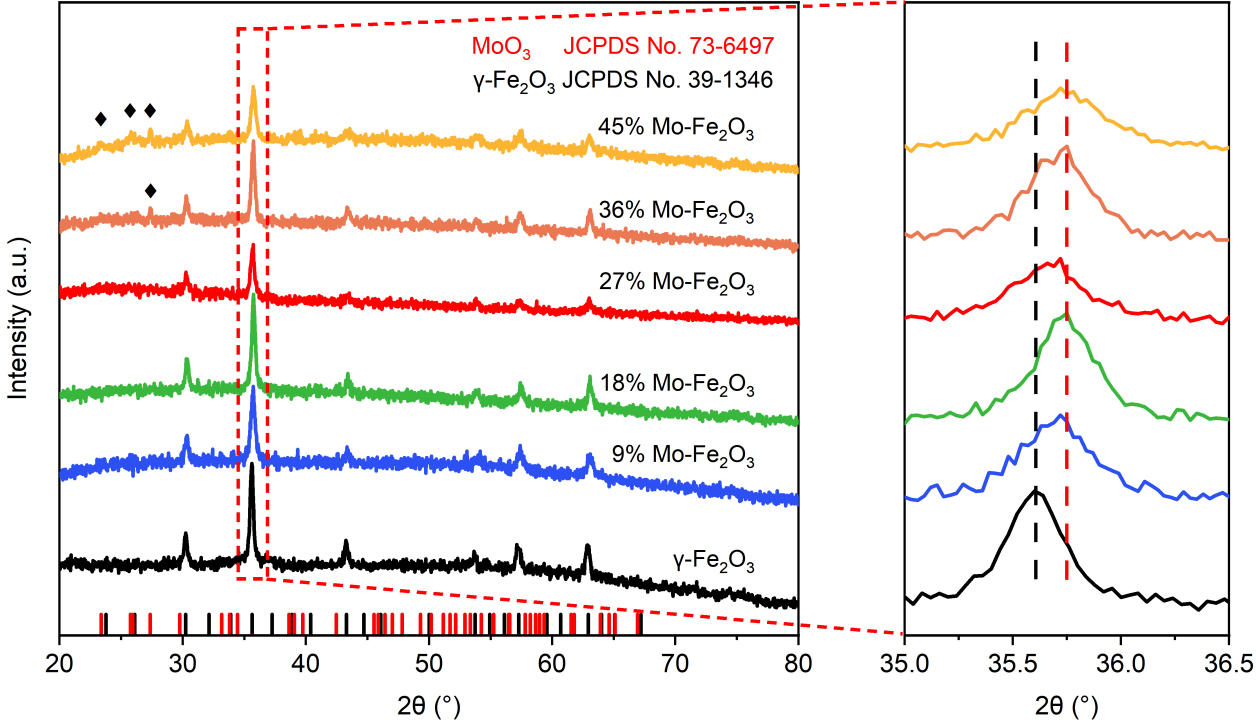
Figure 1. XRD patterns of γ-Fe2O3, 9% Mo-Fe2O3, 18% Mo-Fe2O3, 27% Mo-Fe2O3, 36% Mo-Fe2O3, and 45% Mo-Fe2O3, respectively. In the panel, the diffraction peaks (♦) are assigned to the α-MoO3 phase, and the short vertical lines below the XRD patterns mark all Bragg positions of γ-Fe2O3 (black) and MoO3 (red). XRD patterns on the right are the magnified of the (311) peak of the γ-Fe2O3 phase in the dashed box. XRD: X-ray diffraction.
AC-HAADF-STEM was used to directly observe the surface Mo species in the 27% Mo-Fe2O3. As shown in Figure 2A, the measured interplanar spacing of 2.5 Å was assigned to the (311) lattice plane of γ-Fe2O3[38], consistent with XRD spectra. In AC-HAADF-STEM imaging mode, the image intensity is approximately proportional to the square of the atomic number (Z2). There are some bright flecks detected in the uniform arrangement of the γ-Fe2O3 crystallites, attributed to isolated Mo atoms highlighted by red dashed circles. Therefore, Mo atoms coordinated with O are doped into the lattice of γ-Fe2O3 successfully. As further analyzed by the selected-area intensity surface plot and the corresponding structural model in Figure 2B and C, there are image intensity variations existed in the γ-Fe2O3 lattice and all the Mo atoms with high contrast are located in the lattice sites, confirming the Mo atoms are anchored on the Fe hollow sites in the γ-Fe2O3 lattice structure. Moreover, no Mo nanoclusters or nanoparticles are observed in multiple regions of the 27% Mo-Fe2O3, and the corresponding Mo elemental maps exhibit a uniform distribution [Supplementary Figure 2]. Combined with the AC-HAADF-STEM image in Figure 2, these results indicate that the Mo species are incorporated into the γ-Fe2O3 lattice as dopants.
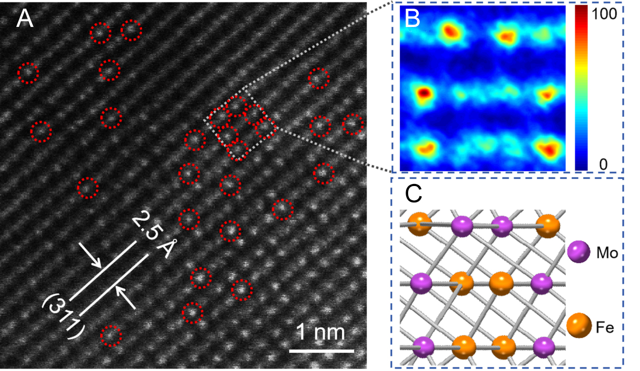
Figure 2. (A) AC-HAADF-STEM image of 27% Mo-Fe2O3; (B) Surface intensity profile and (C) the corresponding structural model of the selected area (blue dashed square area in A). Insets in (C): The purple and orange balls represent Mo and Fe atoms, respectively. AC-HAADF-STEM: Aberration-corrected high-angle annular dark-field scanning transmission electron microscopy.
We investigate the surface chemical states of γ-Fe2O3 and 27% Mo-Fe2O3 by XPS [Figure 3 and Supplementary Table 2]. Figure 3A shows the high-resolution Fe 2p XPS spectra with two spin-splitting bands of Fe 2p3/2 and 2p1/2, both of which are fitted into two peaks: 710.7 (Fe2+ 2p3/2), 713.0 (Fe3+ 2p3/2), 724.0 (Fe2+ 2p1/2) and 726.3 eV (Fe3+ 2p1/2)[39]. By estimating the area ratio (S) of the characteristic peaks of Fe3+, S(Fe3+)/S(Fe2+ + Fe3+), we calculated the relative contents of Fe for both samples [Supplementary Table 2]. Deconvolution of the Fe 2p XPS of γ-Fe2O3 and 27% Mo-Fe2O3 [Figure 3A and Supplementary Table 2] demonstrates that the molar ratio of Fe3+ increases obviously after Mo doping, which is beneficial for improving the structural stability of γ-Fe2O3[40]. Figure 3B shows the O 1s spectra of γ-Fe2O3 and 27% Mo-Fe2O3, which are fitted into three characteristic peaks at 532.5, 530.9 and 529.4 eV, corresponding to adsorbed oxygen species (Oγ), oxygen vacancies (Oβ), and lattice oxygen (Oα), respectively[41]. According to the area ratio of the characteristic peaks provided in Supplementary Table 2, the increase of Oα concentration proves that Mo atoms have successfully occupied the Fe sites in the γ-Fe2O3 lattice[42]. The decrease of Oβ concentration is conducive to capturing photogenerated electrons and inhibiting the recombination of photogenerated carriers[43]. The concentration of Oγ, increasing from 14.62% to 16.62% after Mo doping, is beneficial for the generation of superoxide anion radicals (·O2-) at the photoelectrode and promoting the PEC degradation of TC[44]. Furthermore, the electronic states of the Mo species of MoO3 and 27% Mo-Fe2O3 were also determined by Mo 3d XPS [Supplementary Figure 3]. Obviously, the Mo species of 27% Mo-Fe2O3 are dominantly Mo5+, as judged by the binding energies of the Mo 3d peaks of MoO3 and 27% Mo-Fe2O3, due to the same ionic radius (in Supplementary Table 3) of Mo5+ and Fe2+. By combining the above XRD and HAADF-STEM results, it is convincing that the Mo species are incorporated into the lattice of γ-Fe2O3, leading to the strong electron interaction between Fe and Mo.
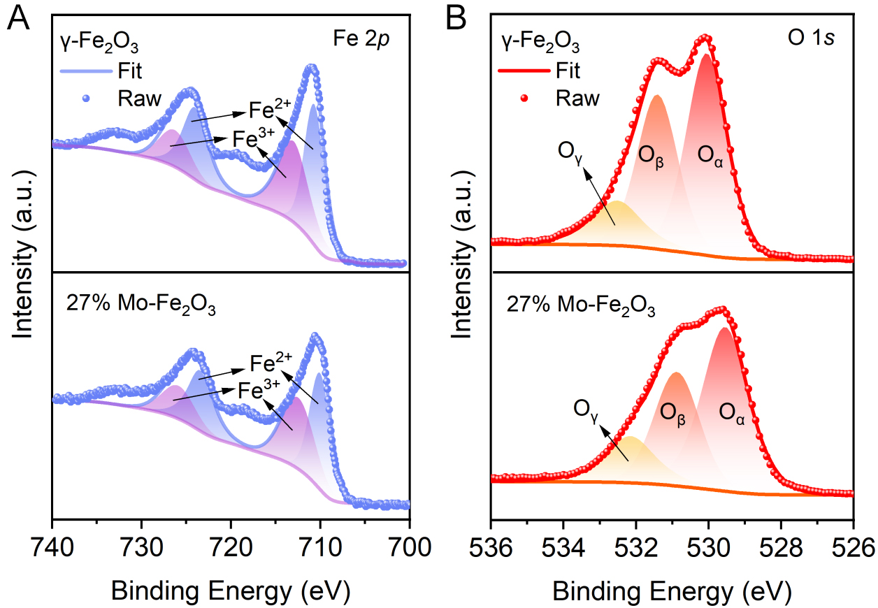
Figure 3. High-resolution (A) Fe 2p and (B) O 1s XPS spectra of γ-Fe2O3 and 27% Mo-Fe2O3. XPS: X-ray photoelectron spectra.
Figure 4 displays the Nyquist plots of photocathodes, which further reveal the influence of the doping amount of Mo on the charge transfer process. The smaller diameter of capacitive loop in the Nyquist plot means the lower charge transfer resistance (Rct) and the higher separation efficiency of photogenerated charges[45]. As shown in Figure 4A, 27% Mo-Fe2O3 shows the smallest arc radius, meaning it has the smallest resistance at the electrolyte interface and the best photoelectric properties, owing to the interaction between Mo and Fe, which promotes the charge transfer at the electrolyte interface. Transient photocurrent testing was employed to further evaluate the superiority of charge transfer on the Mo-Fe2O3 electrode. In Figure 4B, the photocurrent density of 27% Mo-Fe2O3 is -3.45 μA·cm-2, 2.35 times higher than that of γ-Fe2O3
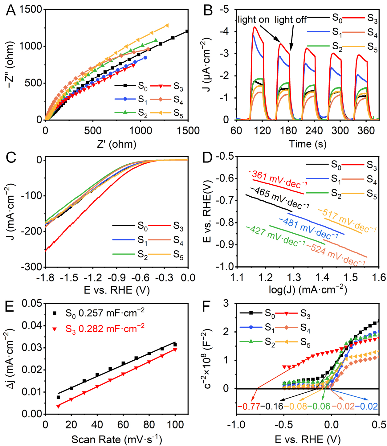
Figure 4. (A) Nyquist plots under AM 1.5 G illumination, (B) photocurrent responses, (C) LSV curves under AM 1.5 G illumination, (D) Tafel plots, (E) Cdl, (F) Mott-Schottky plots of S0, S1, S2, S3, S4 and S5. LSV: Linear sweep voltammogram; AM 1.5 G: air mass 1.5 global solar spectrum; RHE: reversible hydrogen electrode.
As shown in Supplementary Figure 5, UV-vis diffuse reflectance spectroscopy reveals that the optical band gaps of γ-Fe2O3 and the Mo-Fe2O3 samples with different Mo doping amounts are 1.98, 1.94, 1.95, 1.98, 2.01, and 2.02 eV, respectively, indicating that the introduction of Mo atoms does not significantly alter the intrinsic light absorption edge of γ-Fe2O3. Combined with the results of EIS and photocurrent density, the 27% Mo-Fe2O3 exhibits optimized surface photoelectrochemical properties after Mo doping, which is beneficial for promoting the PEC process.
The valence band edges of γ-Fe2O3 and 27% Mo-Fe2O3 were measured by valence-band XPS [Supplementary Figure 6A]. The VBM was determined by linear extrapolation of the leading edge of the spectra. The VBM values are 0.70 and 0.60 eV for γ-Fe2O3 and 27% Mo-Fe2O3, respectively. The work functions of the two samples were further characterized by UPS, as shown in Supplementary Figure 6B and C. The work function decreases from 3.56 eV for γ-Fe2O3 to 3.45 eV upon Mo doping. The concurrent decrease in both VBM and work function indicates that the incorporation of Mo elevates the surface Fermi level of γ-Fe2O3, inducing a downward band bending of approximately 0.1 eV and forming an electron-accumulation layer at the surface. This electronic modification lowers the thermodynamic barrier for photoexcited electrons to migrate from the bulk to the surface and further into vacuum[33], thereby facilitating charge transfer to the surface. In combination with the LSV results, the observed band structure engineering well explains why 27% Mo-Fe2O3 exhibits a significantly higher photocurrent density than γ-Fe2O3.
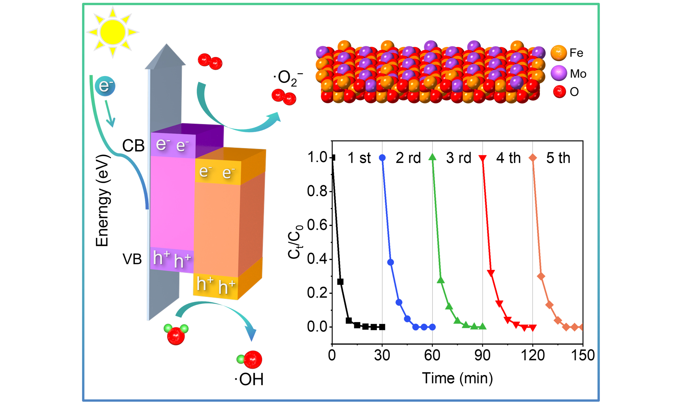
Finally, the PEC degradation of TC using the as-prepared photocathodes were evaluated. As shown in Figure 5A, 27% Mo-Fe2O3 reaches the removal efficiency of 100% in 25 min, while other catalysts achieve the removal efficiency of 100% in 30 min. The first-order rate constant (k) for the reaction was determined using the equation ln(C0/Ct) = kt, where C0 is the initial TC concentration and Ct is the concentration at time t[47]. All degradation experiments were performed three times, and the reported k values are the averages of three independent measurements. The k values of γ-Fe2O3, 9% Mo-Fe2O3, 18% Mo-Fe2O3, 27% Mo-Fe2O3, 36% Mo-Fe2O3, 45% Mo-Fe2O3 are 0.245, 0.214, 0.174, 0.294, 0.199 and 0.172 min-1, respectively [Figure 5B]. The recycling PEC degradation experiments of TC by 27% Mo-Fe2O3 are shown in Figure 5C, quite important for the catalysts. The 27% Mo-Fe2O3 still achieved the removal efficiency of 100% in 30 min after 5 consecutive cycles. In addition, the PEC degradation of TC with high initial concentration by 27% Mo-Fe2O3 were also investigated [Supplementary Figure 7]. When TC initial concentration increased to 50 and 100 mg·L-1, the removal efficiency of TC after 30 min decreased to 86.32% and 67.76%, respectively, which may be attributed to competitive adsorption and accumulation of intermediate products occupying the active sites. The superior PEC property of 27% Mo-Fe2O3 could be attributed to the strong interaction between Mo and Fe, which can promote the separation and migration of photogenerated carriers, and then react with the adsorbed reactants effectively. To elucidate the reaction mechanism, the main active groups in the photocatalytic degradation of TC using 27% Mo-Fe2O3 were conducted by radical capture experiment [Figure 5D]. Four different quenchers (5 mmol·L-1 each) were used to target specific active species: PBQ can capture superoxide anion (·O2-), IPA can capture hydroxyl radical (·OH), EDTA-2Na can capture holes (h+), and KBrO3 can capture electron (e-). The removal rate of TC decreases significantly in the presence of PBQ, suggesting that ·O2- is the primary active species. Secondly, the removal rate of TC decreases moderately in the presence of EDTA-2Na and IPA, implying that h+ and ·OH are the secondary active species. In contrast, KBrO3 has little impact on the removal rate, indicating that e- is not the main active species.
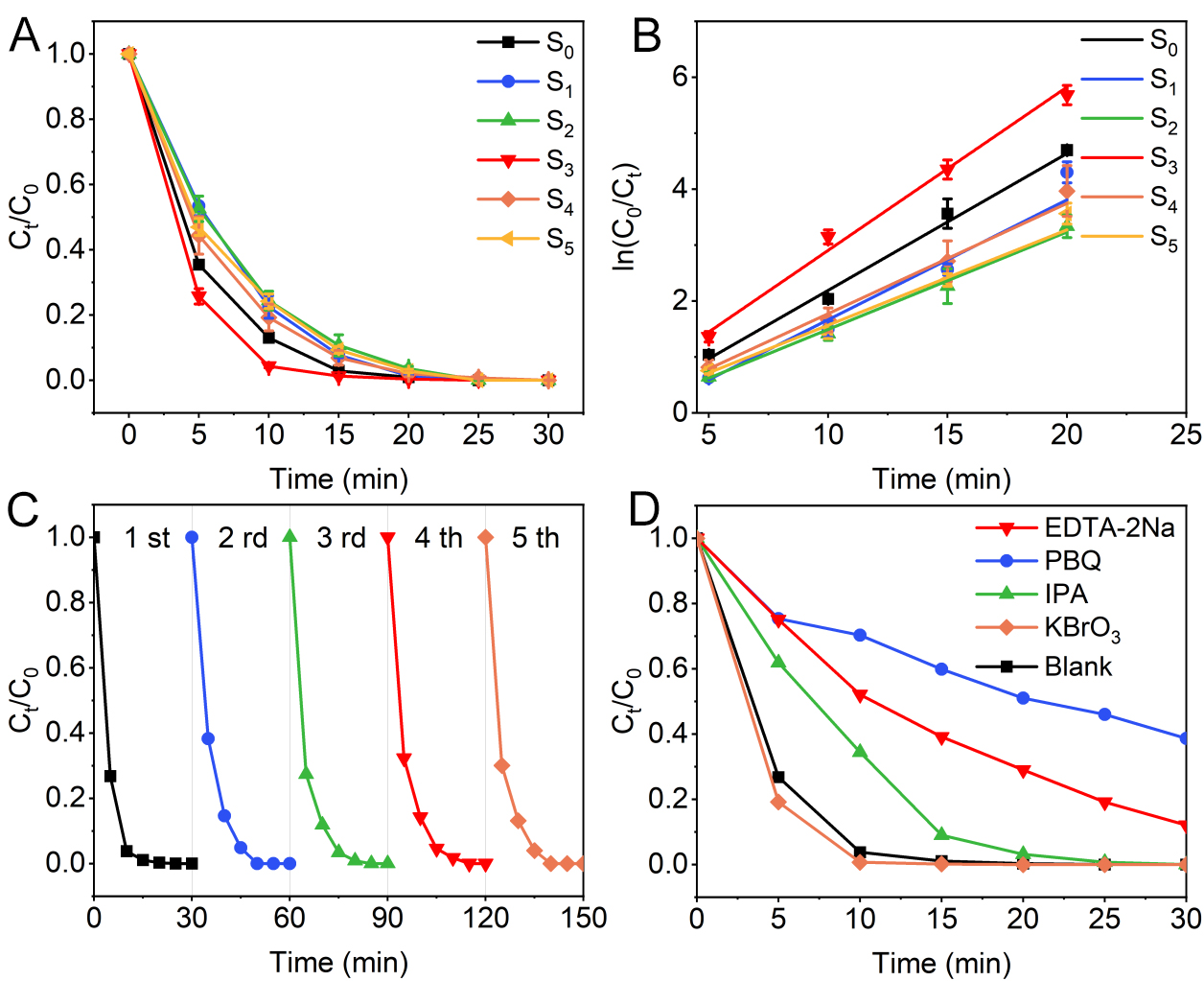
Figure 5. (A) PEC degradation of TC under simulated sunlight by catalysts and (B) the corresponding kinetic linear simulation curves; (C) Recycling PEC degradation of TC by 27% Mo-Fe2O3; (D) The influence of different quenchers on the PEC degradation of TC by 27% Mo-Fe2O3. Error bars in the corresponding figures represent the SD of three independent measurements. TC: Tetracycline; PEC: photoelectrocatalytic; SD: standard deviation; PBQ: p-benzoquinone; IPA: isopropanol; EDTA-2Na: ethylenediaminetetraacetic acid disodium salt dihydrate.
To shed light on the possible degradation pathways of TC by 27% Mo-Fe2O3, we conducted a test on the reaction intermediates employing LC-MS analysis [Supplementary Figure 8] and proposed two possible degradation pathways mainly driven by the synergistic action of ·O2- and h+ according to Figure 6. The TC molecule (m/z = 445) initially is attacked by h+, yielding demethylation intermediate P1 (m/z = 417)[20]. Subsequently, intermediate P1 undergoes a ring-opening reaction to generate intermediate P2 (m/z = 291) and at the same time undergoes demethylation to form intermediate P5 (m/z = 403), respectively, which correspond respectively to reaction pathways I and II. In pathway I, intermediate P2 transforms into P3 (m/z = 227) after undergoing simultaneous elimination of -CH3, -OH and -CH2OH groups, then P3 undergoes a ring-opening reaction to generate P4 (m/z = 218). While in pathway II, intermediate P5 (m/z = 403) loses -OH and -NH2 under the concerted action of ·O2- and h+ to yield P6 (m/z = 349). P6 undergoes elimination of a -COOH mediated by ·O2- to form P7 (m/z = 291). Subsequently, P7 loses -OH to form P8 (m/z = 275). Thereafter, P8 undergoes ring-opening with ·O2- to yield P9 (m/z = 262). The intermediates (P4 and P9) progressively convert into smaller molecular intermediates (P10, P11, P12, P13) under the continuous action of ·O2- and h+, and finally generate harmless substances CO2 and H2O.
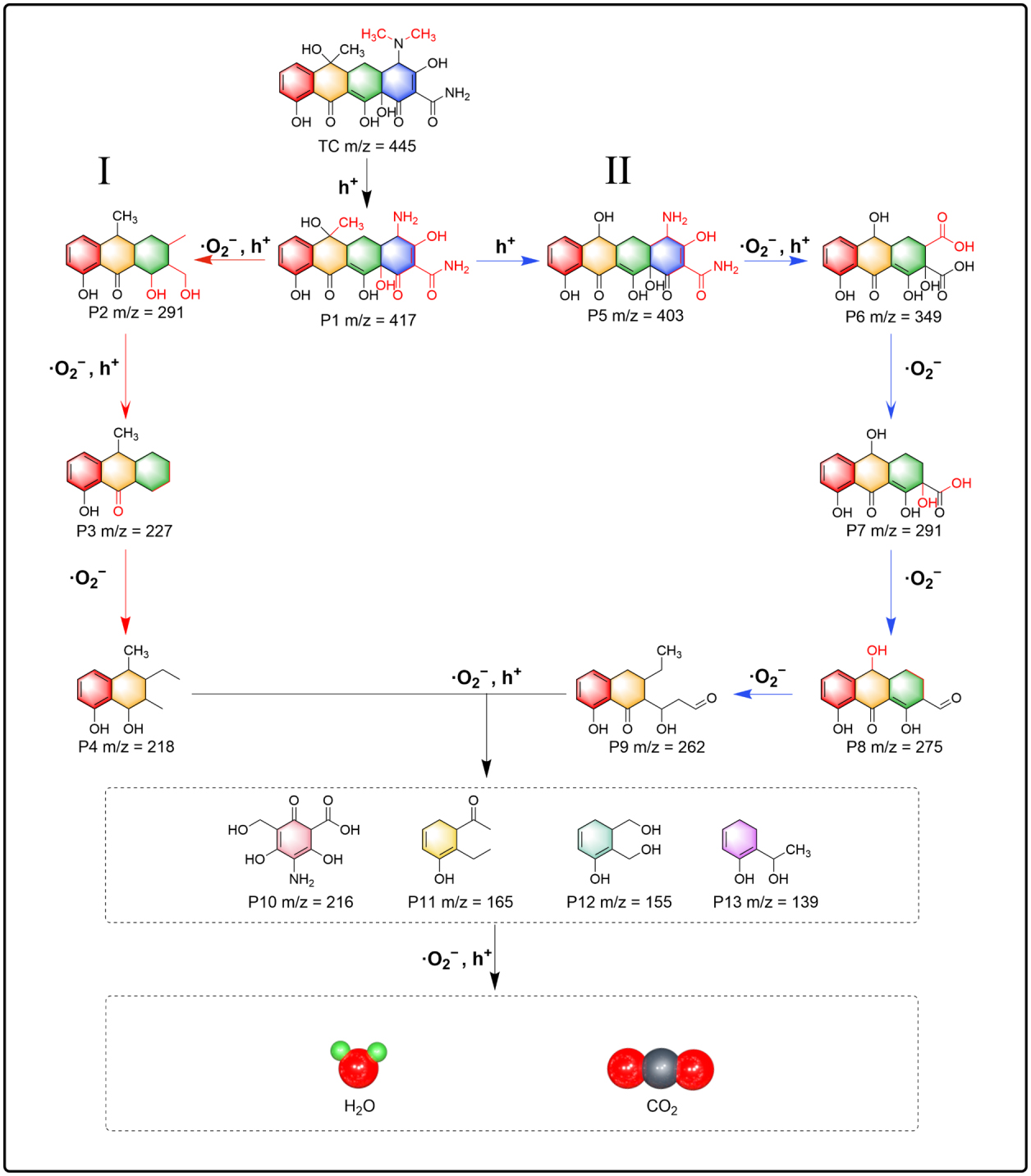
Figure 6. Possible degradation pathways of TC by 27% Mo-Fe2O3. TC: Tetracycline.
Based on the above results, we propose the possible PEC mechanism of 27% Mo-Fe2O3 [Figure 7], including the oxidation reaction at the anode and the reduction reaction at the cathode. When the photocathode absorbs solar energy, photogenerated electrons (e-) transition to the CB, leaving holes (h+) in the valence band (VB) at the same time. Under an applied external bias potential, e- flow to the cathode through the external circuit, while h+ migrate to the surface of the photocathode[48]. A portion of the h+ recombine with electrons, while the remaining h+ participate in reactions. On the one hand, h+ can directly undergo oxidation reactions with the TC molecules adsorbed on the surface of the photocathode, thereby decomposing TC. On the other hand, h+ can also react with the electrolytes in the solution, generating corresponding active free radicals (such as ·OH) and participating in the oxidation of TC indirectly[49]. The dissolved O2 in the solution can also capture e- to generate active species such as ·O2-, thereby participating in the degradation process of TC. This path effectively separates the photogenerated electrons and holes and meanwhile inhibits their recombination, resulting in excellent PEC activity.
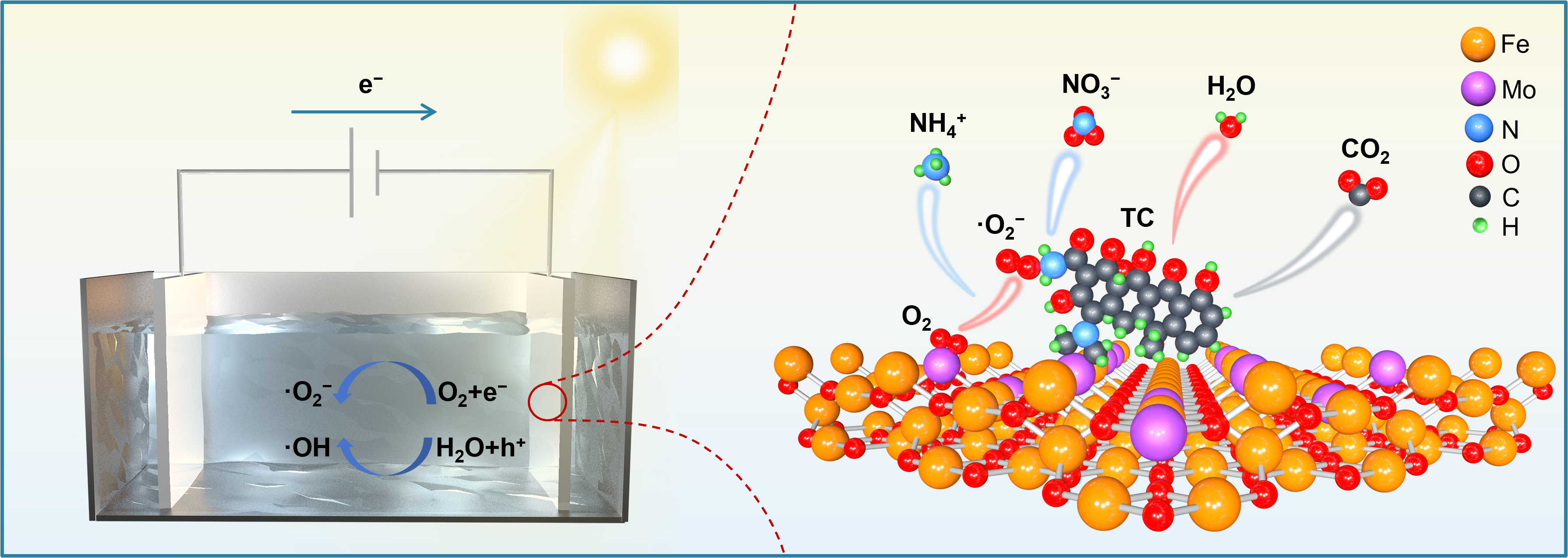
Figure 7. Schematic description of the PEC mechanism of 27% Mo-Fe2O3. PEC: Photoelectrocatalytic.
CONCLUSIONS
In summary, Mo-doped γ-Fe2O3 was successfully synthesized via a facile co-precipitation combined with impregnation method. Combined with the analysis of XRD, XPS, TEM-EDS mapping and HAADF-STEM, Mo atoms are doped into the lattice of γ-Fe2O3. And the modification of Mo doping has an impact on the electronic structure of γ-Fe2O3, facilitating the separation and migration of photogenerated carriers, and resulting in superior PEC activity. Herein, we provide a feasible strategy for developing doped catalysts with superior PEC performance by enhancing the electronic interactions.
DECLARATIONS
Authors’ contributions
Chemical syntheses and characterizations: Xia, X.; Zhang, Z.; Jia, P.
Data analysis and interpretation: Xia, X.; Liu, X.
Topic selection, manuscript review: Liu, X.; Liu, Z.
Editing and supervision: Wang, H.; Liu, W.
All authors contributed to the discussion of results and the manuscript writing.
Availability of data and materials
Some results of supporting the study are presented in the Supplementary Materials. Other raw data that support the findings of this study are available from the corresponding author upon reasonable request.
AI and AI-assisted technologies statement
Not applicable.
Financial support and sponsorship
This research was funded by the National Natural Science Foundation of China (No. 22006082 and 52172147), Major scientific research special projects for the construction of national key laboratories (No. 2025ZDGZ02), the Natural Science Foundation of Shandong Province (No. ZR2022MB095 and ZR2021MB035), and Development Project of Youth Innovation Teams in Colleges and Universities of Shandong Province (Grant No. 2022KJ293).
Conflict of interest
Liu, Z. is affiliated with Shandong Environmental Protection Development Group Co., Ltd., Jinan 250101, China, but this affiliation did not influence the research or its outcomes. The other authors declared that there are no conflicts of interest.
Ethical approval and consent to participate
Not applicable.
Consent for publication
Not applicable.
Copyright
© The Author(s) 2026.
Supplementary Materials
REFERENCES
1. Xu, M.; Wang, Q.; Maqbool, T.; Guo, B.; Lu, H.; Jiang, D. Contrasting responses of antibiotic resistance genes (ARGs) to biocathodic and bioanodic treatment of broad-spectrum tetracycline and narrow-spectrum fidaxomicin. Chem. Eng. J. 2025, 515, 163878.
2. Li, Q.; Zheng, Y.; Guo, L.; et al. Microbial degradation of tetracycline antibiotics: mechanisms and environmental implications. J. Agric. Food. Chem. 2024, 72, 13523-36.
3. Zong, Z.; Huang, Y.; Kwan, J.; Hankins, N. P. Standardized benchmarking of advanced oxidation processes for tetracycline degradation with life cycle assessment and economic evaluation. Chem. Eng. J. 2025, 525, 170664.
4. Zeng, S.; Chen, Y.; Zhang, N.; et al. Tetracycline effects on arsenate reduction in agricultural soils under different soil water contents. Environ. Pollut. 2026, 389, 127398.
5. Daghrir, R.; Drogui, P. Tetracycline antibiotics in the environment: a review. Environ. Chem. Lett. 2013, 11, 209-27.
6. Fan, Y.; Liu, D.; Wan, H.; Liu, X.; Wang, J.; Xiong, K. Enhanced double-exchange interactions enable the Co and Mn co-doped zinc ferrite rapid and efficient removal of tetracycline. J. Environ. Chem. Eng. 2025, 13, 117070.
7. Liang, S.; Zhang, H.; Dai, H.; et al. Efficient, rapid and simple adsorption method by polydopamine polystyrene nanofibers mat for removal of multi-class antibiotic residues in environmental water. Chemosphere 2022, 288, 132616.
8. Scaria, J.; Anupama, K.; Nidheesh, P. Tetracyclines in the environment: an overview on the occurrence, fate, toxicity, detection, removal methods, and sludge management. Sci. Total. Environ. 2021, 771, 145291.
9. Song, C.; Sun, X.; Xia, P.; Wang, Y.; Wang, S. Investigation of fate and behavior of tetracycline in nitrifying sludge system. RSC. Adv. 2015, 5, 87333-40.
10. Zhang, Y.; Huang, X.; Zheng, Q.; Yang, J.; Zhai, L.; Song, Y. Spherical Bi4O5I2/MoS2 QDs nanocomposites activate persulfate to degrade tetracycline under visible light. Adv. Sustain. Syst. 2023, 8, 2300439.
11. Kumar, A.; Choudhary, P.; Kumar, A.; Camargo, P. H. C.; Krishnan, V. Recent advances in plasmonic photocatalysis based on TiO2 and noble metal nanoparticles for energy conversion, environmental remediation, and organic synthesis. Small 2021, 18, 2101638.
12. Wang, Z.; Meng, S.; Li, J.; et al. Oxygen Vacancy engineering and constructing built‐in electric field in Fe‐g‐C3N4/Bi2MoO6 Z‐scheme heterojunction for boosting photo‐fenton catalytic degradation performance of tetracycline. Small 2024, 20, 2406125.
13. Li, J.; Cai, L.; Shang, J.; Yu, Y.; Zhang, L. Giant Enhancement of internal electric field boosting bulk charge separation for photocatalysis. Adv. Mater. 2016, 28, 4059-64.
14. Li, J.; Zhang, X.; Xiong, X.; Wu, C.; Jin, Y.; Lv, K. Breaking Type-I heterojunction limitations: harnessing an ohmic-like/S-scheme cascade charge transfer mechanism for enhanced photocatalytic H2 evolution. Sep. Purif. Technol. 2025, 354, 129444.
15. Yu, H.; Xu, J.; Liu, H.; et al. Defect synergy enabled by spin-polarization-tunable engineering for enhanced photocatalysis: carrier migration mechanisms. Chem. Eng. J. 2025, 523, 168394.
16. Li, C.; Wu, M.; Lei, J.; et al. Work-function-driven built-in electric field in flame-synthesized BiVO4 QDs/TiO2 composites: a high-efficiency photoelectrocatalytic platform for continuous flow degradation of tetracycline in wastewater. J. Environ. Chem. Eng. 2025, 13, 118655.
17. Adak, D.; Chakrabarty, P.; Majumdar, P.; et al. Pd nanoparticle-decorated hydrogen plasma-treated TiO2 for photoelectrocatalysis-based solar energy devices. ACS. Appl. Electron. Mater. 2020, 2, 3936-45.
18. Wu, H.; Liu, Q.; Zhang, L.; Tang, Y.; Wang, G.; Mao, G. Novel nanostructured WO3@prussian blue heterojunction photoanodes for efficient photoelectrochemical water splitting. ACS. Appl. Energy. Mater. 2021, 4, 12508-14.
19. Wang, R.; Chen, S.; Ng, Y. H.; et al. ZnO/CdS/PbS nanotube arrays with multi-heterojunctions for efficient visible-light-driven photoelectrochemical hydrogen evolution. Chem. Eng. J. 2019, 362, 658-66.
20. He, S.; Yan, C.; Chen, X.; et al. Construction of core-shell heterojunction regulating α-Fe2O3 layer on CeO2 nanotube arrays enables highly efficient Z-scheme photoelectrocatalysis. Appl. Catal. B. Environ. 2020, 276, 119138.
21. Chen, Y.; Lin, M.; Peng, Z.; et al. Fabrication of piezotronic ZnO p-n homojunction via metal/oxygen defects modulation for efficient photoelectrocatalysis. Chem. Eng. Sci. 2024, 295, 120174.
22. Li, Y.; Yu, L.; Xie, J.; et al. Efficient photoelectrocatalytic degradation of tetracycline coupled with hydrogen production by N-doped carbon layer-wrapped TiO2 nanorods. J. Environ. Chem. Eng. 2025, 13, 115153.
23. Yu, J.; Su, Q.; Huang, J.; Teng, W.; Song, Y.; Zhang, H. Synergistic interfacial engineering of S-scheme 2H-MoSe2/NiFe-LDH architecture for dual functional photoelectrocatalysis: multi-pollutant mineralization and visible light driven hydrogen evolution. Chem. Eng. J. 2025, 519, 164914.
24. Liu, J.; Liang, R.; Hu, Z.; Zhang, X.; Zhou, M. Photoelectrocatalysis/photoelectro-fenton system based on cone-like TiO2/nickel foam photoanode for efficient degradation of carbamazepine: comparison with DSA. Chem. Eng. J. 2024, 491, 152088.
25. Li, B.; Lv, M.; Zhang, Y.; et al. Single-particle imaging photoinduced charge transfer of ferroelectric polarized heterostructures for photocatalysis. ACS. Nano. 2024, 18, 25522-34.
26. Vaghasiya, J. V.; Mayorga‐martinez, C. C.; Zelenka, J.; Sharma, S.; Ruml, T.; Pumera, M. Magnetic soft centirobot to mitigate biological threats. SmartMat 2024, 5, e1289.
27. Hu, H.; Zhang, C.; Qi, H.; et al. Seawater electrosynthesis of hydrogen peroxide at industrial‐level current densities enabled by pentagonal defect‐rich nanocarbon with chlorine doping. Angew. Chem. Int. Ed. 2025, 65, e12138.
28. Wong, K. J.; Foo, J. J.; Siang, T. J.; Ong, W. J. Transition metal carbide‐based photocatalysts for artificial photosynthesis. SmartMat 2023, 5, e1238.
29. Kahng, S.; Kim, J. H. Heterojunction photoanode of SnO2 and Mo-doped BiVO4 for boosting photoelectrochemical performance and tetracycline hydrochloride degradation. Chemosphere 2022, 291, 132800.
30. Zhang, X.; Xu, C.; Yu, C.; et al. Oxygen vacancy defect engineering in MoO2/Mo-doped BiOCl ohmic junctions for enhanced photocatalytic antibiotic elimination. J. Alloys. Compd. 2024, 1005, 176220.
31. Qu, W.; Chen, Y.; Huang, Z.; et al. Active tetrahedral iron sites of γ-Fe2O3 catalyzing NO reduction by NH3. Environ. Sci. Technol. Lett. 2017, 4, 246-50.
32. Zheng, J.; Xu, S.; Sun, J.; et al. Boosting efficient C-N bonding toward photoelectrocatalytic urea synthesis from CO2 and nitrate via close Cu/Ti bimetallic sites. Appl. Catal. B. Environ. 2023, 338, 123056.
33. De La Fuente, B.; Bomnuter, J.; Del Moro, M.; et al. On the combination of ultraviolet photoelectron spectroscopy with optical absorption studies to investigate Cu2O||TiO2 direct Z-scheme junctions with different Cu2O loading. Appl. Surf. Sci. 2024, 657, 159796.
34. Han, S.; Hu, X.; Wang, J.; Fang, X.; Zhu, Y. Novel route to Fe‐based cathode as an efficient bifunctional catalysts for rechargeable Zn-air battery. Adv. Energy. Mater. 2018, 8, 1800955.
35. Cheng, L.; Jiang, T.; Yan, K.; Gong, J.; Zhang, J. A dual-cathode photoelectrocatalysis-electroenzymatic catalysis system by coupling BiVO4 photoanode with hemin/Cu and carbon cloth cathodes for degradation of tetracycline. Electrochim. Acta. 2019, 298, 561-9.
36. Al Huwayz, M.; Tahir, A.; Alomairy, S.; et al. RETRACTED: Harnessing the interfacial charge transfer in Mn-doped γ-Fe2O3@ZnO heterojunction for broad spectrum photocatalytic degradation of organic dyes. Opt. Mater. 2024, 157, 116348.
37. Shannon, R. D. Revised effective ionic radii and systematic studies of interatomic distances in halides and chalcogenides. Acta. Cryst. A. 1976, 32, 751-67.
38. Liu, J.; Hu, J.; Han, X.; et al. Simple synthesis of low-load γ-Fe2O3/C/CNT for efficient building absorber. Mater. Today. Commun. 2025, 46, 112529.
39. Chen, Y.; Liu, L.; Zhang, L.; et al. Construction of Z-type heterojunction BiVO4/Sm/α-Fe2O3 photoanode for selective degradation: efficient removal of bisphenol A based on multifunctional Sm-doped modification. Appl. Catal. B. Environ. Energy. 2023, 333, 122775.
40. Hsu, J. H.; Kuo, P. C.; Hsu, C. W. Improved thermal stability of Mo-modified γ-Fe2O3 particles. J. Appl. Phys. 1990, 67, 5152-4.
41. Lu, L.; Xin, Z.; Wang, X.; et al. KOH-modified Ni/LaTiO2N Schottky junction efficiently reducing CO2 to CH4 under visible light irradiation. Appl. Catal. B. Environ. 2019, 244, 786-94.
42. Zheng, Y.; Hussain, G.; Zheng, C.; et al. Impact of the rhenium substitution on the oxygen evolution reaction of spinel CoFe2O4. J. Mater. Chem. A. 2024, 12, 19521-31.
43. Wei, X.; Yang, Y.; Li, S.; Meng, Y.; Yuan, W.; Ni, H. Dual role of oxygen vacancies in TiO2/Bi2O3 for enhanced carrier separation and PDS activation: a combined experimental and DFT study. Environ. Res. 2025, 282, 122042.
44. Li, J.; Tang, H.; Liu, C.; et al. Selective generation of superoxide radical for de-NOx via fly ash/H2O2 slurry: unveiling the role of surface hydroxyls for H2O2 activation. Sep. Purif. Technol. 2025, 379, 135081.
45. Guardiano, M. G.; Gonzaga, I. M. D.; Ribeiro, L. K.; Almeida, C. V. D. S.; Mascaro, L. H. Gd-BiVO4: an efficient photoanode for pharmaceuticals degradation in contaminated waters via solar photoelectrocatalysis. Chem. Eng. J. 2025, 503, 158463.
46. Ma, L.; Lu, M.; Li, K.; et al. Photocatalytic degradation of octadecylamine and 4-dodecylmorpholine over titanium based photocatalyst: activity and mechanism insights. Chem. Eng. J. 2023, 472, 144782.
47. Zhong, X.; Li, L.; Xiang, W.; et al. Stable and rapid degradation of tetracycline using a photoelectrochemical system comprising a Ag/N-TiO2 photoanode and P-Pd cathode. J. Environ. Chem. Eng. 2023, 11, 109875.
48. Ren, M.; Lu, H.; Liu, Z.; et al. Synergistic antibiotic degradation by polydopamine/BiOBr-modified photocathode in self-powered photo-bio-electrochemical system. Bioresour. Technol. 2026, 442, 133669.
Cite This Article
How to Cite
Download Citation
Export Citation File:
Type of Import
Tips on Downloading Citation
Citation Manager File Format
Type of Import
Direct Import: When the Direct Import option is selected (the default state), a dialogue box will give you the option to Save or Open the downloaded citation data. Choosing Open will either launch your citation manager or give you a choice of applications with which to use the metadata. The Save option saves the file locally for later use.
Indirect Import: When the Indirect Import option is selected, the metadata is displayed and may be copied and pasted as needed.
About This Article
Special Topic
Copyright

Data & Comments
Data
0


Comments
Comments must be written in English. Spam, offensive content, impersonation, and private information will not be permitted. If any comment is reported and identified as inappropriate content by OAE staff, the comment will be removed without notice. If you have any queries or need any help, please contact us at [email protected].