

Artificial intelligence in interventional cardiology: present landscape and future directions
 0

Graphical Abstract
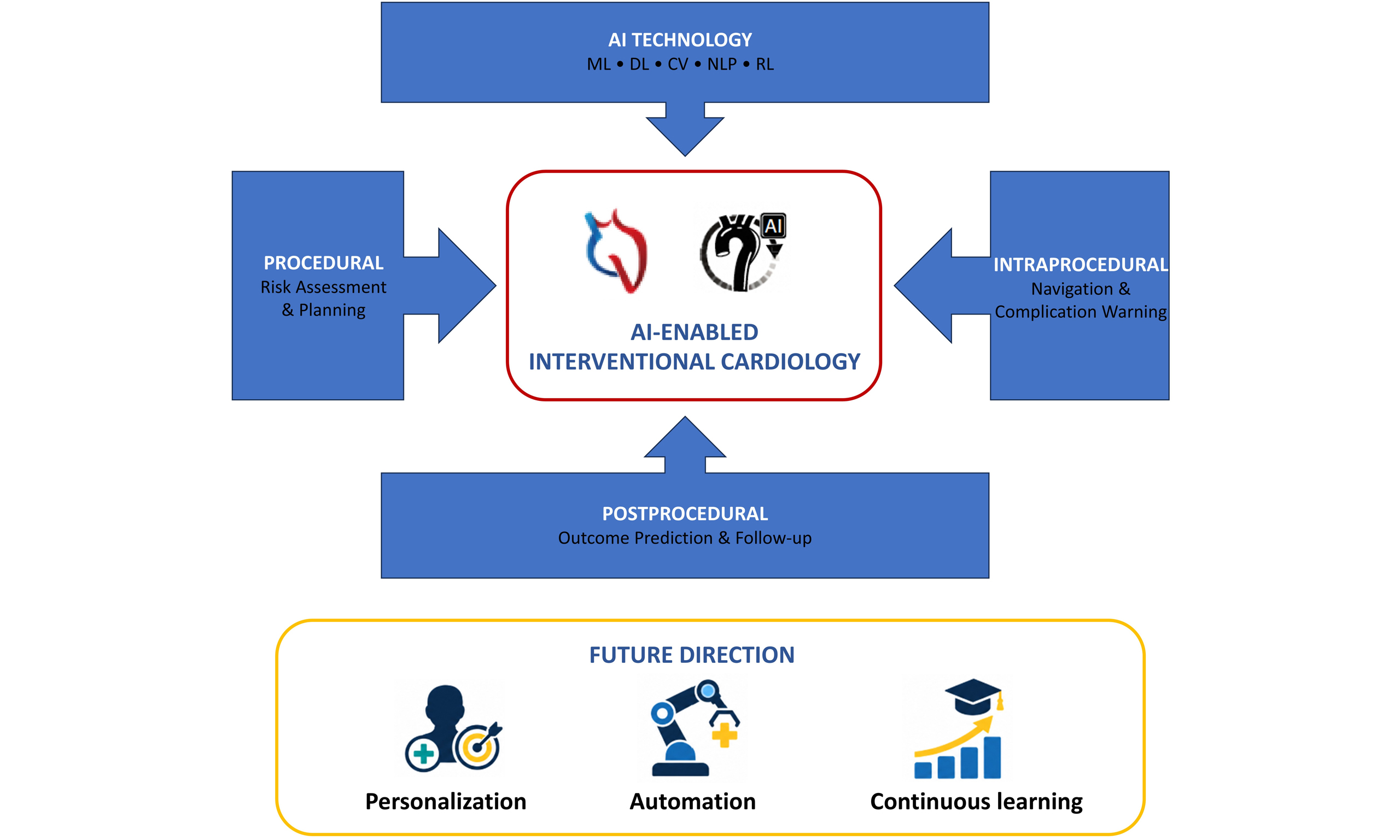
Abstract
With population aging and the rising incidence of cardiac disease, interventional cardiology, as a cornerstone of treatment for conditions such as coronary and structural heart disease, faces persistent challenges, including stringent precision requirements, heavy reliance on operator experience, and variability in diagnostic and therapeutic efficiency. With robust capabilities in data mining, image recognition, real-time analysis, and predictive modeling, artificial intelligence (AI) offers a novel pathway to address the aforementioned challenges and support the intelligent transformation of interventional cardiology. This narrative review briefly outlines the background to the application of AI in interventional cardiology, focuses on core supporting technologies including deep learning, machine learning, and computer vision, and summarizes current research and clinical applications of AI across preprocedural risk assessment and planning, intraprocedural real-time navigation and early warning of complications, and postprocedural follow-up and prognosis prediction. It also analyzes the principal challenges currently facing AI in this field, such as data quality and security issues, insufficient algorithm interpretability, barriers to clinical translation, and constraints from ethical and regulatory frameworks. Finally, in light of technological advances and clinical needs, this review outlines future directions, including the integration of AI with interventional devices, multimodal data integration and modeling, and the development of personalized interventional strategies, thereby providing a reference for promoting the standardized and large-scale application of AI technology in interventional cardiology.
Keywords


 Download PDF
INTRODUCTION
Cardiac disease remains a leading global cause of morbidity and mortality, imposing an enormous burden on healthcare systems worldwide[1], and interventional cardiology - a cardiovascular subspecialty focused on catheter-based treatment - has become indispensable in its management, evolving from simple balloon angioplasties to advanced procedures such as percutaneous coronary intervention (PCI) and transcatheter aortic valve replacement (TAVR) that significantly improve patient outcomes[2].
However, traditional interventional cardiology practice still faces considerable challenges: the anatomical complexity of the heart, such as chronic total occlusions and bifurcation lesions, poses great technical difficulties[3]; individual differences in patients’ physiology and genetic makeup limit the applicability of standardized treatment protocols[4]; TAVR is now established for appropriately selected patients, although the durability of contemporary devices in younger populations and lifetime valve-management strategies require continued evaluation. Transcatheter mitral valve replacement remains an evolving therapy for selected anatomies, whereas transcatheter tricuspid interventions, including replacement, are now available for selected high-risk patients but require longer-term comparative evidence[5]; endoleaks and aneurysm expansion persist in peripheral endovascular therapies; and the uneven global distribution of specialized interventional care, exacerbated by shortages of trained specialists, leads to geographical disparities.
The emergence of artificial intelligence (AI) and machine learning has created new opportunities in healthcare, with the ability to process large volumes of data, identify subtle patterns, and generate actionable insights, showing great potential in optimizing diagnosis, treatment planning, and risk stratification in interventional cardiology[6].
This review aims to comprehensively explore the integration of AI in interventional cardiology, including its current applications, challenges, and future directions [Figure 1]. Structurally, it will first analyze the current status of AI application in interventional cardiology, then discuss existing limitations, and finally outline future development trends to inform clinical practice and research.
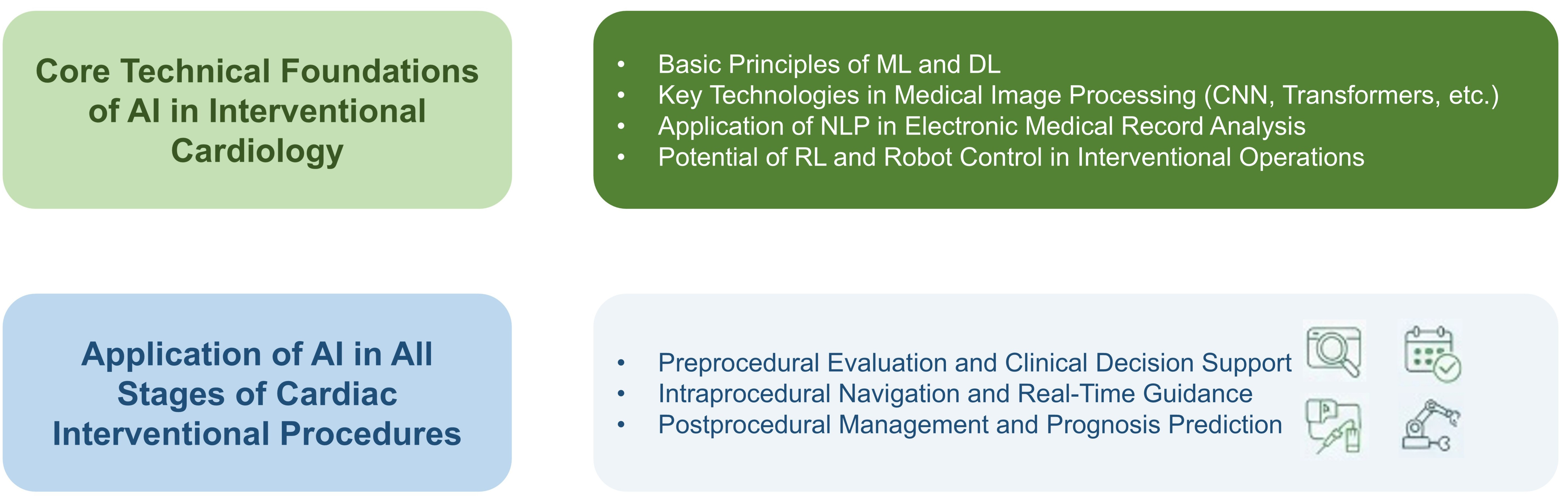
Figure 1. Schematic of Core Technical Foundations and Clinical Applications of AI in Cardiac Interventional Procedures. ML: Machine learning; DL: deep learning; CNN: convolutional neural network; RL: reinforcement learning; NLP: natural language processing; AI: artificial intelligence.
CLINICALLY RELEVANT AI APPROACHES IN INTERVENTIONAL CARDIOLOGY
Rather than providing an extensive description of algorithmic mechanisms, this section briefly introduces the artificial intelligence approaches most relevant to clinical workflows in interventional cardiology. Their specific applications and supporting clinical evidence are discussed according to the preprocedural, intraprocedural, and postprocedural stages in the following sections and are summarized in Table 1.
Representative clinical studies of machine learning and deep learning for preprocedural evaluation and decision support in interventional cardiology
| Application | AI method | Dataset and study design | Main performance or clinical finding | References |
| Non-invasive functional assessment of coronary stenosis using CCTA-derived FFR | Supervised ML-based CT-FFR computation | Retrospective multicenter cohort; 74 patients and 91 vessels; invasive FFR used as reference | Per-vessel AUC, 0.907; per-patient diagnostic accuracy improved from 66% with CCTA alone to 85% after CT-FFR; mean processing time, 16.4 ± 7.5 min | [18] |
| CT-FFR-guided management of stable CAD (TARGET trial) | On-site ML-based CT-FFR | Six-center randomized trial; 1,216 patients with 30%-90% stenosis on CCTA | Low-yield ICA was reduced versus standard care (28.3% vs. 46.2%, P < 0.001); revascularization increased, whereas 1-year MACE was similar | [19] |
| Automated coronary calcium detection and risk stratification using IVUS | Support vector classifier applied to individual IVUS A-lines | Algorithm validation: 105 IVUS pullbacks from three imaging systems; prognostic cohort: 408 patients with approximately 6-year follow-up | Testing accuracy was 0.87-0.89 across three systems; an automated IVUS calcium score ≥ 85 was associated with POCE (adjusted HR, 1.51) | [20,21] |
| Prediction of OCT-defined TCFA from IVUS | ANN, SVM, and naive Bayes using geometry- and texture-derived IVUS features | 517 patients; 414 training and 103 test lesions; 40,908 co-registered IVUS-OCT frames | Five-fold cross-validation AUCs: ANN 0.80 ± 0.08, SVM 0.74 ± 0.05, naive Bayes 0.77 ± 0.04 | [22] |
| Automated TCFA classification from IVUS | FNN, KNN, random forest, and CNN | 12,325 IVUS images labeled using corresponding OCT images | AUCs: FNN 0.859, KNN 0.848, random forest 0.844, CNN 0.911 | [23] |
| Automated characterization of coronary plaque composition using OCT | Pseudo-3D encoder-decoder deep CNN | 509 OCT pullbacks from 391 patients; 10,517 training and 1,156 testing cross-sections; external validation on 300 cross-sections | External diagnostic accuracy was 97.6% for fibrous plaque, 90.5% for lipid plaque, and 88.5% for calcium; median analysis time, 21.4 s per pullback | [26] |
| Automated coronary-segment and lesion-morphology recognition from angiography | Two-stage DNN architecture (DeepDiscern) | Single-center retrospective dataset; 20,612 angiograms from 10,073 patients | Segment-recognition accuracy was 98.4%; lesion-morphology F1 scores ranged from 0.802 to 0.854; processing required < 2 s | [31] |
| Fully automated coronary angiography interpretation and stenosis estimation | Sequential task-specific deep neural networks (CathAI) | 13,843 angiographic studies comprising 195,195 videos from 11,972 patients; additional external validation cohorts | AUC for detecting ≥ 70% stenosis was 0.862 internally and 0.869 in external validation | [30] |
Machine learning and deep learning for clinical prediction and decision support
Machine learning (ML) and deep learning (DL) enable quantitative analysis of clinical, imaging, and procedural data[7]. Conventional ML remains useful for structured variables and moderately sized datasets[8,9], whereas DL is particularly suitable for complex imaging data[10]. These approaches are increasingly used across interventional cardiology workflows[11], including preprocedural risk stratification, lesion and plaque characterization, non-invasive hemodynamic assessment, procedural planning, complication prediction, and postprocedural outcome assessment. Their clinical value depends not only on predictive accuracy but also on representative training data, external validation, model calibration, and integration into physician-led decision-making.
AI-based cardiovascular image analysis
Medical imaging represents one of the most mature applications of AI in interventional cardiology[11]. Convolutional neural networks are widely used for automated image segmentation and characterization[12], while attention-based architectures provide complementary capabilities for complex medical image analysis[13]. Across coronary computed tomography angiography (CCTA), coronary angiography, intravascular ultrasound (IVUS), and optical coherence tomography (OCT), these models can identify stenoses, characterize plaque composition, quantify calcification, and recognize relevant anatomical or procedural features. These functions may shorten interpretation time, reduce interobserver variability, and provide standardized quantitative information for treatment planning. Nevertheless, performance may decline in the presence of poor image quality, severe calcification, device-related artifacts, or variations in imaging equipment and acquisition protocols, emphasizing the importance of multicenter and multi-device validation.
Natural language processing for clinical data integration
Natural language processing (NLP) can extract clinically relevant information from unstructured electronic medical records, procedure reports, and follow-up documentation[14]. In cardiology, this includes extraction of clinically meaningful functional-status information from routine documentation[15]. For patients undergoing cardiovascular interventions, NLP may help identify comorbidities, prior procedures, complications, medication changes, and follow-up outcomes. By reducing the burden of manual data abstraction, NLP can support the construction of structured clinical datasets and the integration of textual information into risk-prediction and decision-support systems. However, differences in documentation practices and terminology across institutions remain important barriers to reliable clinical implementation.
Reinforcement learning and robotic assistance
Reinforcement learning (RL) is an AI approach in which an agent learns behavior through repeated interaction with an environment[16]. In interventional cardiology, early work has demonstrated RL-based guidewire navigation in a coronary artery phantom[17]. Potential applications include robotic catheter and guidewire navigation, path planning, force control, and simulation-based procedural training. By learning from repeated interactions in virtual or experimental environments, RL-based systems may support more consistent and precise device manipulation, particularly in anatomically complex procedures. However, current evidence is predominantly derived from simulations, vascular phantoms, and other early-stage experimental settings. Further validation under realistic clinical conditions is required before autonomous or semi-autonomous control can be routinely adopted, and interventional physicians should retain oversight of all safety-critical decisions.
Because the clinical relevance of AI depends more on validated performance and workflow integration than on algorithmic complexity alone, the following section organizes the available evidence by application across the interventional care pathway. Figure 2 summarizes this clinically oriented pathway from multimodal data inputs and curation through AI model development, stage-specific applications, and supervised clinical translation.
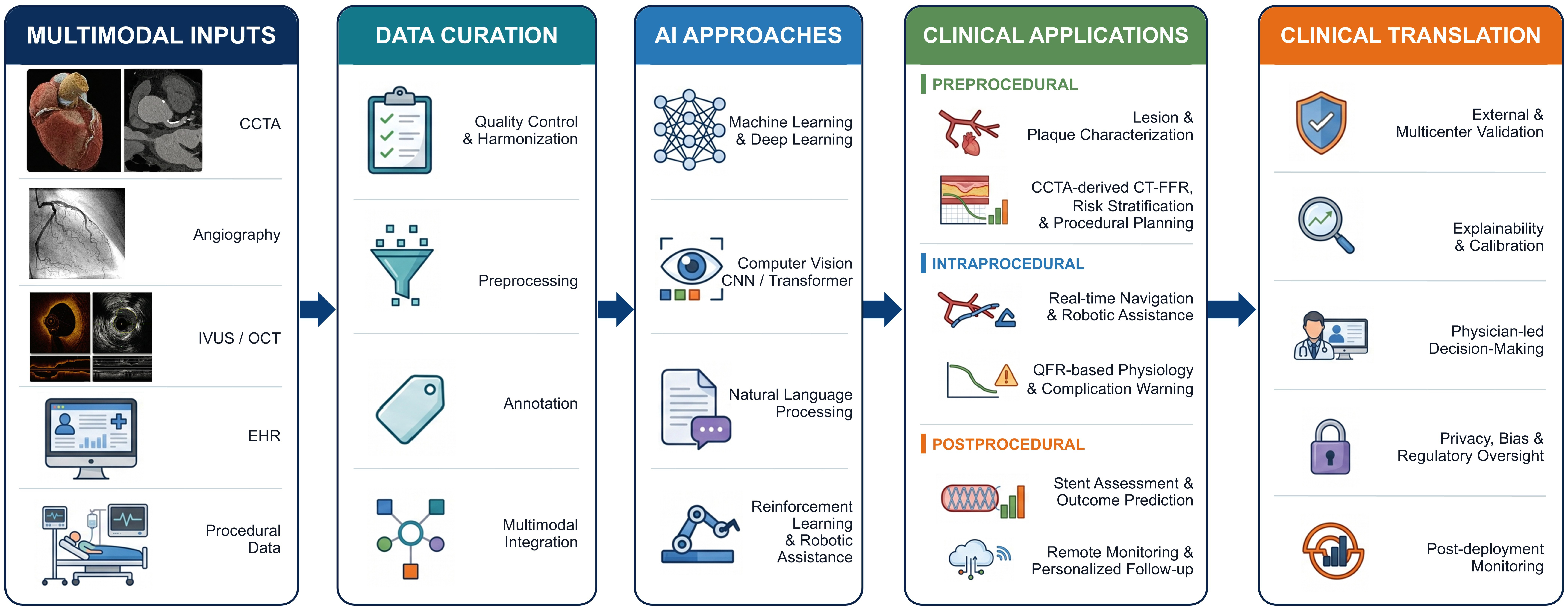
Figure 2. Clinically oriented workflow of artificial intelligence in interventional cardiology. Multimodal imaging, clinical, and procedural data are curated and analyzed using machine learning, deep learning, computer vision, natural language processing, and reinforcement learning. Representative applications include CCTA-derived CT-FFR for preprocedural functional assessment, IVUS/OCT image analysis, lesion and plaque characterization, procedural planning, angiography-derived QFR, intraprocedural navigation and complication warning, and postprocedural stent assessment, outcome prediction, and remote follow-up. Clinical translation requires external validation, calibration, explainability, physician-led decision-making, regulatory oversight, and post-deployment monitoring. AI: Artificial intelligence; CCTA: coronary computed tomography angiography; CNN: convolutional neural network; EHR: electronic health record; IVUS: intravascular ultrasound; OCT: optical coherence tomography; QFR: quantitative flow ratio; CT-FFR: computed tomography-derived fractional flow reserve.
CLINICAL APPLICATIONS OF AI ACROSS THE INTERVENTIONAL CARE PATHWAY
Preprocedural evaluation and clinical decision support
Automated lesion identification and imaging-based risk stratification are central to preprocedural assessment. The integration of multimodal imaging technologies such as CCTA, IVUS, and OCT with AI algorithms provides robust support for precise risk stratification. Studies have demonstrated that machine learning algorithms applied to CCTA data can estimate fractional flow reserve (FFR) non-invasively, supporting functional assessment of coronary stenoses and potentially reducing unnecessary invasive evaluation[18,19]. For IVUS image analysis, an algorithm developed in 2020 automatically detected and quantitatively assessed coronary calcification, with a validation accuracy exceeding 0.87 across three different IVUS systems. Notably, the calculated calcium index showed a significant correlation with adverse cardiovascular events during approximately 6 years of follow-up[20,21]. Two related studies have respectively utilized IVUS data to train machine learning models for predicting coronary thin-cap fibroatheromas and to achieve automatic detection of vulnerable plaques in IVUS images[22,23]. Additionally, a classification-based approach has been proposed to automatically identify the media-adventitia border in IVUS images and rapidly compute key vascular parameters[24]. A 2022 study combined near-infrared spectroscopy with IVUS imaging to classify atherosclerotic tissue components using machine learning, with validation through histology[25]. For OCT imaging, software developed from OCT data from 391 patients achieved diagnostic accuracies of 97.6%, 90.5%, and 88.5% for fibrous plaques, lipid plaques, and calcifications, respectively, with a median analysis time of 21.4 seconds per pullback[26]. Using optical frequency domain imaging (OFDI), a frequency-domain form of intravascular OCT, a deep-learning model achieved validation and test F-scores of 0.63 and 0.66 and intersection-over-union values of 0.49 and 0.52, respectively; section-level areas under the receiver operating characteristic curves (AUCs) ranged from 0.85 to 0.91 across four histological plaque categories[27]. A multimodule algorithm achieved a precision of 88.84% and a recall of 95.02% for the detection of “unstable” atherosclerotic plaques[28], while multi-view contour-constrained Transformer networks, incorporating both polar and Cartesian coordinate OCT images, have further improved the recognition performance of thin-cap fibroatheromas[29].
AI-assisted diagnostic systems have shown remarkable advantages in diagnosing coronary heart disease, structural heart disease, and other cardiovascular conditions. In the field of coronary heart disease diagnosis, the developed CathAI system achieves fully automated interpretation of coronary angiography based on neural networks, encompassing coronary artery identification, vascular segmentation, stenosis localization, and severity prediction[30]. A constructed deep learning architecture integrates coronary segmentation and lesion morphology detection functions, boasting a lesion recognition accuracy of 98.4% and F1 scores ranging from 0.802 to 0.854 for various lesions[31]. A study proposed an intensity-based multi-level classification method to analyze coronary plaque features in IVUS images[32], while two other studies have achieved automatic classification of atherosclerotic tissue in OCT images[33,34].
Personalized treatment recommendations rely on AI-based integration of multidimensional data, providing an evidence base for clinical decision-making. A recent study developed a machine-learning framework that integrates coronary angiography and clinical data to support classification of PCI versus coronary artery bypass grafting (CABG) treatment decisions in patients with multivessel coronary artery disease; external validation and prospective outcome evaluation remain necessary[35]. Deep-learning analysis of pre-TAVR computed tomography can automate measurements of the aortic annulus, coronary ostial height, and calcification and can assist in identifying selected anatomical risk features[36]. For complex lesions, novel indicators such as the lipid-to-cap ratio and optical flow coefficient derived from OCT data have proven more effective than traditional parameters in predicting adverse target vessel events[37]. Moreover, the combination of IVUS and OCT data with random forest algorithms for predicting plaque fibrous cap thickness index and stress/strain index yields an accuracy exceeding 83.3%[38], providing useful information to support treatment decision-making in complex lesions.
Representative clinical studies applying machine learning and deep learning to preprocedural evaluation and decision support in interventional cardiology are summarized in Table 1.
Intraprocedural navigation and real-time guidance
AI-assisted image overlay, segmentation, landmark tracking, and image registration have shown technical feasibility for supporting intraprocedural visualization. Dynamic coronary roadmapping has reduced contrast use in selected studies, whereas automated PCI guidance, TAVR landmark tracking, and three-dimensional registration remain at protocol or technical-validation stages[39-44]. These approaches should therefore be described as workflow-support technologies rather than as systems with established clinical outcome benefit.
Advancements in real-time hemodynamic prediction technologies have improved the efficiency and accuracy of intraprocedural parameter assessment. The Functional Assessment by Virtual Online Reconstruction (FAVOR) II Europe-Japan study validated the diagnostic performance of intraprocedurally derived quantitative flow ratio (QFR) from angiography. The computational QFR approach derives hemodynamic parameters from angiography to support assessment of whether revascularization is warranted[45]. The Angiography-derived Fractional Flow Reserve versus Intravascular Ultrasound to Guide Percutaneous Coronary Intervention in Patients with Coronary Artery Disease (FLAVOUR) II study subsequently evaluated angiography-derived fractional flow reserve (AngioFFR) in clinical practice. This multicenter, randomized, non-inferiority trial conducted at 22 centers in China demonstrated that 12-month clinical outcomes of AngioFFR-guided PCI were non-inferior to those of IVUS-guided PCI[46]. Proposed mesh neural networks for the special Euclidean group SE(3)-equivariant hemodynamic estimation of the arterial wall offer a novel approach for blood flow parameter calculation[47]. In selected settings, these technologies may avoid pressure-wire instrumentation and adenosine administration while supporting standardized physiological assessment and reducing operator dependence.
Robotic PCI and AI-enabled device guidance may improve operator ergonomics, reproducibility, and procedural support; however, evidence supporting AI-controlled intervention in complex coronary lesions remains limited[48,49]. Dedicated clinical studies are required before such systems can be considered for routine use.
AI-based analysis of imaging data before and during procedures may support risk assessment and procedural safety. A deep-learning model based on preprocedural IVUS data from 618 patients achieved an area under the curve of 0.94 for predicting postprocedural stent underexpansion, potentially supporting predeployment planning[50]. Specific deep learning architectures can detect lesions such as dissections and thrombi in coronary angiography, with F1 scores ranging from 0.802 to 0.854, facilitating early intraprocedural identification of complications[31]. A convolutional model integrated with pseudo-3D images achieved automatic detection and segmentation of stent struts in OCT, with a Dice coefficient of 0.907 and a precision of 0.943, enabling timely detection of issues such as stent malapposition[51] and providing support for the timely adjustment of procedural strategies.
Postprocedural management and prognosis prediction
AI-assisted postprocedural assessment includes outcome prediction and automated analysis of stent coverage, apposition, and vascular healing. A deep-learning survival model using post-PCI OCT images from 418 patients with acute coronary syndrome predicted target vessel failure. During a median follow-up of 961 days, 60 patients experienced target vessel failure, and the ensemble model achieved a C-index of 0.796[52]. A highly automated intravascular OCT (IVOCT) analysis method classified uncovered stent struts with a sensitivity of 94%, a specificity of 90%, and an AUC of 0.97[53]. Another study developed an automated method for detecting vascular lumens and stent struts in OCT, enabling effective evaluation of stent apposition and neointimal coverage[54]. An automated algorithm developed for patients with varying endothelialization thicknesses and multiple layers of stents achieved a precision of 0.932 ± 0.009, supporting automated assessment of stent status[55]. Additionally, a method for delineating coronary stents in IVUS pullback images provides technical support for postprocedural stent status assessment[56].
Importantly, the follow-up framework, and therefore the translational value, differed across these studies. In the outcome-prediction study, the median clinical follow-up was 961 days, during which 60 patients experienced target vessel failure[52], providing direct time-to-event evidence for prognostic use. The stent-coverage study analyzed OCT pullbacks acquired 2-5 months after stent implantation[53], supporting automated assessment at an early healing time point. By contrast, the lumen and stent-strut detection study did not specify a standardized longitudinal follow-up interval and was validated against manual image interpretation[54]. The thick-tissue and multilayer stent analysis study used follow-up OCT cases but did not report the interval from implantation[55], while the IVUS stent delineation work was a technical segmentation study without longitudinal patient follow-up[56]. Accordingly, only the outcome-prediction model was directly linked to subsequent clinical events; the remaining models primarily demonstrate feasibility for scalable follow-up imaging analysis and require prospective external validation with prespecified follow-up periods before long-term clinical utility can be established.
Although direct evidence in patients after cardiovascular interventions remains limited, AI-driven follow-up planning and remote monitoring may offer useful approaches to postprocedural management. AI may support personalized follow-up schemes based on postprocedural risk stratification, including more frequent imaging review for high-risk patients and longer intervals for low-risk patients, which could improve follow-up efficiency and resource utilization[48,57]. Reviews have suggested that natural language processing technologies can assist AI in analyzing symptom descriptions remotely reported by patients after an interventional procedure, while large language models such as ChatGPT may support rapid summarization of follow-up information[58], helping physicians obtain a comprehensive overview of patients’ postprocedural status.
Evidence from broader heart-failure and digital-health settings suggests that longitudinal physiological data and machine-learning alerts may support remote monitoring and medication management[59-62]. The Artificial Intelligence Mobile Health Trial of a Digital Platform to Optimize Guideline-Directed Heart Failure Therapy Using Wearable Sensors (AIM-POWER) trial was designed to evaluate a digital intervention for medication optimization in patients with heart failure with reduced ejection fraction; however, this evidence should not be interpreted as demonstrating benefit after coronary or structural heart intervention. Dedicated prospective studies in post-interventional populations are still required.
Broader cardiovascular and digital-health research suggests that AI may support medication adherence and personalized lifestyle interventions during long-term follow-up. A retrospective modeling preprint explored how machine-learning algorithms could use laboratory and clinical data to predict adverse drug-related events and support post-PCI medication planning[63]. More broadly, AI may support personalized rehabilitation and lifestyle coaching, although direct evidence demonstrating improved post-interventional outcomes remains limited[64,65].
CURRENT CHALLENGES AND LIMITATIONS
Despite the significant progress of AI technology in the field of cardiac intervention, it still faces several challenges and limitations in clinical application [Table 2]. Specifically, data-related issues such as fragmented data silos, selection bias in training datasets, and the lack of high-quality labeled data limit model generalizability[66-69], while technical limitations include inconsistent performance in complex lesions, risks of misdiagnosis, and the limited interpretability of “black-box” algorithms[70-72]. Furthermore, clinical application is hindered by the lack of harmonized industry standards, variable clinician acceptance, and high deployment costs[73-76]. Finally, evolving regulatory frameworks, risks to patient-data security and privacy, and the unclear allocation of medical liability pose significant regulatory and ethical risks[76-78].
Challenges and limitations of AI application in interventional cardiology
| Category | Core challenges and limitations |
| Data-related issues | 1. Fragmented data silos and limited integration of multimodal clinical data[66,67] 2. Selection bias in training datasets and limited generalizability[67,68] 3. Limited high-quality labeled data and time-consuming manual annotation with interobserver variability[68,69] |
| Technical and diagnostic limitations | 1. Inconsistent performance in complex lesions 2. Risk of misdiagnosis[70] 3. Limited interpretability of “black-box” algorithms[71,72] |
| Clinical application barriers | 1. Lack of harmonized industry standards[73,74] 2. Variable clinician acceptance of AI[75,76] 3. High deployment and maintenance costs that limit broad adoption[73,74] |
| Regulatory and ethical risks | 1. Evolving regulatory frameworks for AI medical devices[77] 2. Risks to patient data security and privacy[78] 3. Unclear allocation of medical liability[76] |
Data-related issues
Data-related challenges remain a major barrier to the development and application of AI in interventional cardiology. First, clinical data remain fragmented across isolated systems, and AI platforms often cannot seamlessly integrate multimodal data from different sources, including preprocedural imaging data, intraprocedural real-time angiography results, and detailed patient medical history, which limits the comprehensiveness of input data for AI models[79]. Second, the training datasets used by most AI systems are subject to substantial selection bias, as they are mostly derived from large, well-equipped tertiary hospitals with standardized diagnosis and treatment processes, lacking representative data from primary medical institutions and diverse populations (such as elderly patients with multiple comorbidities and underrepresented racial and ethnic groups), leading to poor generalization ability of the models in clinical deployment[80]. In addition, high-quality labeled data are extremely scarce; manual labeling of cardiac intervention images, hemodynamic parameters, and other indicators is not only time-consuming and labor-intensive but also subject to interobserver variability, which further affects model performance and diagnostic accuracy[81]. Relevant studies have confirmed that the shortage of high-quality data and fragmentation across data sources have become important factors hindering the clinical translation of AI in the field of cardiac intervention.
Technical and diagnostic limitations
Technical limitations may restrict the application of AI algorithms in complex interventional settings. On the one hand, AI performance may be inconsistent in complex coronary lesions, such as chronic total occlusion (CTO), severely calcified lesions, and complex multivessel lesions; models may have difficulty identifying the proximal cap, the course of the occluded segment, and lesion boundaries and may not reliably evaluate the functional significance of lesions, which may limit their value in guiding clinical interventions. On the other hand, AI systems may produce erroneous classifications; models developed from limited input modalities may fail to capture relevant clinical context, increasing the risk of misclassification in atypical or subtle lesions. In addition, limited interpretability of some “black-box” algorithms remains a concern; the basis for model-generated diagnostic results and interventional suggestions may be difficult to trace and interpret, complicating clinicians’ verification of results and accountability for clinical decisions and limiting trust in AI systems.
Clinical application barriers
Despite its technical advantages, AI still faces practical barriers to clinical implementation and broader adoption[82]. First, there is a lack of unified industry standards and norms for AI systems in the field of cardiac intervention, including unified technical parameters, standardized performance evaluation criteria and clear clinical application paths; this leads to uneven quality of AI products on the market, and it is difficult for clinicians to effectively select and apply AI tools that meet clinical needs. Second, the acceptance of AI by clinicians varies greatly; some clinicians have doubts about the reliability and stability of AI results and are reluctant to rely on AI systems to make key clinical intervention decisions, which prevents AI tools from being fully integrated into clinical workflows. In addition, the deployment and maintenance costs of AI systems are relatively high; the purchase of equipment, software updates, technical training, and daily maintenance all require substantial financial and human resources, which limits the broader deployment of AI in primary medical institutions with limited medical resources, and also affects the equity of the application of AI technology in interventional cardiology.
Regulatory and ethical risks
Evolving regulatory frameworks and ethical risks remain important barriers to timely clinical translation of AI technology in interventional cardiology. In terms of regulation, regulatory frameworks for AI medical devices are not yet fully harmonized; the approval cycle of AI products is long, and regulatory standards may not fully accommodate the rapid pace of AI development and iteration, which may delay clinical translation[83]. In terms of ethics and data security, AI training and clinical application involve large amounts of patients’ personal health information, and there are risks of data leakage and misuse, which may compromise patient privacy[83]. More importantly, the attribution of medical liability is unclear; when adverse medical events occur due to AI system errors, it is difficult to clarify the liability among AI developers, clinicians, and medical institutions, which not only brings legal disputes but also increases the risk of clinical application of AI technology. Relevant studies have pointed out that improving the regulatory system and clarifying the ethical norms and liability attribution are important prerequisites for the healthy development of AI in the field of cardiac intervention.
FUTURE DEVELOPMENT DIRECTIONS AND OUTLOOK
The future development of artificial intelligence in cardiovascular intervention is likely to involve multidimensional integration and systematic innovation. Multimodal integrated AI, which synthesizes vascular imaging, genomics, clinical data, and dynamic information from wearable devices, may support personalized diagnostic and therapeutic decision-making systems[16]. The real-time application of edge computing and embedded AI in catheterization laboratories is expected to enable low-latency intraprocedural navigation and early risk detection, thereby enhancing procedural precision and safety[84]. The use of generative AI and large language models in case simulation and physician training may reshape interventional medical education, while the continued development of human-machine collaborative systems may improve procedural standardization while maintaining clinician oversight[85]. Furthermore, the establishment of standardized cardiovascular AI datasets and open-source platforms, along with the formulation of clinical guidelines and healthcare policies for AI-assisted diagnosis and treatment, may provide institutional support for the scalable development of this field[86]. Together, these directions point toward a transition in cardiovascular intervention from digital assistance to intelligent integration.
Future clinical translation must also be aligned with evolving international governance frameworks. World Health Organization (WHO) guidance for large multimodal models emphasizes transparency, stakeholder participation, independent post-deployment auditing, and impact assessment[87], while the European Union (EU) Artificial Intelligence Act imposes risk-management, human-oversight, and post-market-monitoring obligations on qualifying high-risk medical AI systems[88]. Medical testing of prominent large language models, including ChatGPT and DeepSeek, should move beyond general-purpose benchmarks toward specialty-specific clinical evaluation. In a recent comparative study, DeepSeek-R1 was comparable to ChatGPT-o1 for several diagnostic and management tasks but performed less well in imaging-report summarization[89]. These task-dependent findings support external validation, prospective multicenter evaluation, and continuous performance monitoring before such models are used in cardiovascular intervention, with clinicians retaining final responsibility.
CONCLUSION
Artificial intelligence technology has made substantial advances in the field of cardiovascular intervention, with its application spanning the entire clinical workflow from preprocedural evaluation through intraprocedural assistance to postprocedural management, providing innovative solutions to address long-standing challenges in traditional interventional practice[10]. From a technical perspective, synergistic advancements in machine learning, deep learning, and medical image processing have enabled precise lesion identification, non-invasive hemodynamic assessment, and AI-assisted procedural guidance. Some cardiovascular AI tools have entered regulatory review or clinical evaluation, but their indications, jurisdictions, and levels of clinical validation vary substantially[90]. Particularly for extending high-quality care to primary healthcare settings and training junior interventionalists, AI may offer meaningful potential.
Nevertheless, persistent challenges - including inconsistent data quality and annotation standards, insufficient model interpretability, lagging regulatory and ethical frameworks, and difficulties in clinical integration - remain critical barriers to the widespread adoption of AI in cardiovascular intervention[91]. Addressing these issues cannot be accomplished by any single discipline alone. It requires close interdisciplinary collaboration among clinical medicine, computer science, bioinformatics, and health policy, aiming to establish standardized data platforms, develop explainable AI algorithms, and refine regulatory and ethical frameworks to promote the seamless integration of technology and clinical practice.
Looking ahead, with the iterative advancement of technologies such as multimodal AI, embedded edge-computing systems, and generative large language models, alongside the gradual refinement of standardized guidelines and healthcare reimbursement policies, AI is poised to evolve from an “assistive tool” toward an integrated component of cardiovascular intervention, contributing to the establishment of a precise, efficient, and accessible model of intelligent interventional care. Only by maintaining a clinically driven approach, strengthening multicenter collaborative validation, and balancing technological innovation with medical safety can we realize the sustainable integration of AI in cardiovascular intervention, ultimately delivering higher-quality care to patients with cardiovascular diseases worldwide.
DECLARATIONS
Authors’ contributions
Conceived and designed the review, performed the literature search and data collection, and drafted the initial manuscript: Li J
Assisted in drafting the manuscript, contributed to the organization and synthesis of the literature, and provided revisions to the text: Li Y
Critically revised the manuscript for important intellectual content, provided expert input on key sections, and approved the final version to be published: Zhu Q, Liu X
All authors have read and approved the final manuscript and agree to be accountable for all aspects of the work.
Availability of data and materials
Not applicable.
AI and AI-assisted tools statement
During the preparation of this manuscript, the AI tool Google Gemini 2.5 Flash Image (Nano Banana; model version gemini-2.5-flash-image-preview, released 2025-08-26) was used solely for generating some graphical elements in Figure 2. The generated elements were subsequently edited and integrated by the authors using Microsoft PowerPoint (Microsoft 365, version 2608 Build 20326.20158). The AI tool did not influence the study design, data collection, analysis, interpretation, scientific conclusions, or generation of scientific data. All authors have reviewed and verified Figure 2 and take full responsibility for the accuracy, integrity, and final content of the manuscript.
Financial support and sponsorship
This research was funded by Zhejiang Province Science and Technology Innovation Leading Talents Project (No. 2023R5236 for XBL); State Key Laboratory of Transvascular Implantation Devices; The National Natural Science Foundation of China (No. 82271606 for XBL); National Science Fund for Distinguished Young Scholars of China (No. 82425007 for XBL); Noncommunicable Chronic Diseases-National Science and Technology Major Project (No. 2024ZD0527100 for XBL).
Conflicts of interest
Liu X is a Guest Editor of the Special Topic: Interventional Cardiology: Recent Advances in Clinical and Translational Research of the journal Vessel Plus. Liu X was not involved in any steps of editorial processing, including reviewers’ selection, manuscript handling, and decision-making. The other authors declare no conflicts of interest.
Ethical approval and consent to participate
Not applicable.
Consent for publication
Not applicable.
Copyright
© The Author(s) 2026.
REFERENCES
1. Qu C, Liao S, Zhang J, et al. Burden of cardiovascular disease among elderly: based on the Global Burden Of Disease Study 2019. Eur Heart J Qual Care Clin Outcomes. 2024;10:143-53.
2. Faxon DP, Williams DO. Interventional cardiology: current status and future directions in coronary disease and valvular heart disease. Circulation. 2016;133:2697-711.
3. Ullah M, Wahab A, Khan SU, et al. Stent as a novel technology for coronary artery disease and their clinical manifestation. Curr Probl Cardiol. 2023;48:101415.
4. Safdar M, Ullah M, Wahab A, et al. Genomic insights into heart health: exploring the genetic basis of cardiovascular disease. Curr Probl Cardiol. 2024;49:102182.
5. Avvedimento M, Tang GHL. Transcatheter aortic valve replacement (TAVR): recent updates. Prog Cardiovasc Dis. 2021;69:73-83.
6. Beyar R, Davies JE, Cook C, Dudek D, Cummins PA, Bruining N. Robotics, imaging, and artificial intelligence in the catheterisation laboratory. EuroIntervention. 2021;17:537-49.
7. Arfat Y, Mittone G, Esposito R, Cantalupo B, DE Ferrari GM, Aldinucci M. Machine learning for cardiology. Minerva Cardiol Angiol. 2022;70:75-91.
8. Hou L, Zhao J, He T, Su K, Li Y. Machine Learning-Based Prediction of In-Stent Restenosis Risk Using Systemic Inflammation Aggregation Index Following Coronary Stent Placement. Risk Manag Healthc Policy. 2024;17:1779-86.
9. Shameer K, Johnson KW, Glicksberg BS, Dudley JT, Sengupta PP. Machine learning in cardiovascular medicine: are we there yet? Heart. 2018;104:1156-64.
10. Johnson KW, Torres Soto J, Glicksberg BS, et al. Artificial intelligence in cardiology. J Am Coll Cardiol. 2018;71:2668-79.
11. Rudnicka Z, Pręgowska A, Glądys K, Perkins M, Proniewska K. Advancements in artificial intelligence-driven techniques for interventional cardiology. Cardiol J. 2024;31:321-41.
12. Li Z, Liu F, Yang W, Peng S, Zhou J. A survey of convolutional neural networks: analysis, applications, and prospects. IEEE Trans Neural Netw Learn Syst. 2022;33:6999-7019.
13. Papanastasiou G, Dikaios N, Huang J, Wang C, Yang G. Is attention all you need in medical image analysis? A review. IEEE J Biomed Health Inform. 2024;28:1398-411.
14. Reading Turchioe M, Volodarskiy A, Pathak J, Wright DN, Tcheng JE, Slotwiner D. Systematic review of current natural language processing methods and applications in cardiology. Heart. 2022;108:909-16.
15. Adejumo P, Thangaraj PM, Dhingra LS, et al. Natural language processing of clinical documentation to assess functional status in patients with heart failure. JAMA Netw Open. 2024;7:e2443925.
16. Jiang F, Jiang Y, Zhi H, et al. Artificial intelligence in healthcare: past, present and future. Stroke Vasc Neurol. 2017;2:230-43.
17. Kweon J, Kim K, Lee C, et al. Deep reinforcement learning for guidewire navigation in coronary artery phantom. IEEE Access. 2021;9:166409-22.
18. Kurata A, Fukuyama N, Hirai K, et al. On-site computed tomography-derived fractional flow reserve using a machine-learning algorithm-clinical effectiveness in a retrospective multicenter cohort. Circ J. 2019;83:1563-71.
19. Yang J, Shan D, Wang X, et al. On-Site Computed tomography-derived fractional flow reserve to guide management of patients with stable coronary artery disease: the TARGET randomized trial. Circulation. 2023;147:1369-81.
20. Liu S, Neleman T, Hartman EMJ, et al. Automated quantitative assessment of coronary calcification using intravascular ultrasound. Ultrasound Med Biol. 2020;46:2801-9.
21. Neleman T, Liu S, Tovar Forero MN, et al. The prognostic value of a validated and automated intravascular ultrasound-derived calcium score. J Cardiovasc Transl Res. 2021;14:992-1000.
22. Bae Y, Kang SJ, Kim G, et al. Prediction of coronary thin-cap fibroatheroma by intravascular ultrasound-based machine learning. Atherosclerosis. 2019;288:168-74.
23. Jun TJ, Kang SJ, Lee JG, et al. Automated detection of vulnerable plaque in intravascular ultrasound images. Med Biol Eng Comput. 2019;57:863-76.
24. Wang YY, Qiu CH, Jiang J, Xia SR. Detecting the media-adventitia border in intravascular ultrasound images through a classification-based approach. Ultrason Imaging. 2019;41:78-93.
25. Bajaj R, Eggermont J, Grainger SJ, et al. Machine learning for atherosclerotic tissue component classification in combined near-infrared spectroscopy intravascular ultrasound imaging: validation against histology. Atherosclerosis. 2022;345:15-25.
26. Chu M, Jia H, Gutiérrez-Chico JL, et al. Artificial intelligence and optical coherence tomography for the automatic characterisation of human atherosclerotic plaques. EuroIntervention. 2021;17:41-50.
27. Shibutani H, Fujii K, Ueda D, et al. Automated classification of coronary atherosclerotic plaque in optical frequency domain imaging based on deep learning. Atherosclerosis. 2021;328:100-5.
28. Liu R, Zhang Y, Zheng Y, Liu Y, Zhao Y, Yi L. Automated detection of vulnerable plaque for intravascular optical coherence tomography images. Cardiovasc Eng Technol. 2019;10:590-603.
29. Liu S, Xin J, Wu J, et al. Multi-view contour-constrained transformer network for thin-cap fibroatheroma identification. Neurocomputing. 2023;523:224-34.
30. Avram R, Olgin JE, Ahmed Z, et al. CathAI: fully automated coronary angiography interpretation and stenosis estimation. NPJ Digit Med. 2023;6:142.
31. Du T, Xie L, Zhang H, et al. Training and validation of a deep learning architecture for the automatic analysis of coronary angiography. EuroIntervention. 2021;17:32-40.
32. Kim GY, Lee JH, Hwang YN, Kim SM. A novel intensity-based multi-level classification approach for coronary plaque characterization in intravascular ultrasound images. Biomed Eng Online. 2018;17:151.
33. Zhou P, Zhu T, He C, Li Z. Automatic classification of atherosclerotic tissue in intravascular optical coherence tomography images. J Opt Soc Am A Opt Image Sci Vis. 2017;34:1152-9.
34. Lee J, Prabhu D, Kolluru C, et al. Automated plaque characterization using deep learning on coronary intravascular optical coherence tomographic images. Biomed Opt Express. 2019;10:6497-515.
35. Kim B, Kim J, Kim YH, Jun TJ, Ahn JM. Deep learning-based treatment decision support framework for multi-vessel coronary artery disease using integrated coronary angiography and clinical data. BMC Med Inform Decis Mak. 2026:26.
36. Wang M, Niu G, Chen Y, et al. Development and validation of a deep learning-based fully automated algorithm for pre-TAVR CT assessment of the aortic valvular complex and detection of anatomical risk factors: a retrospective, multicentre study. EBioMedicine. 2023;96:104794.
37. Hong H, Jia H, Zeng M, et al. Risk stratification in acute coronary syndrome by comprehensive morphofunctional assessment with optical coherence tomography. JACC Asia. 2022;2:460-72.
38. Lv R, Wang L, Maehara A, et al. Combining IVUS + OCT data, biomechanical models and machine learning method for accurate coronary plaque morphology quantification and cap thickness and stress/strain index predictions. J Funct Biomater. 2023;14:41.
39. Piayda K, Phinicarides R, Afzal S, et al. Dynamic coronary roadmap in percutaneous coronary intervention: results from an open-label, randomized trial. JACC Cardiovasc Interv. 2021;14:2523-5.
40. Piayda K, Kleinebrecht L, Afzal S, et al. Dynamic coronary roadmapping during percutaneous coronary intervention: a feasibility study. Eur J Med Res. 2018;23:36.
41. Ploscaru V, Popa-Fotea NM, Calmac L, et al. Artificial intelligence and cloud based platform for fully automated PCI guidance from coronary angiography-study protocol. PLoS One. 2022;17:e0274296.
42. Danilov VV, Klyshnikov KY, Gerget OM, et al. Aortography keypoint tracking for transcatheter aortic valve implantation based on multi-task learning. Front Cardiovasc Med. 2021;8:697737.
43. Park J, Kweon J, Kim YI, et al. Selective ensemble methods for deep learning segmentation of major vessels in invasive coronary angiography. Med Phys. 2023;50:7822-39.
44. Park T, Khang S, Jeong H, et al. Deep learning segmentation in 2D X-ray images and non-rigid registration in multi-modality images of coronary arteries. Diagnostics. 2022;12:778.
45. Westra J, Andersen BK, Campo G, et al. Diagnostic performance of in-procedure angiography-derived quantitative flow reserve compared to pressure-derived fractional flow reserve: the FAVOR II Europe-Japan Study. J Am Heart Assoc. 2018:7.
46. Hu X, Zhang J, Yang S, et al.; FLAVOUR II study group. Angiography-derived fractional flow reserve versus intravascular ultrasound to guide percutaneous coronary intervention in patients with coronary artery disease (FLAVOUR II): a multicentre, randomised, non-inferiority trial. Lancet. 2025;405:1491-504.
47. Suk J, de Haan P, Lippe P, Brune C, Wolterink JM. Mesh neural networks for SE(3)-equivariant hemodynamics estimation on the artery wall. Comput Biol Med. 2024;173:108328.
48. Benjamins JW, Hendriks T, Knuuti J, Juarez-Orozco LE, van der Harst P. A primer in artificial intelligence in cardiovascular medicine. Neth Heart J. 2019;27:392-402.
49. Jeelani S, Dany A, Anand B, Vandana S, Maheswaran T, Rajkumar E. Robotics and medicine: a scientific rainbow in hospital. J Pharm Bioallied Sci. 2015;7:S381-3.
50. Min HS, Ryu D, Kang SJ, et al. Prediction of coronary stent underexpansion by pre-procedural intravascular ultrasound-based deep learning. JACC Cardiovasc Interv. 2021;14:1021-9.
51. Wu P, Gutiérrez-Chico JL, Tauzin H, et al. Automatic stent reconstruction in optical coherence tomography based on a deep convolutional model. Biomed Opt Express. 2020;11:3374-94.
52. Hamana T, Nishimori M, Shibata S, et al. Deep-learning-driven optical coherence tomography analysis for cardiovascular outcome prediction in patients with acute coronary syndrome. Eur Heart J Digit Health. 2024;5:692-701.
53. Lu H, Lee J, Ray S, et al. Automated stent coverage analysis in intravascular OCT (IVOCT) image volumes using a support vector machine and mesh growing. Biomed Opt Express. 2019;10:2809-28.
54. Nam HS, Kim CS, Lee JJ, Song JW, Kim JW, Yoo H. Automated detection of vessel lumen and stent struts in intravascular optical coherence tomography to evaluate stent apposition and neointimal coverage. Med Phys. 2016;43:1662.
55. Yang G, Mehanna E, Li C, et al. Stent detection with very thick tissue coverage in intravascular OCT. Biomed Opt Express. 2021;12:7500-16.
56. Wissel T, Riedl K, Schaefers K, et al. Delineation of coronary stents in intravascular ultrasound pullbacks. In: Medical Imaging 2021: Image-Guided Procedures, Robotic Interventions, and Modeling; 2021 Feb 15-19; Online, California, USA. 2021.
58. Sallam M. ChatGPT utility in healthcare education, research, and practice: systematic review on the promising perspectives and valid concerns. Healthcare. 2023;11:887.
59. Stehlik J, Schmalfuss C, Bozkurt B, et al. Continuous wearable monitoring analytics predict heart failure hospitalization: the LINK-HF Multicenter Study. Circ Heart Fail. 2020;13:e006513.
60. Devore AD, Xie J, Etters L, et al. A digital platform to optimize guideline-directed heart failure therapy: rationale and design of the AIM-POWER trial. J Card Fail. 2025;31:1837-46.
61. McDonagh TA, Metra M, Adamo M, et al.; ESC Scientific Document Group. 2021 ESC Guidelines for the diagnosis and treatment of acute and chronic heart failure: developed by the Task Force for the diagnosis and treatment of acute and chronic heart failure of the European Society of Cardiology (ESC) With the special contribution of the Heart Failure Association (HFA) of the ESC. Eur Heart J. 2021;42:3599-726.
62. Stevenson LW, Ross HJ, Rathman LD, Boehmer JP. Remote monitoring for heart failure management at home. J Am Coll Cardiol. 2023;81:2272-91.
63. Iftikhar H. AI-driven personalization of dual antiplatelet therapy duration post-PCI: a novel approach balancing ischemic and bleeding risks in the UAE population. medRxiv. 2025:2025.03.16.25324067.
64. Johnson KW, Shameer K, Glicksberg BS, et al. Enabling precision cardiology through multiscale biology and systems medicine. JACC Basic Transl Sci. 2017;2:311-27.
65. Yan Y, Zhang JW, Zang GY, Pu J. The primary use of artificial intelligence in cardiovascular diseases: what kind of potential role does artificial intelligence play in future medicine? J Geriatr Cardiol. 2019;16:585-91.
66. Topol EJ. High-performance medicine: the convergence of human and artificial intelligence. Nat Med. 2019;25:44-56.
68. Willemink MJ, Koszek WA, Hardell C, et al. Preparing medical imaging data for machine learning. Radiology. 2020;295:4-15.
69. Kelly CJ, Karthikesalingam A, Suleyman M, Corrado G, King D. Key challenges for delivering clinical impact with artificial intelligence. BMC Med. 2019;17:195.
70. Liu X, Faes L, Kale AU, et al. A comparison of deep learning performance against health-care professionals in detecting diseases from medical imaging: a systematic review and meta-analysis. Lancet Digit Health. 2019;1:e271-97.
71. Rudin C. Stop explaining black box machine learning models for high stakes decisions and use interpretable models instead. Nat Mach Intell. 2019;1:206-15.
72. Reyes M, Meier R, Pereira S, et al. On the interpretability of artificial intelligence in radiology: challenges and opportunities. Radiol Artif Intell. 2020;2:e190043.
73. He J, Baxter SL, Xu J, Xu J, Zhou X, Zhang K. The practical implementation of artificial intelligence technologies in medicine. Nat Med. 2019;25:30-6.
74. Sendak MP, D’Arcy J, Kashyap S, et al. A path for translation of machine learning products into healthcare delivery. EMJ Innov. 2020.
76. Vayena E, Blasimme A, Cohen IG. Machine learning in medicine: addressing ethical challenges. PLoS Med. 2018;15:e1002689.
77. Gerke S, Minssen T, Cohen G. Ethical and legal challenges of artificial intelligence-driven healthcare. In: Bohr A, Memarzadeh K, Editors. Artificial Intelligence in Healthcare. Academic Press; 2020. pp. 295-336.
79. Bajwa J, Munir U, Nori A, Williams B. Artificial intelligence in healthcare: transforming the practice of medicine. Future Healthc J. 2021;8:e188-94.
80. Mihan A, Pandey A, Van Spall HGC. Artificial intelligence bias in the prediction and detection of cardiovascular disease. NPJ Cardiovasc Health. 2024:1.
81. Gong Y, Liu G, Xue Y, Li R, Meng L. A survey on dataset quality in machine learning. Inf Softw Technol. 2023;162:107268.
82. Olawade DB, Fidelis SC, Marinze S, Egbon E, Osunmakinde A, Osborne A. Artificial intelligence in clinical trials: a comprehensive review of opportunities, challenges, and future directions. Int J Med Inform. 2026;206:106141.
83. Youssef A, Nichol AA, Martinez-Martin N, et al. Ethical considerations in the design and conduct of clinical trials of artificial intelligence. JAMA Netw Open. 2024;7:e2432482.
84. Choksi S, Szot S, Zang C, et al. Bringing artificial intelligence to the operating room: edge computing for real-time surgical phase recognition. Surg Endosc. 2023;37:8778-84.
85. Haque MA, Siddique HR. Generative artificial intelligence and large language models in smart healthcare applications: current status and future perspectives. Comput Biol Chem. 2026;120:108611.
86. Finlayson SG, Subbaswamy A, Singh K, et al. The clinician and dataset shift in artificial intelligence. N Engl J Med. 2021;385:283-6.
87. World Health Organization. Ethics and governance of artificial intelligence for health: Guidance on large multi-modal models. 2025. Available from: https://www.who.int/publications/i/item/9789240084759 [accessed 13 October 2026].
88. Aboy M, Minssen T, Vayena E. Navigating the EU AI act: implications for regulated digital medical products. NPJ Digit Med. 2024;7:237.
89. Tordjman M, Liu Z, Yuce M, et al. Comparative benchmarking of the DeepSeek large language model on medical tasks and clinical reasoning. Nat Med. 2025;31:2550-5.
90. Samant S, Panagopoulos AN, Wu W, Zhao S, Chatzizisis YS. Artificial intelligence in coronary artery interventions: preprocedural planning and procedural assistance. J Soc Cardiovasc Angiogr Interv. 2025;4:102519.
Cite This Article
 Open Access
How to Cite
Li J, Li Y, Zhu Q, Liu X. Artificial intelligence in interventional cardiology: present landscape and future directions. Vessel Plus. 2026;10:56. https://dx.doi.org/10.20517/2574-1209.2026.12
Download Citation
If you have the appropriate software installed, you can download article citation data to the citation manager of your choice. Simply select your manager software from the list below and click on download.
Export Citation File
Type of Import
Tips on Downloading Citation
Citation Manager File Format
Type of Import
Direct Import: When the Direct Import option is selected (the default state), a dialogue box will give you the option to Save or Open the downloaded citation data. Choosing Open will either launch your citation manager or give you a choice of applications with which to use the metadata. The Save option saves the file locally for later use.
Indirect Import: When the Indirect Import option is selected, the metadata is displayed and may be copied and pasted as needed.
About This Article
Special Topic
Copyright

Data & Comments
Data
0









Comments
Comments must be written in English. Spam, offensive content, impersonation, and private information will not be permitted. If any comment is reported and identified as inappropriate content by OAE staff, the comment will be removed without notice. If you have any queries or need any help, please contact us at [email protected].