

Gut microbiota in hepatocellular carcinoma: pathogenic mechanisms and clinical translation
 0
0 Graphical Abstract
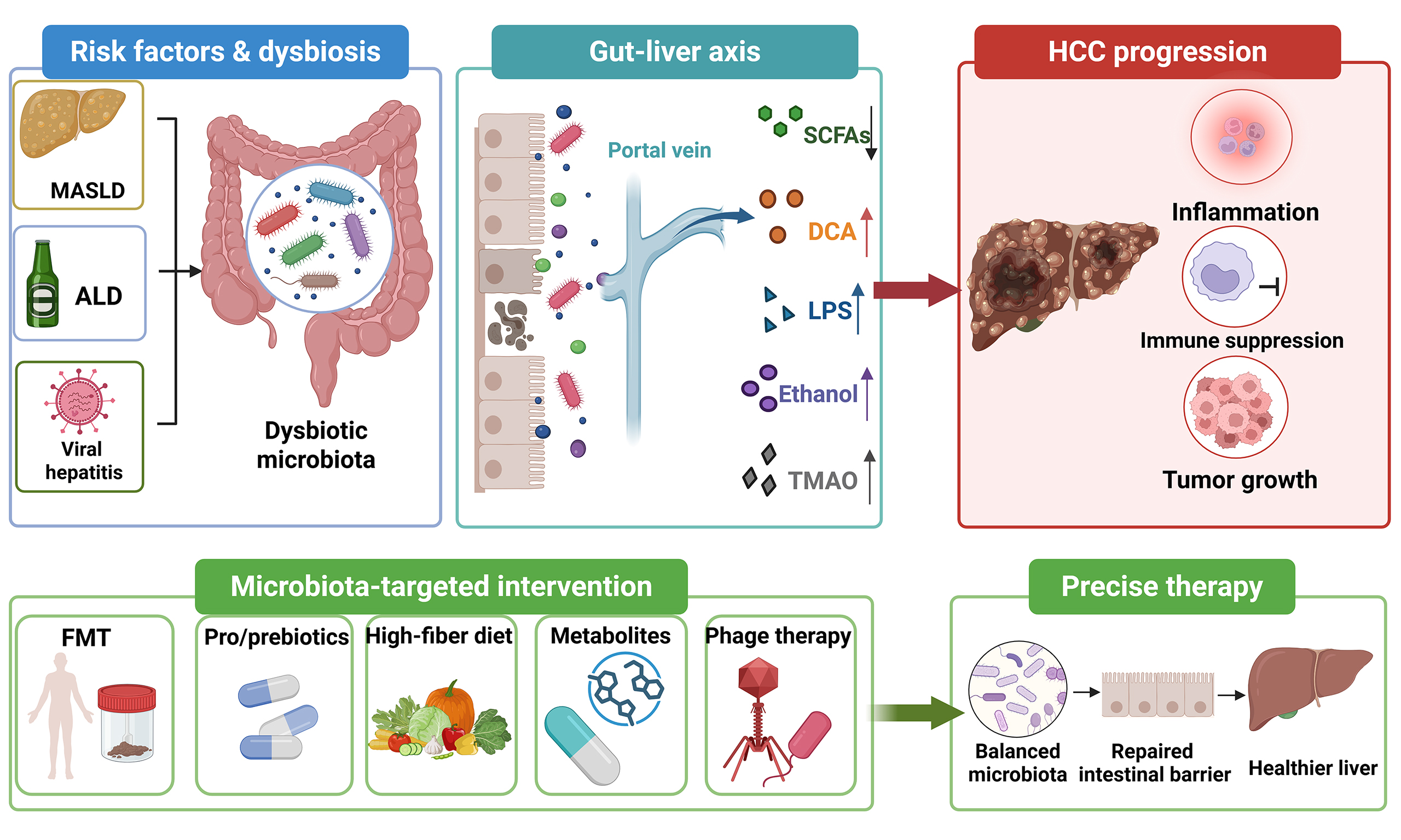
Abstract
The gut microbiota is an important modulator of host metabolism and immunity. In hepatocellular carcinoma (HCC), the most common form of primary liver cancer, dysbiosis, impaired intestinal barrier integrity, and dysregulated host-microbial immune signaling are considered contributing factors to disease progression. These perturbations may contribute to hepatocarcinogenesis across disease trajectories arising from metabolic dysfunction-associated steatohepatitis, alcohol-associated liver disease, and chronic viral hepatitis, many but not all of which involve progression through advanced fibrosis and cirrhosis. This narrative review synthesizes evidence indicating that microbiota-derived metabolites and microbial products, including short-chain fatty acids, bile acids, lipopolysaccharide and the host-microbial co-metabolite trimethylamine N-oxide, interact with host receptors and inflammatory pathways implicated in hepatocarcinogenesis. Preclinical studies have provided strong mechanistic support for microbiota modulation; however, clinical evidence in HCC remains preliminary, heterogeneous, and largely exploratory. This disparity underscores the need to elucidate the contributions of bacterial, fungal, viral, host genetic, sex-specific, and intratumoral microbial factors before their efficacy, safety and clinical utility in HCC can be rigorously assessed. Accordingly, microbiota-targeted strategies, such as probiotics, prebiotics, fecal microbiota transplantation, dietary modulation, and bacteriophage-based approaches, should be viewed as potential adjunctive or preventive interventions rather than as established HCC therapies.
Keywords


 Download PDF
INTRODUCTION
Hepatocellular carcinoma (HCC) is the predominant histological subtype of primary liver cancer and remains a leading cause of cancer-related death worldwide[1,2]. In this review, the broader term “liver cancer” is used only for epidemiological or conceptual statements that include multiple primary liver malignancies, whereas HCC is used when the evidence specifically concerns HCC. The etiology of HCC is undergoing a notable shift, as metabolic dysfunction-associated steatotic liver disease (MASLD) has emerged as one of the fastest-growing contributors to HCC in Europe and the United States[3]. Obesity and diabetes are established metabolic risk factors for HCC, whereas gut microbiota dysbiosis is an associated feature under active investigation[4]. This epidemiological shift points to the gut-liver axis as a biologically plausible target for HCC risk stratification, prevention research, and treatment strategies.
In recent years, the gut microbiota is increasingly recognized as a functional microbial ecosystem with immunometabolic effects on the host metabolism, immune homeostasis, and barrier function. The microbiota communicates with host receptors [such as G protein-coupled receptor 41 (GPR41)/43, peroxisome proliferator-activated receptor gamma (PPARγ), aryl hydrocarbon receptor (AhR), and Toll-like receptors (TLRs)] through a diverse array of microbial metabolites such as short-chain fatty acids (SCFAs), host-microbial co-metabolites such as secondary bile acids and trimethylamine N-oxide (TMAO), and related microbial products. Through these interactions, the microbiota participates in the development and progression of obesity, type 2 diabetes, MASLD and cancer[5]. Dysbiosis disrupts the intestinal epithelium and vascular barriers, promotes bacterial translocation, and increases portal exposure to the pathogen-associated molecular patterns (PAMPs), such as lipopolysaccharide (LPS). These signals activate Toll-like receptor 4 (TLR4)/nuclear factor kappa B (NF-κB) signaling and recruit myeloid-derived suppressor cells (MDSCs), forming a pro-tumor fibrotic-inflammatory microenvironment that may accelerate progression from steatohepatitis to fibrosis and HCC[6]. The microbiota may also influence hepatic natural killer T (NKT) cell activity and immune checkpoint-associated T-cell states, such as programmed cell death protein 1 (PD-1) and cytotoxic T-lymphocyte-associated protein 4 (CTLA-4)[7]. Clinically, fecal microbiota transplantation (FMT) and responder-derived microbial consortia have entered exploratory cancer-immunotherapy trials, including studies relevant to HCC, but their efficacy, safety, donor selection, and durability remain unresolved[8]. Therefore, targeting the gut-liver axis should currently be framed as an investigational strategy to reshape the immune microenvironment and enhance anti-tumor immune surveillance, rather than as an established therapy.
This review synthesizes evidence on plausible molecular mechanisms by which the gut microbiota may promote the progression of MASLD, alcohol-associated liver disease (ALD), and viral hepatitis to HCC through metabolic, immune, and microenvironmental pathways. It integrates preclinical and early clinical evidence, highlights the translational potential and limitations of microbiota-targeted interventions, and identifies key gaps that must be addressed before these strategies can be incorporated into precision prevention or treatment of HCC.
GUT MICROBIOTA DYSBIOSIS AND RISK FACTORS FOR HCC
MASLD and HCC
With the global rise of obesity and diabetes, MASLD has become one of the fastest-growing etiological contributors to HCC in the United States and Europe. In this context, gut microbiota dysbiosis and the resulting immunosuppressive microenvironment may synergistically increase the risk of hepatocarcinogenesis[9]. Following intestinal barrier dysfunction, pro-inflammatory microbial products such as LPS and potentially deleterious metabolites such as TMAO can enter the liver through the portal circulation; LPS activates TLR4/NF-κB signaling, whereas TMAO contributes to oxidative and inflammatory stress[10]. The microbiota can also influence hepatic metabolic homeostasis by regulating bile acid metabolism and intestinal hormones such as glucagon-like peptide 1 (GLP-1) and fibroblast growth factor 15/19 (FGF15/19)[11].
SCFAs, especially acetate, propionate, and butyrate, which are the primary metabolites of dietary fiber fermentation by the intestinal microbiota, have been implicated in regulating hepatic lipid metabolism and inflammation. Experimental and translational studies suggest that SCFAs may reduce hepatic steatosis by activating G protein-coupled receptors in the liver and intestine, inhibiting lipid synthesis, enhancing fatty acid oxidation, and reducing oxidative stress and the release of inflammatory factors; depletion of SCFA-producing bacteria is commonly associated with MASLD progression[12]. In preclinical studies, the polysaccharide extracted from Ganoderma lucidum promoted the growth of beneficial intestinal bacteria, repaired the intestinal barrier, and increased the production of SCFAs, changes that were associated with improvements in metabolic and inflammatory phenotypes, and FMT experiments provided additional support for a microbiota-dependent component[13].
The intestinal microbiota may also affect hepatic lipid metabolism in the host, inflammation, and fibrosis by regulating bile acid metabolism. Experimental evidence indicates that reducing intestinal bacterial bile salt hydrolase (BSH) activity can alter the conjugated bile-acid pool and may mitigate selected metabolic and inflammatory abnormalities[14]. Bile acids regulate hepatic lipid metabolism by activating the nuclear receptor farnesoid X receptor (FXR); dysbiosis in MASLD models has been reported to increase secondary bile acids such as deoxycholic acid (DCA), which may activate the TLR4-inflammasome pathway in hepatocytes through the gut-liver axis, stimulate hepatic stellate cells (HSCs) to produce fibrogenic factors, and interact with DNA damage and immune suppression to favor hepatocarcinogenesis even in non-cirrhotic settings[15]. FXR agonists have reduced triglyceride levels in experimental settings by inhibiting key genes of hepatic lipid synthesis, such as Scd1, Lpin1 and Dgat2, and by reducing intestinal lipid absorption[16]. These findings provide a rationale for investigating the gut microbiota-liver axis and its metabolites as potential targets for prevention research in MASLD-related HCC.
In MASLD-HCC, the broadest consensus is that metabolic dysfunction, barrier impairment, reduced SCFA-producing taxa, and altered bile acid signaling coexist and may reinforce hepatic inflammation. The central controversy is whether dysbiosis is an upstream driver, a marker of metabolic liver injury, or both, because most human data remain observational and many mechanistic claims are derived from diet- or toxin-based animal models [Table 1].
Relationships between gut microbiota, microbial products, and major HCC risk factors
| Risk factor | Microbiota and metabolite changes | Gut-liver axis relevance | Biomarker or intervention implication |
| MASLD | Reduced Faecalibacterium/Eubacterium and other SCFA producers; enriched Proteobacteria, Streptococcus/Clostridium in advanced disease; altered bile acids, TMAO, acetate and butyrate balance[9-16] | Barrier leakage, DCA/TLR4-NF-κB, FXR/FGF19 signaling and chronic steatohepatitis create a carcinogenic inflammatory niche | Fecal taxonomic panels plus SCFAs, bile acids and TMAO may support risk stratification |
| ALD | Lower diversity with loss of Ruminococcaceae/Faecalibacterium and enrichment of Enterobacteriaceae, cytolysin-positive Enterococcus faecalis, and Candida-related mycobiome signals[20] | Ethanol and acetaldehyde injury disrupt barrier integrity; bacterial and fungal products drive TLR/NLRP3 activation, hepatocyte death and fibrosis | Enterococcus/fungal signatures may identify high-risk ALD populations; phage-based or mycobiome-targeted strategies remain preclinical |
| HBV/HCV and cirrhosis | Increased Enterobacteriaceae, Veillonella, Klebsiella, and Streptococcus with loss of butyrate producers and altered secondary bile acids[28-30] | Microbial products reinforce T-cell exhaustion, Treg expansion and checkpoint expression, facilitating persistent inflammation and immune escape | Longitudinal stool-metabolite panels could complement antiviral response, fibrosis staging and AFP surveillance |
| Diet exposure | Low-fiber/high-fat patterns and antibiotics can deplete SCFA producers, enrich pathobionts and perturb bile acid conversion[87-91] | These exposures confound microbiome-HCC associations and may modify ICI/TKI toxicity and response | Microbiome studies should record diet |
ALD and HCC
Alcohol-associated liver disease and HCC (ALD-HCC) is a major contributor to HCC and has been estimated to account for approximately 19-30% of HCC deaths globally[17]. Thus, ALD has become a major public health concern, increasingly contributing to the global burden of disease and premature mortality, and requiring a combination of metabolic risk-factor intervention and screening of high-risk populations.
The pathogenesis of ALD-related HCC is a multifactorial, multistage process involving oxidative stress-induced DNA damage, lipid metabolism disorders, gut microbiota dysbiosis, immune-inflammatory responses, and programmed cell death pathways such as ferroptosis, pyroptosis, and necroptosis[18]. Ethanol is oxidized to acetaldehyde in the liver, which not only forms complexes with DNA and proteins but also triggers inflammatory and fibrotic signaling; CYP2E1-mediated reactive oxygen species (ROS) release further induces lipid peroxidation and cell death[19]. Patients with ALD, particularly severe alcohol-associated hepatitis, have been reported to exhibit reduced microbial diversity and enrichment of potentially pathogenic taxa, including cytolysin-positive Enterococcus faecalis; whether these features constitute an ALD-related HCC-specific signature remains unclear[20]. In addition, fungal dysbiosis (the gut mycobiome), especially expansion of Candida species and exposure to fungal products such as β-glucan and candidalysin, can worsen alcohol-associated liver injury by activating pattern-recognition receptors and inflammatory pathways, providing a plausible link between ALD, barrier failure, and hepatocarcinogenesis[20-22]. Bacterial toxins, fungal products, DCA, and other microbial molecules may enter the portal circulation, activate NOD-like receptor protein 3 (NLRP3) inflammasome signaling and hepatocyte death via TLR/NF-κB/mitogen-activated protein kinase (MAPK) signaling, and reinforce a pro-fibrotic niche[23]. Apoptotic hepatocytes release exosomes rich in miR-155, miR-122, and miR-27a, which remodel the microbiota and strengthen M2 polarization and TGF-β/Smad-driven fibrosis, suggesting an ethanol-microbiota-exosome feedback loop that may promote fibrosis and malignant transformation[24]. In animal models, bacteriophage therapy targeting cytolysin-positive Enterococcus faecalis reduced ethanol-induced liver injury[25].
For ALD-HCC, there is broad agreement that alcohol, barrier disruption, bacterial and fungal products, and inflammatory-fibrotic signaling interact along the gut-liver axis to promote disease progression. However, pathogen-specific interventions such as phage therapy are supported mainly by ALD models rather than HCC trials, so their clinical relevance for preventing ALD-HCC should be interpreted with caution [Table 1].
Viral hepatitis and HCC
Viral hepatitis, particularly hepatitis B virus (HBV) and hepatitis C virus (HCV) infection, is a major global cause of HCC[26]. With broader HBV vaccination and increasingly effective antiviral therapy for HBV and HCV, the aging of chronically infected individuals and metabolic-viral dual hits are emerging as new epidemiological features of virus-associated HCC[27].
Chronic hepatitis virus infection not only triggers local immune disorders in the liver but also indirectly affects viral replication and immune response by disrupting the intestinal microbial homeostasis. In the progression of virus-associated HCC, human studies frequently report a “pro-inflammatory, low-diversity” microbial signature, characterized by a significant reduction in protective butyrate-producing bacteria, such as Lachnospiraceae, Butyricimonas, and Faecalibacterium, and an increase in pro-inflammatory bacteria, such as Enterobacteriaceae, Veillonella, Klebsiella, and Streptococcus, accompanied by a decrease in SCFA levels, an increase in plasma LPS, and an imbalance of secondary bile acids[28]. These changes are associated with altered peripheral T-cell composition and increased expression of inhibitory receptors, including PD-1, T-cell immunoreceptor with Ig and ITIM domains (TIGIT), and T-cell immunoglobulin and mucin domain-containing protein 3 (TIM-3); these findings are compatible with dysfunctional or exhaustion-associated T-cell states[29,30]. In HBV-associated chronic liver disease, microbiota-associated features and bacterial products have been linked to alterations in peripheral immune responses, including T helper 1 (Th1)/Th17-related pathways[31]. The intestinal microbiota may participate in immune and metabolic processes associated with viral persistence and liver injury, although its independent causal contribution to viral hepatocarcinogenesis remains uncertain. Targeted probiotics, FMT, or metabolite regulators are therefore candidate interventions for restoring barrier function and reshaping the immune microenvironment, providing possible avenues for precision prevention and treatment research in virus-associated HCC. However, the unresolved issue is directionality: antiviral treatment, cirrhosis severity, diet, and metabolic comorbidities may shape the microbiota, making it difficult to prove that microbial changes independently accelerate viral hepatocarcinogenesis [Table 1].
PROPOSED MECHANISMS BY WHICH GUT MICROBIOTA MAY CONTRIBUTE TO HCC DEVELOPMENT
Action of metabolites
Gut microbiota-derived metabolites are increasingly implicated in cancer pathogenesis. SCFAs are core metabolites generated by bacterial fermentation of dietary fiber and mainly include acetate, propionate, and butyrate. These metabolites sustain intestinal epithelial energy supply, strengthen barrier integrity, induce regulatory T-cell (Treg) differentiation, and suppress excessive inflammation by activating G protein-coupled receptors such as GPR41 (FFAR3), GPR43 (FFAR2) and GPR109A (HCAR2), inhibiting histone deacetylases (HDACs), or acting as ligands of transcription factors[32]. Accumulating evidence suggests that SCFAs may indirectly regulate metabolism-related HCC through the gut-liver axis; in experimental settings, butyrate has been shown to reshape tumor-cell chromatin, upregulate C-X-C motif chemokine ligand 11 (CXCL11), recruit natural killer (NK) cells and inhibit HCC progression[33]. Song et al. reported in MASLD-HCC models that acetate produced by Bifidobacterium pseudolongum reached the liver via the portal vein, activated GPR43 in hepatocytes, blocked IL-6/Janus kinase 1 (JAK1)/signal transducer and activator of transcription 3 (STAT3) carcinogenic signaling, and inhibited both metabolic dysfunction-associated steatohepatitis (MASH) and tumor formation; GPR43 knockout or acetate depletion abolished this protective effect in that model[34]. In MASLD, intestinal barrier damage and bile acid metabolic disorders reduce SCFA-producing bacteria, including butyrate producers such as Faecalibacterium prausnitzii and Eubacterium rectale, and propionate-associated taxa, including members of Bacteroidetes and genera such as Prevotella and Phascolarctobacterium; acetate-producing Bifidobacterium may initially increase and later decrease, but the direction and mechanisms of these changes remain uncertain[35]. Mcbrearty et al. further found in HBV X protein (HBx) transgenic mice that oral supplementation with a physiological mixture of sodium acetate, propionate and butyrate upregulated the tumor suppressor DAB2, inhibited Ras-GTP activity, and downregulated HBx-associated pathways including phosphoinositide 3-kinase (PI3K), NF-κB, and vascular endothelial growth factor (VEGF), thereby delaying dysplastic changes and liver-tumor development in HBx-transgenic mice[36]. Taken together, these findings suggest that SCFAs are key metabolic messengers linking the gut microbiota to HCC-prevention biology, although therapeutic translation still requires clinical validation.
Bile acid diversity results from host-microbial co-metabolism, and disruption of bile acid-microbiota homeostasis may contribute to HCC development[37]. Shen et al. found that gut BSH-producing bacteria, such as Bifidobacteriales, were markedly depleted in HCC patients and DEN-induced hepatocarcinoma mice, resulting in a sharp decline in secondary bile acids, especially the conjugated deoxycholic acids glycodeoxycholic acid (GDCA) and taurodeoxycholic acid (TDCA); antibiotic-mediated removal of these bacteria further reduced GDCA levels and promoted tumor growth[38]. Moreover, abnormal bile acid metabolism has been shown in experimental models to promote HCC development through Yes-associated protein (YAP) activation and FXR inhibition, forming a positive feedback loop associated with cholestasis, inflammation, and hepatocarcinogenesis; targeting the FXR-HDAC1-YAP axis can reverse this process in experimental models[39]. TMAO, a host-microbial co-metabolite generated predominantly by hepatic flavin-containing monooxygenase 3 (FMO3) oxidation of microbiota-derived trimethylamine (TMA), has been reported to induce oxidative stress, inflammation, epithelial-mesenchymal transition (EMT), and insulin resistance by activating pathways such as MAPK, PERK-FoxO1, and NLRP3, and may promote HCC cell proliferation while inhibiting apoptosis[40]. In HCC models, TMAO has been linked to tumor-promoting inflammatory signaling, including POSTN-associated pathways[41].
In summary, SCFAs often appear protective, whereas dysregulated bile acids, LPS-related signaling, TMAO, and selected microbial oncometabolites may promote inflammation and tumor growth. Therefore, targeting the intestinal microbiota and its metabolites represents a promising research direction for HCC prevention and treatment. However, questions regarding dosage, timing, host conditions, and the ability of metabolite manipulation to reproduce animal-model effects in heterogeneous HCC patients still need to be resolved [Figure 1].
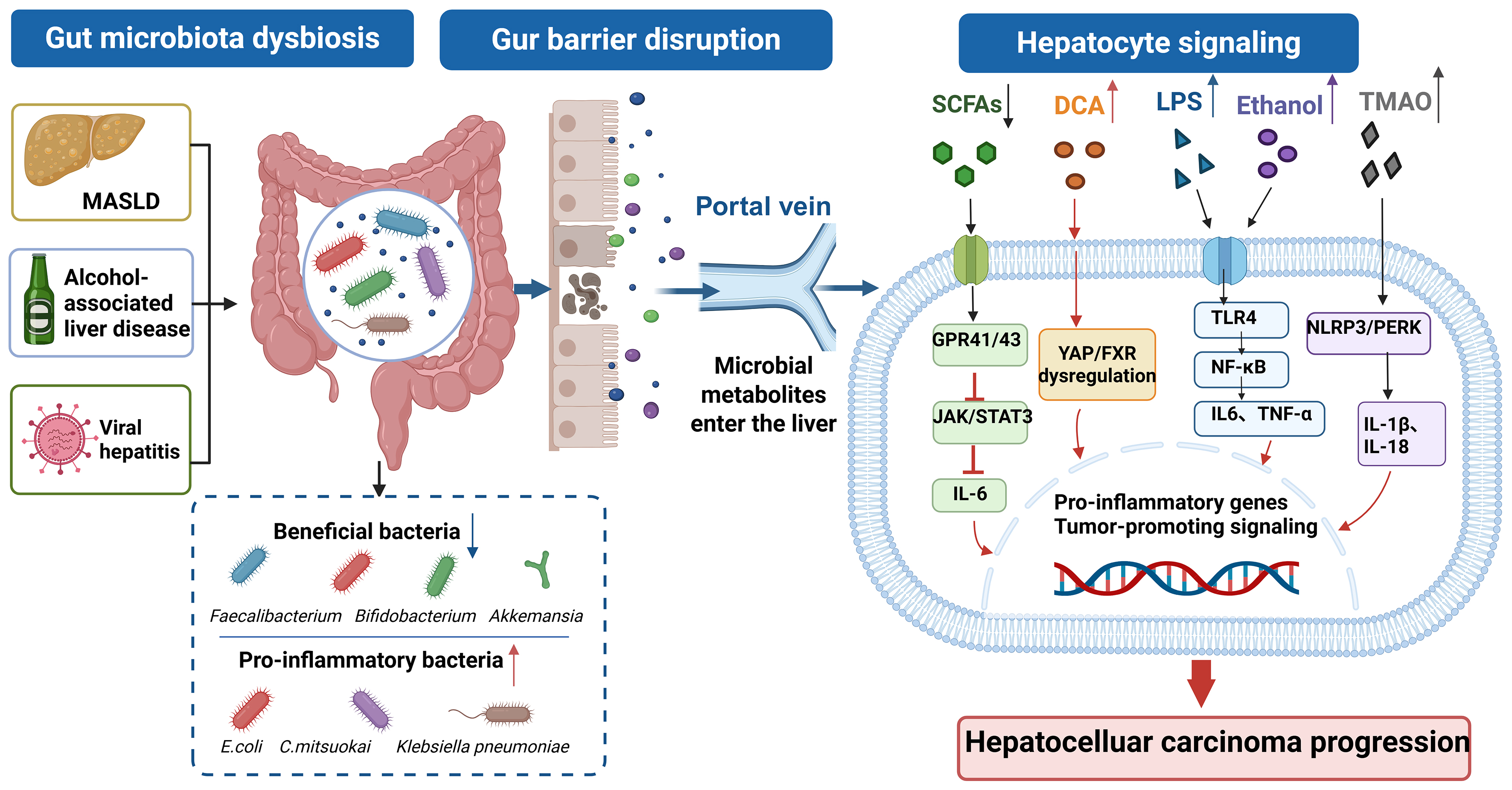
Figure 1. Mechanisms by which gut microbiota-derived metabolites may promote hepatocarcinogenesis. Underlying liver diseases such as MASLD, ALD, and viral hepatitis are associated with gut microbiota dysbiosis, characterized by depletion of butyrate-producing bacteria and enrichment of pro-inflammatory taxa. Reduced SCFA availability weakens epithelial barrier integrity and antitumor immune support; conversely, acetate, propionate, and butyrate can activate GPR41/43-related pathways, restrain excessive inflammation, and support NK/CD8+ T-cell activity. In contrast, these factors act through partly distinct pathways, including DCA-YAP/FXR dysregulation, LPS-TLR4/NF-κB signaling and TMAO-associated inflammatory and metabolic pathways, thereby promoting HCC development. Created in BioRender. Zhang, K. (2026) https://BioRender.com/rc7m51p. The upward arrow indicates an increase; The downward arrow indicates an decrease. ALD: Alcohol-associated liver disease; CD8: cluster of differentiation 8; DCA: deoxycholic acid; FXR: farnesoid X receptor; GPR41/43: G protein-coupled receptor 41/43; HCC: hepatocellular carcinoma; IL-6: interleukin-6; JAK/STAT3: Janus kinase/signal transducer and activator of transcription 3; LPS: lipopolysaccharide; MASLD: metabolic dysfunction-associated steatotic liver disease; NF-κB: nuclear factor kappa-light-chain-enhancer of activated B cells; NK: natural killer; NLRP3: NOD-like receptor protein 3; PERK: protein kinase R-like endoplasmic reticulum kinase; SCFA: short-chain fatty acid; TLR4: Toll-like receptor 4; TMAO: trimethylamine N-oxide; TNF-α: tumor necrosis factor-α; YAP: Yes-associated protein.
Immune regulation and chronic inflammation
The role of the intestinal microbiota and its metabolites in HCC pathogenesis through the gut-liver axis has been extensively investigated in recent years. A decrease in butyrate-producing and bile acid-transforming bacteria, together with ethanol-acetaldehyde exposure in ALD and selected MASLD models, may contribute to barrier breakdown, PAMP translocation, and viable bacterial translocation[42]. Subsequently, microbial products and metabolites reach the liver via the portal vein and act on Kupffer cells, HSCs, and recruited MDSCs. Endotoxin- or ethanol-related signals activate the TLR4-NF-κB/MAPK pathway, inducing Kupffer cells to secrete TNF-α and IL-6 and promoting fibrotic activation of HSCs[43]. In experimental models, DCA together with selected microbial products such as lipoteichoic acid (LTA) has been linked to HSC senescence and SASP-associated immunosuppression. Activated HSCs can recruit MDSCs through the CXCL12 (SDF-1)/C-X-C chemokine receptor type 4 (CXCR4) axis in an orthotopic HCC model[44]. Consequently, gut-derived signals may reshape the hepatic immune network, contributing to an inflammatory-fibrotic-immunosuppressive microenvironment and potentially promoting progression from cirrhosis to HCC. Across different experimental systems, SCFAs, bile-acid receptor agonists and other gut-liver-axis interventions have each been reported to affect selected barrier or immune endpoints, including Treg/Th17 balance, macrophage polarization and lymphocyte recruitment[45,46] [Figure 2].
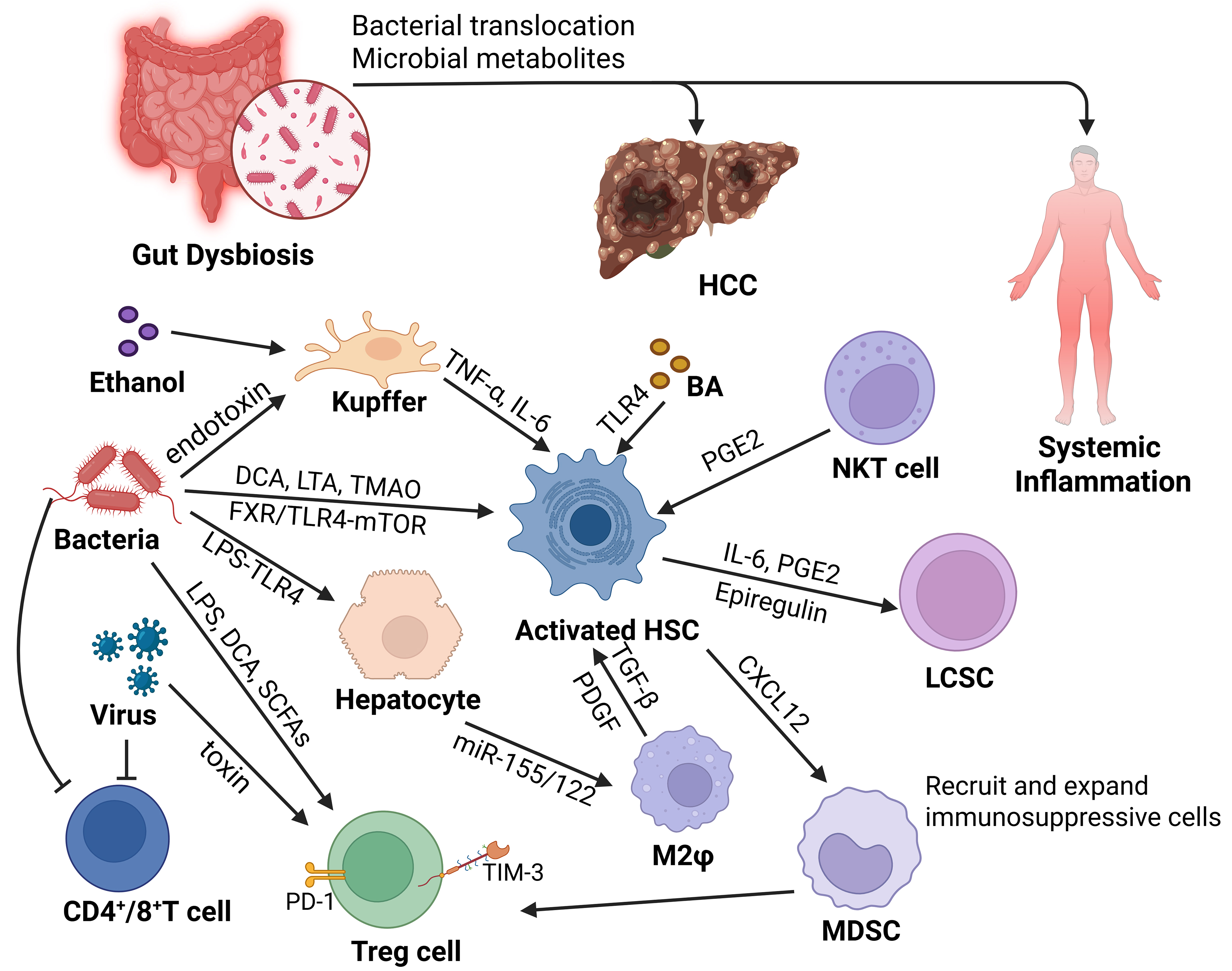
Figure 2. Intestinal microbiota dysbiosis and the immune microenvironment of HCC. Gut-derived LPS, DCA, TMAO, ethanol-related products, bacterial toxins, and fungal products can enter the liver through the gut-liver axis, activate Kupffer cells and HSCs, and shape an inflammatory and immunosuppressive microenvironment. Activated HSCs may promote the self-renewal of LCSCs through the FXR/TLR4-mTOR axis. HSC-derived CXCL12 can recruit CXCR4-expressing MDSCs. PGE2 is displayed separately in the NKT-cell/HSC interaction. Together with M2 macrophage polarization, these signals contribute to CD4+/CD8+ T-cell exhaustion and Treg expansion, forming an immunosuppressive microenvironment that promotes HCC development. Created in BioRender. Zhang, K. (2026) https://BioRender.com/bflwt7r. BA: Bile acid; CD4/CD8: cluster of differentiation 4/8; CXCL12: C-X-C motif chemokine ligand 12; CXCR4: C-X-C chemokine receptor type 4; DCA: deoxycholic acid; FXR: farnesoid X receptor; HCC: hepatocellular carcinoma; HSC: hepatic stellate cell; IL-6: interleukin-6; LCSC: liver cancer stem cell; LPS: lipopolysaccharide; LTA: lipoteichoic acid; MDSC: myeloid-derived suppressor cell; mTOR: mechanistic target of rapamycin; NF-κB: nuclear factor kappa-light-chain-enhancer of activated B cells; NKT: natural killer T; PD-1: programmed cell death protein 1; PGE2: prostaglandin E2; SCFA: short-chain fatty acid; TMAO: trimethylamine N-oxide; TNF-α: tumor necrosis factor-α; TLR4: Toll-like receptor 4; Treg: regulatory T cell; TIM-3: T-cell immunoglobulin and mucin domain-containing protein 3; M2φ:macrophage.
Immune-regulation studies converge on barrier failure, PAMP translocation, Kupffer-cell/HSC activation, myeloid-cell recruitment, and T-cell suppression as plausible links between dysbiosis and HCC. Yet most pathway-level evidence remains experimental, and clinical studies have not yet established which immune nodes represent actionable biomarkers or therapeutic targets in routine HCC care.
HCC stem-like cells and tumor microenvironment
In recent years, the mechanisms by which intestinal microbiota and its metabolites regulate the self-renewal of liver cancer stem cells (LCSCs) through the “gut-liver axis” have attracted increasing attention. Experimental studies indicate that bile-acid and microbial-product signaling can activate HSCs and alter secretion of cytokines and mitogens, including IL-6, prostaglandin E2 (PGE2) and epiregulin, and enhance sphere formation and serial-transplantation tumorigenicity of LCSCs[47]. Microbiota-derived genotoxins have been shown to induce DNA damage and accelerate the accumulation of mutations in tumor-initiating cells[48]. In contrast, SCFAs such as butyrate may enhance CD8+ T-cell IFN-γ secretion and weaken the maintenance of stemness[49]. In addition, urolithin A has been reported to activate silent mating type information regulation 2 homolog 1 (SIRT1)/3, block Wnt/β-catenin signaling and decrease the proportion of CD133+ EpCAM+ stem cells[50] [Figure 2].
The microbiota-LCSC concept offers a testable hypothesis for how microbial metabolites and immune signaling might contribute to recurrence and treatment resistance, especially through bile-acid, genotoxin, and immune-mediated pathways. The current controversy is that LCSC assays and tumor-microenvironment models remain mostly preclinical, so evidence for direct microbiota control of stem-like cells in human HCC remains preliminary.
INTERACTIONS BETWEEN GUT MICROBIOTA AND HCC TREATMENT
Immunotherapy
A growing body of evidence has revealed an association between the gut microbiota, HCC development, and immunotherapy response. Preclinical studies and early clinical datasets from HCC or other solid tumors suggest that probiotics, defined microbial consortia, or fecal microbiota products may influence PD-1/programmed death-ligand 1 (PD-L1) efficacy, but these interventions remain investigational[51-56]. In HCC models and in studies of other solid tumors, taxa including Akkermansia, Bifidobacterium and Faecalibacterium have been associated with immune features such as enhanced antigen presentation and CD8+ T-cell activity. Conversely, enrichment of Proteobacteria and Veillonellaceae and accumulation of harmful secondary bile acids may sustain TLR4 signaling, IL-6 and TNF-α production, M2 tumor-associated macrophage (TAM) and Treg expansion, and suppression of the CXCL16-CXCR6 axis, thereby promoting immune escape. Probiotics, prebiotics, and FMT should currently be regarded as investigational adjuncts rather than validated HCC treatments [Figure 3].
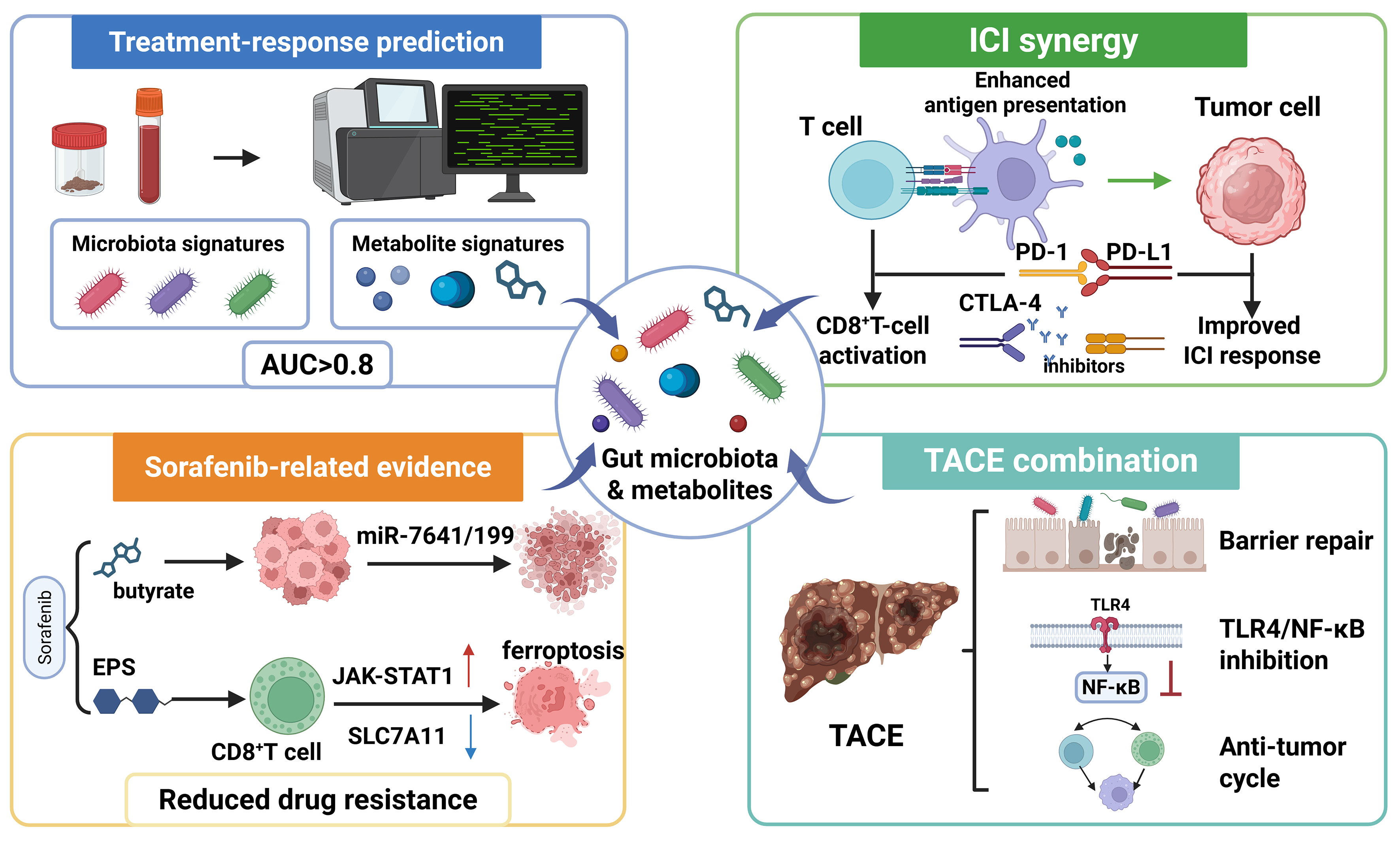
Figure 3. Potential roles of the gut microbiota in modulating HCC treatment response. Microbiota and metabolite signatures may have potential value in predicting treatment response; response-associated commensal bacteria may influence ICI efficacy by regulating antigen presentation and antitumor immunity. Sorafenib-specific preclinical evidence suggests that butyrate may reduce drug resistance by miRNAs, whereas Enterococcus faecium-derived exopolysaccharides may enhance sorafenib-associated ferroptosis through IFN-γ+CD8+ T-cell activation and JAK-STAT1-mediated suppression of SLC7A11. Microbiota remodeling following TACE therapy may contribute to intestinal barrier restoration and attenuation of TLR4/NF-κB-related inflammatory signaling. These applications remain exploratory and require prospective clinical validation. Created in BioRender. Zhang, K. (2026) https://BioRender.com/o8qwnjw. The upward arrow indicates activation of JAK-STAT1; The downward arrow indicates inhibition of SLC7A11. AUC: Area under the curve; CD8: cluster of differentiation 8; CTLA-4: cytotoxic T-lymphocyte-associated protein 4; HCC: hepatocellular carcinoma; ICI: immune checkpoint inhibitor; IFN-γ: interferon-γ; JAK-STAT1: Janus kinase-signal transducer and activator of transcription 1; miRNA: microRNA; NF-κB: nuclear factor kappa-light-chain-enhancer of activated B cells; PD-1: programmed cell death protein 1; PD-L1: programmed death-ligand 1; SLC7A11: solute carrier family 7 member 11; TACE: transarterial chemoembolization; TLR4: Toll-like receptor 4.
Recent HCC therapy has shifted from tyrosine kinase inhibitor (TKI) monotherapy to immune-based combinations. Atezolizumab plus bevacizumab and the STRIDE regimen of tremelimumab plus durvalumab are widely used first-line options for eligible patients with advanced HCC[57,58]. This shift makes microbiome-response research clinically more relevant: a prospective multi-kingdom study of 80 immune checkpoint inhibitor (ICI)-treated patients with HCC identified response-associated differences in bacterial, fungal and metabolite profiles, developed an 18-bacterial-species model for durable clinical benefit, and identified bacterial-metabolite signatures associated with survival[59]. These data support fecal metagenomics, mycobiome profiling, and metabolomics as candidate companion biomarkers, while underscoring that antibiotics, diet, and liver function reserve must be controlled in future trials.
The strongest clinical signal for microbiota-treatment interactions currently comes from immunotherapy cohorts, where baseline microbial diversity and specific taxa correlate with response. Consensus is limited to association and biomarker potential; controversy remains over causality, confounding by antibiotics, diet, and liver function, and whether microbiota modulation improves ICI outcomes in HCC.
Microbiota modulation of sorafenib response
Although immune-based combinations have become the preferred first-line systemic therapies for advanced HCC, sorafenib and lenvatinib remain alternative options for selected patients who are ineligible for these regimens. Notably, most available evidence concerning interactions between the gut microbiota and targeted therapy in HCC is sorafenib-specific and predominantly preclinical; these findings should therefore not be extrapolated directly to lenvatinib or other TKIs. Prohep, a probiotic mixture, has been reported to enhance the antitumor activity of low-dose sorafenib in experimental models by increasing SCFA production, activating AMPK signaling, and suppressing PI3K/mechanistic target of rapamycin (mTOR) signaling[60]. In vitro, sodium butyrate reduced the viability of sorafenib-resistant HCC cells, increased apoptosis, and modulated sorafenib-resistance-associated miRNAs, including miR-7641 and miR-199[61]. However, this study did not directly demonstrate that microbiota-derived butyrate induces ferroptosis. A separate nanomedicine study used butyrate as a surface-modifying ligand to improve the intestinal absorption and tumor targeting of nanoparticles co-loaded with sorafenib and salinomycin[62]. In this system, ferroptosis was primarily induced by the released sorafenib and salinomycin rather than by butyrate itself. More direct microbiota-related evidence came from a study showing that Enterococcus faecium-derived exopolysaccharides promoted IFN-γ-producing CD8+ T cells, activated JAK-STAT1 signaling, suppressed solute carrier family 7 member 11 (SLC7A11) expression, and thereby enhanced sorafenib-associated ferroptosis in experimental HCC models[63] [Figure 3].
Targeted-therapy research should also extend beyond sorafenib. Lenvatinib, regorafenib, cabozantinib, and ramucirumab act through partly distinct VEGF, fibroblast growth factor receptor (FGFR), MET, AXL receptor tyrosine kinase (AXL) or VEGFR2-related axes, so microbiome findings from sorafenib should therefore not be generalized across targeted agents. For future studies, baseline fecal microbiota, bile acid profiles, SCFA levels, and antibiotic exposure should be prospectively collected in patients receiving TKIs alone or in TKI-ICI combinations, because diarrhea, anorexia, bile acid disruption, and systemic inflammation may both reflect and reshape the intestinal ecosystem.
Transarterial chemoembolization (TACE)
TACE is a standard treatment for intermediate-stage HCC and is also used in selected advanced or combination settings. Its efficacy depends on local ischemia and chemotherapy-induced tumor killing, and may also relate to microecological remodeling along the gut-liver axis. Clinical studies suggest that TACE treatment is associated with dynamic microbiota changes in HCC patients: in the early post-procedural period (1-5 days), dysbiosis may be temporarily aggravated by ischemia-reperfusion injury and chemotherapy toxicity; however, by approximately 10 days after TACE, some studies have reported decreases in intestinal-permeability markers and circulating LPS together with increases in butyrate-producing bacteria[64]. These observations have led to the hypothesis that post-TACE microbiota changes may be associated with attenuation of TLR4/NF-κB-related inflammatory signaling[64]. Clinical studies have shown that baseline Limosilactobacillus reuteri and indole-3-lactic acid (ILA) levels were associated with post-TACE liver injury and clinical outcomes[65]. TACE-TKI-ICI has shown clinical activity in unresectable HCC[66]. However, no prospective longitudinal microbiome study has demonstrated that this combination reproducibly remodels the gut microbiota or that microbiota changes mediate treatment efficacy; these questions remain to be established. Future prospective studies could evaluate microbiota-targeted interventions alongside TACE, with prespecified safety, biological and efficacy endpoints [Figure 3].
Chemotherapy-related microbiome effects in HCC are most relevant in locoregional regimens such as TACE, hepatic arterial infusion chemotherapy, because conventional systemic cytotoxic chemotherapy has limited use in cirrhosis-associated HCC. Locoregional regimens may themselves alter intestinal and systemic physiology, making it difficult to distinguish treatment-induced microbiome changes from changes secondary to tumor response or liver injury. A 2026 prospective TACE microbiome study reported that lower Prevotella and higher Bacteroides/Faecalibacterium were associated with better progression-free survival, while higher Phascolarctobacterium or Veillonella correlated with worse overall survival[67].
For TACE and other locoregional therapies, available studies suggest that microbiota features may track treatment response, liver injury, and prognosis. The controversy is whether observed remodeling is a cause of response, a consequence of embolization-induced tumor and liver changes, or a composite marker of host reserve.
THE POTENTIAL OF GUT MICROBIOTA AS A BIOMARKER FOR HCC
Recent multi-omics evidence indicates that the intestinal microbiota and its metabolites may serve as candidate non-invasive biomarkers for early HCC diagnosis. Cross-sectional studies comparing HBV- or MASLD-associated disease stages have reported lower abundance of selected butyrate-producing taxa in cirrhosis and HCC, whereas taxa and microbial functions associated with secondary bile-acid transformation and increased endotoxin exposure are enriched; corresponding dysbiosis indices and LPS/DCA levels correlate with clinical stage, suggesting potential value as multi-omics biomarkers for early HCC risk detection[68]. Intestinal epithelial hypoxia, increased mobile genetic elements, and elevated serum LPS and zonula occludens-1 (ZO-1), together with fecal calprotectin, indicate that dysbiosis may worsen alongside intestinal barrier leakage[69]. In a prospective multicenter fecal microbiome study, a 30-microbial-marker random-forest classifier distinguished early HCC from non-HCC controls with an area under the curve (AUC) of 0.806 in the discovery cohort and showed diagnostic potential in independent geographic validation cohorts; however, its performance was not sufficient for stand-alone clinical diagnosis and requires broader prospective validation[70]. Integrated microbiome-metabolome studies have also reported altered intestinal microbiota and serum metabolite profiles in HCC patients, supporting combined microbial-metabolic biomarker exploration, but the specific claim that DCA/butyrate changes predict HCC several years before diagnosis should be treated cautiously unless supported by longitudinal pre-diagnostic cohorts[71].
Likewise, gut microbiota biomarkers have shown promising but preliminary potential for predicting postoperative recurrence and metastasis of HCC. A 2023 original study of HBV-related HCC integrated stool 16S rRNA sequencing with tumor-tissue metabolomics and found that several taxa, including Dialister, Veillonella, the Eubacterium coprostanoligenes group, Lactobacillus, Streptococcus pneumoniae and Bifidobacterium faecale, were associated with early recurrence; a nomogram combining microbial species and clinical indicators achieved an AUC of 0.780 for early-recurrence prediction[72]. Moreover, a recent study constructed a prognostic model based on eight butyrate-metabolism genes [butyrate-metabolism genes (BMGs): LCAT, G6PD, SPP1, etc.], which may help stratify prognosis and therapy response in public HCC datasets, although it is not a validated preoperative blood test and requires clinical validation[73]. Review-level evidence further suggests that serum TMAO, LPS-related markers and fecal SCFAs are biologically plausible adjunctive biomarkers[74]. Therefore, longitudinal microbiome and metabolite profiling may be explored as a complementary research tool for HCC risk assessment, treatment-response stratification and prognosis, with each intended use requiring separate prospective validation [Figure 3].
An updated biomarker framework should progress from genus-level descriptions to integrated microbial functions. Candidate diagnostic and prognostic signals include loss of butyrate-producing bacteria, enrichment of pathobionts, fecal or serum SCFAs, secondary bile acids, TMAO, and indole derivatives; quinolinic acid (QA) is better described at present as a mechanistically supported HCC-promoting microbial metabolite rather than an established clinical biomarker[59,75-77]. In unresectable HCC treated with anti-PD-1-based combination therapy, specific baseline microbiota signatures have been associated with clinical efficacy and survival, suggesting a possible non-invasive route for immunotherapy stratification[77]. However, these signatures should be treated as complementary to alpha-fetoprotein (AFP), protein induced by vitamin K absence-II (PIVKA-II), imaging, liver function, and tumor burden, rather than as stand-alone diagnostic tests. At present, the consensus is that microbiota-based biomarkers are complementary research tools; the controversy is reproducibility across geography, sequencing pipelines, etiologies, and prospective screening settings.
CANDIDATE STRATEGIES FOR HCC PREVENTION AND TREATMENT TARGETING THE INTESTINAL MICROBIOTA
FMT
FMT is being explored as a microbiota-modulating strategy for solid tumors and chronic liver diseases, but its role in HCC remains investigational[74]. Compared with single-strain probiotic supplementation, FMT may alter gut-derived inflammatory signaling, including exposure to microbial products such as LPS, and may offer therapeutic potential in selected chronic liver diseases[78]. In HCC-related models or early translational settings, FMT may improve dysbiosis and the immune microenvironment by restoring butyrate-producing bacteria, inhibiting pro-inflammatory metabolites, enhancing CD8+ T-cell function, regulating Treg balance, and potentially synergizing with PD-1/PD-L1 blockade[79,80]. In animal models and preliminary clinical trials of MASLD or liver fibrosis, FMT has been reported to improve steatosis, inflammation, and fibrosis by restoring intestinal barrier function, modulating the bile acid-FXR axis, and reducing systemic inflammation, with possible heterogeneity between lean and obese MASLD populations[81,82]. Therefore, FMT is conceptually attractive because it can restore ecological function rather than targeting one organism at a time. Nonetheless, donor variability, engraftment unpredictability, infection risk, and the lack of HCC-specific randomized data mean that FMT should remain a trial-based intervention rather than routine prevention or treatment [Figure 4].
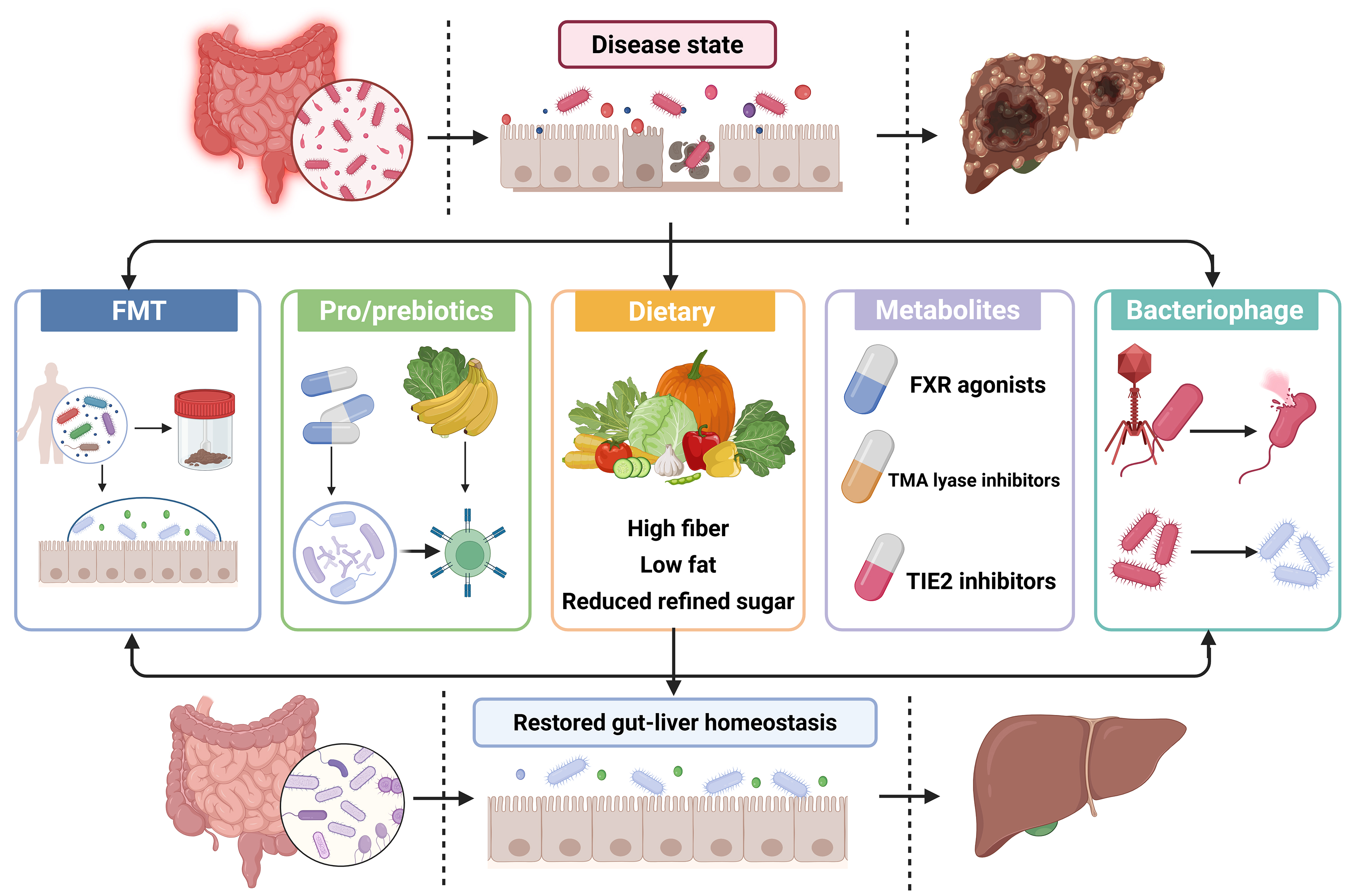
Figure 4. Candidate intervention strategies targeting the gut microbiota in HCC-related settings: FMT can broadly reshape the microbial ecosystem by introducing donor microbiota; probiotic or prebiotic supplementation may selectively promote beneficial bacteria and enhance the intestinal barrier; dietary intervention (high-fiber, low-fat, and low-refined-sugar diets) may improve microbiota composition; metabolite-targeted therapies, such as FXR agonists or TMA lyase inhibitors, may regulate microbiota-derived signaling molecules; bacteriophage therapy may specifically remove toxin-producing pathogens. These strategies are hypothesized to act through the gut-liver axis and modify microbial or metabolic function; most remain preclinical or at an early clinical stage in HCC. Created in BioRender. Zhang, K. (2026) https://BioRender.com/3p4nage. FMT: Fecal microbiota transplantation; FXR: farnesoid X receptor; HCC: hepatocellular carcinoma; TIE2: TEK tyrosine kinase; TMA: trimethylamine.
Probiotics and prebiotics
Probiotics may remodel the intestinal microbiota and attenuate inflammatory-carcinogenic signaling, offering multi-targeted and relatively low-toxicity prospects for HCC prevention research and adjuvant-therapy development[83,84]. Probiotics may enhance anticancer immune responses by strengthening the intestinal barrier, promoting SCFA production, and activating CD8+ T-cells, whereas prebiotics may indirectly improve host immune status by selectively promoting beneficial bacteria; this statement is supported mainly by review-level evidence and should not be attributed to a single HCC-specific clinical trial[75]. Lau et al. reported that Lactobacillus acidophilus supplementation suppressed tumorigenesis in a MASLD-HCC model; produced pentanoic acid, which engaged GPR41/43 on hepatocytes, inhibited carcinogenic Rho-GTPase signaling, and induced cell-cycle arrest and apoptosis[85]. Song et al. found that Bifidobacterium was significantly depleted in MASLD-HCC mouse models and patients, and Bifidobacterium pseudolongum supplementation suppressed tumorigenesis in this MASLD-HCC model through acetate-dependent signaling[34]. Moreover, prebiotics, as non-digestible dietary components that selectively promote beneficial bacteria, have been shown in animal models to restore intestinal microecological balance, strengthen the intestinal barrier, reduce systemic inflammation, and inhibit the development of HCC[86]. Probiotics and prebiotics have the practical advantage of scalability and established safety, and animal models support immune and barrier effects. The key controversies concern strain specificity, dose, durability, and whether benefits observed in MASLD-HCC models translate to patients with established cirrhosis or advanced HCC [Figure 4].
Dietary interventions
Diet is closely linked to HCC risk. Several systematic reviews and prospective studies have shown that Western dietary patterns are associated with higher HCC risk, chronic inflammation, insulin resistance and microbiota dysbiosis are plausible mediators of this association, whereas mediterranean, plant-based, high-fiber, and low-glycemic-load diets may reduce risk by improving metabolic status, enhancing intestinal barrier function, and regulating immune responses[87]. Mechanistically, monounsaturated fatty acids (MUFAs), vitamin E, folic acid, and β-carotene may interfere with the chronic hepatitis-fibrosis-carcinogenesis axis through antioxidant activity, suppression of HSC activation, and effects on DNA methylation; conversely, dietary patterns rich in saturated fat, sugar and processed meats may increase exposure to N-nitroso compounds (NOCs) and may influence host insulin-like growth factor 1 (IGF-1) signaling, thus may increase carcinogenesis risk[88]. Experimentally, mice fed a high-fat, high-cholesterol diet show increased abundance of mucin-producing genera, Desulfovibrio, and Anaerostipes, reduced Deltaproteobacteria or Bacteroides, and increased hepatic lipid accumulation, inflammation, and cell proliferation[89]. In experimental settings, phytic acid, a component of high-fiber foods such as soybeans and nuts, ameliorated these changes by reducing hepatic lipid synthesis via SREBP-1c/FAS inhibition, suppressing NF-κB inflammatory signaling, and limiting harmful bacteria[90]. Similarly, dietary carbohydrate restriction (DCR) improved the hepatic metabolic-inflammatory environment and restored the microbiota-bile acid-immune axis, and was associated with inhibition of HCC progression in experimental settings[91] [Figure 4].
Clinical intervention evidence is also beginning to extend beyond animal models. A 2026 single-center randomized trial evaluated a multicomponent nutritional and microbiota-directed intervention comprising high-fiber dietary advice, probiotics, prebiotics and increased unsaturated-fat intake; the bundled design precludes attribution of clinical effects to any single component[92].
Dietary intervention has the strongest population-level plausibility because diet simultaneously influences obesity, diabetes, inflammation, and microbial metabolism. However, clinical evidence in HCC remains difficult to interpret because bundled lifestyle interventions, treatment adherence, cancer stage, and concurrent therapy can confound microbiota-mediated effects.
Microbial metabolite-targeted therapy
Changes in metabolites during liver disease progression suggest potential targets for small-molecule drug development. For example, interventions that inhibit microbial TMA formation or pharmacologically activate FXR have shown metabolic improvement or antitumorigenic effects in animal models of MASLD and liver fibrosis, although HCC-specific clinical evidence remains limited[93,94]. Butyrate has been reported to reduce the expression of lipid-synthesis enzymes such as fatty acid synthase (FASN), acetyl-CoA carboxylase (ACC), ATP-citrate lyase (ACLY), and stearoyl-CoA desaturase (SCD) by inhibiting the PI3K/Akt/mTOR-SREBP-1 axis, thereby reducing hepatic lipid deposition and inflammatory responses; small-molecule drugs targeting the lipid-metabolism axis, such as the FASN inhibitor TVB-2640 and ACC inhibitor ND-654, have shown anti-HCC potential in animal models or in early clinical development[95]. In addition, Zhang et al. found that the tumor and fecal samples from HCC patients were enriched with Catenibacterium mitsuokai, which was reported to translocate to the liver and adhere to HCC cells through an interaction between its surface protein Gtr1/RagA and host-cell γ-catenin, while secreted QA bound TEK receptor tyrosine kinase (TIE2) and activated oncogenic PI3K/AKT signaling. TIE2 inhibitors blocked the QA-mediated tumor-promoting effect in vivo, suggesting that the microbiota-QA-TIE2 axis is a candidate actionable metabolic-signaling pathway[76]. Metabolite-targeted therapy offers a more druggable route to microbiota translation by focusing on defined enzymes or receptors. However, many metabolites exert dual, context-dependent effects, and most candidate pathways still require pharmacodynamic biomarkers and HCC-specific clinical testing [Figure 4].
Bacteriophages
Bacteriophages, an important component of the intestinal virome, have shown experimental value as precise microbiome-editing tools in liver disease models. The proposed strategy is to eliminate cancer-promoting or liver-injury-promoting pathogens and thereby potentially interrupt gut-liver axis-driven malignant transformation[96,97]. Duan et al. found that Enterococcus faecalis is significantly enriched in the intestinal tract of patients with alcoholic hepatitis and that its secreted cytolysin worsens ethanol-induced liver injury; oral phage cocktail therapy targeting this bacterium reduced its abundance and attenuated liver disease progression in mice[25]. Gan et al. showed that phages targeting high-alcohol-producing Klebsiella pneumoniae inhibited MASLD progression in mice by blocking endogenous ethanol production and reversing hepatic steatosis, inflammation, and fibrotic cascades[98]. Additional translational rationale comes from studies of other inflammatory diseases showing that designed phage consortia can selectively suppress target commensals, but comparable HCC-specific clinical evidence remains unavailable[99]. Bacteriophage therapy may offer greater taxonomic specificity than broad-spectrum antibiotics for selected microbial targets, although resistance, delivery and host immune clearance remain major limitations[100,101]. Phage therapy illustrates the potential for precision microbiome editing, especially when a defined harmful bacterium or toxin is present. The major uncertainty is whether HCC has sufficiently stable pathogen targets for phage design, and whether resistance, delivery, and immune clearance can be managed safely in cirrhosis or cancer therapy settings [Figure 4].
CURRENT CHALLENGES AND FUTURE DIRECTIONS
The clinical translation of microbiota-targeted therapies for HCC faces multiple interconnected hurdles, including mechanistic uncertainty, a lack of validated personalized biomarkers, and practical barriers to implementation in cancer populations[102]. FMT is constrained by heterogeneity in donor selection and screening protocols, uncertain donor-recipient matching, variable engraftment and the risk of transmitting pathogens or antimicrobial-resistance determinants. Conventional FMT often yields unpredictable colonization, with risks of introducing drug-resistance genes or opportunistic pathogens[84]. Phage therapy offers precision in eliminating pathogenic bacteria, but phage resistance, host immune clearance, delivery optimization and the lack of defined HCC-specific bacterial targets remain substantial obstacles[103]. Emerging strategies such as engineered probiotics and postbiotics have yet to undergo systematic validation of manufacturing standards, dose-response relationships, and long-term safety[104]. Second, at the biomarker level, although microbial signatures have achieved AUCs > 0.8 for early HCC diagnosis in selected retrospective datasets, prospective cohort validation remains insufficient, and dynamic monitoring with longitudinal follow-up is lacking[70,105]. Third, inter-individual heterogeneity severely limits universal efficacy: genetic background, sex, age, geography, diet, medication exposure, and disease etiology all contribute to highly variable microbiota compositions, rendering a one-size-fits-all approach unlikely[106]. Finally, at the technical level, considerable heterogeneity arises from differences in DNA extraction methods, sequencing platforms (16S rRNA vs. metagenomics), and bioinformatics pipelines, which can bias results across studies[107].
Addressing these layered obstacles will require a coordinated research agenda. Future efforts should prioritize the functional dissection of key bacterial strains, their genes, and metabolic pathways, with the aim of developing highly specific interventions such as in vivo biopharmaceuticals, engineered bacteria, and metabolic-enzyme inhibitors[108]. Concurrently, well-designed randomized controlled, multi-center, cross-ethnic clinical trials should be conducted to test FMT or probiotics in combination with immunotherapy or targeted therapy, establishing efficacy-predictive markers and optimal dosing regimens[109]. Integrative approaches leveraging artificial intelligence and multi-omics data could further construct individualized flora-metabolite-receptor panoramic maps and derive non-invasive early diagnostic and prognostic models based on metabolite ratios and pathway-activation signatures[22]. Such integrative strategies may support more precise prevention and treatment research tailored to high-risk populations and HCC patients.
However, even these forward-looking plans must contend with several broader conceptual gaps that are often overlooked. Most HCC microbiome studies implicitly equate the gut microbiota with bacteria, neglecting the fungal and viral components as distinct ecological layers. In ALD, fungal dysbiosis, including expansion of Candida species and production of β-glucan and candidalysin, can aggravate barrier injury and hepatic inflammation, yet direct evidence linking the gut mycobiome to human HCC remains sparse[21,110]. The intestinal virome, particularly bacteriophages, may reshape bacterial ecology and offer therapeutic potential (as discussed above), but its diagnostic and mechanistic relevance in HCC still requires systematic metagenomic and longitudinal investigation[98-100]. Sex differences and host genetic variants also deserve explicit consideration: HCC incidence is higher in men, sex hormones may modulate bile acid metabolism, immune tone and microbial composition, and variants such as PNPLA3, TM6SF2, and HSD17B13 influence lipid handling, inflammation, and HCC susceptibility[1,2,111,112]. These host factors should therefore be assessed in studies of MASLD- or ALD-related HCC.
Finally, a critical distinction must be drawn between gut microbiota and intratumoral microbiota. The former refers to microbial communities and products within the intestinal ecosystem that affect the liver via the gut-liver axis; the latter refers to microbial cells, nucleic acids or other microbial signatures detected within tumor tissue. Because HCC tissue is a low-biomass specimen, such signals require aseptic sampling, extensive negative controls, contamination-aware sequencing and orthogonal validation before biological function is inferred[113,114]. Given the methodological and biological differences, this review focuses primarily on gut microbiota; intratumoral microbiota is mentioned only as an emerging, methodologically distinct field that lies beyond the current scope of this review.
The field shares a clear consensus that microbiome-based HCC translation will require standardized sampling, multi-omics integration, longitudinal cohorts, and randomized controlled trials. The leading disputes concern how to handle ecological complexity, low-biomass contamination, host heterogeneity, sex/genetic effects, and the boundary between gut-derived and intratumoral microbial signals.
CONCLUSION
Gut microbiota imbalance may contribute to hepatocarcinogenesis across MASLD, ALD and chronic viral hepatitis, including pathways that involve advanced fibrosis or cirrhosis as well as selected non-cirrhotic settings. Microbial-derived molecules have context-dependent and sometimes opposing effects. SCFAs such as acetate, propionate, and butyrate generally support intestinal barrier integrity, restrain excessive inflammation, and enhance NK/CD8+ T-cell antitumor activity, although their effects may vary with concentration, tissue context, and disease stage. By contrast, dysregulated secondary bile acids, LPS, ethanol-related products, TMAO and selected bacterial or fungal products have been linked to distinct inflammatory, immunosuppressive, genotoxic or stemness-associated pathways. Microbiota-related features may influence, or serve as biomarkers of, responses to ICIs and targeted therapies and of treatment-related changes following TACE; direct therapeutic enhancement remains to be demonstrated clinically. Probiotics, prebiotics, FMT, dietary remodeling, metabolite-targeted therapy, and phage therapy have shown biological activity or preliminary clinical signals, predominantly in preclinical or early-stage studies. Multi-omics microbiota-metabolite models show promise for HCC risk prediction, diagnosis, treatment-response assessment, and recurrence monitoring, but they require prospective validation, standardization, and careful separation of HCC-specific evidence from broader liver-disease or pan-cancer evidence.
Microbiota-targeted interventions face unresolved standardization problems (donor selection, strain identity, dose, route, timing, and manufacturing quality), reproducibility problems (geography, diet, antibiotics, sequencing pipelines, and low-biomass contamination), safety concerns (pathogen transmission, antimicrobial-resistance genes, bacteremia, sepsis risk in cirrhosis or immunosuppression, and uncertain long-term engraftment), and regulatory challenges. Therefore, HCC applications should proceed through regulated clinical trials with GMP-grade products, transparent donor or strain screening, pharmacovigilance, predefined microbiome/metabolite endpoints, and external validation before routine clinical adoption.
DECLARATIONS
Acknowledgments
The Graphical Abstract was created with BioRender.com [Created in BioRender. Zhang, K. (2026) https://BioRender.com/fhcsa1d].
Authors’ contributions
Made substantial contributions to the conception and design of the review: Zhang K, Wang L
Contributed to drafting sections of the manuscript: Zhang K, Xue T, Hu J
Critically revised the manuscript for important intellectual content: Wang L
All authors read and approved the final manuscript.
Availability of data and materials
Not applicable.
AI and AI-assisted tools statement
During the preparation of this manuscript, the AI tool ChatGPT (version 5.5, released 2026-04-24) was used solely for language editing. The tool did not influence the study design, data collection, analysis, interpretation, or the scientific content of the work. All authors take full responsibility for the accuracy, integrity, and final content of the manuscript.
Financial support and sponsorship
This work was supported by a grant from the National Natural Science Foundation of China (No. 82103301).
Conflicts of interest
All authors declared that there are no conflicts of interest.
Ethical approval and consent to participate
Not applicable.
Consent for publication
Not applicable.
Copyright
© The Author(s) 2026.
REFERENCES
1. Toh MR, Wong EYT, Wong SH, et al. Global epidemiology and genetics of hepatocellular carcinoma. Gastroenterology. 2023;164:766-82.
2. Singal AG, Kanwal F, Llovet JM. Global trends in hepatocellular carcinoma epidemiology: implications for screening, prevention and therapy. Nat Rev Clin Oncol. 2023;20:864-84.
3. Koshy A. Evolving global etiology of hepatocellular carcinoma (HCC): insights and trends for 2024. J Clin Exp Hepatol. 2025;15:102406.
4. Motta BM, Masarone M, Torre P, Persico M. From non-alcoholic steatohepatitis (NASH) to hepatocellular carcinoma (HCC): epidemiology, incidence, predictions, risk factors, and prevention. Cancers. 2023;15:5458.
5. De Vos WM, Tilg H, Van Hul M, Cani PD. Gut microbiome and health: mechanistic insights. Gut. 2022;71:1020-32.
6. Schneider KM, Mohs A, Gui W, et al. Imbalanced gut microbiota fuels hepatocellular carcinoma development by shaping the hepatic inflammatory microenvironment. Nat Commun. 2022;13:3964.
7. Hsu CL, Schnabl B. The gut-liver axis and gut microbiota in health and liver disease. Nat Rev Microbiol. 2023;21:719-33.
8. Kim Y, Kim G, Kim S, et al. Fecal microbiota transplantation improves anti-PD-1 inhibitor efficacy in unresectable or metastatic solid cancers refractory to anti-PD-1 inhibitor. Cell Host Microbe. 2024;32:1380-93.e9.
9. Huang DQ, El-serag HB, Loomba R. Global epidemiology of NAFLD-related HCC: trends, predictions, risk factors and prevention. Nat Rev Gastroenterol Hepatol. 2020;18:223-38.
10. Han H, Jiang Y, Wang M, et al. Intestinal dysbiosis in nonalcoholic fatty liver disease (NAFLD): focusing on the gut-liver axis. Crit Rev Food Sci Nutr. 2021;63:1689-706.
11. Song Q, Zhang X. The role of gut-liver axis in gut microbiome dysbiosis associated NAFLD and NAFLD-HCC. Biomedicines. 2022;10:524.
12. Zhou Y, Wang M, Wang Z, et al. Polysaccharides from hawthorn fruit alleviate high-fat diet-induced NAFLD in mice by improving gut microbiota dysbiosis and hepatic metabolic disorder. Phytomedicine. 2025;139:156458.
13. Zhao H, Li Y, Liu N, et al. Ganoderma lucidum polysaccharides alleviate non-alcoholic fatty liver disease by modulating gut microbiota against TLR4/NF-κB/MAPK pathway and activating AMPK pathway. J Ethnopharmacol. 2026;355:120723.
14. Wang S, Sheng F, Zou L, Xiao J, Li P. Hyperoside attenuates non-alcoholic fatty liver disease in rats via cholesterol metabolism and bile acid metabolism. J Adv Res. 2021;34:109-22.
15. Loomba R, Friedman SL, Shulman GI. Mechanisms and disease consequences of nonalcoholic fatty liver disease. Cell. 2021;184:2537-64.
16. Clifford BL, Sedgeman LR, Williams KJ, et al. FXR activation protects against NAFLD via bile-acid-dependent reductions in lipid absorption. Cell Metab. 2021;33:1671-84.e4.
17. Åberg F, Jiang ZG, Cortez-pinto H, Männistö V. Alcohol-associated liver disease - Global epidemiology. Hepatology. 2024;80:1307-22.
18. Hong X, Huang S, Jiang H, et al. Alcohol-related liver disease (ALD): current perspectives on pathogenesis, therapeutic strategies, and animal models. Front Pharmacol. 2024;15:1432480.
19. Hyun J, Han J, Lee C, Yoon M, Jung Y. Pathophysiological aspects of alcohol metabolism in the liver. Int J Mol Sci. 2021;22:5717.
20. Wu X, Fan X, Miyata T, et al. Recent advances in understanding of pathogenesis of alcohol-associated liver disease. Annu Rev Pathol. 2023;18:411-38.
21. Yang A, Inamine T, Hochrath K, et al. Intestinal fungi contribute to development of alcoholic liver disease. J Clin Investig. 2017;127:2829-41.
22. Zuo S, Ma J, Li X, et al. The dual role of gut microbiota and their metabolites in hepatocellular carcinoma: a context-dependent framework. Microorganisms. 2025;14:73.
23. Wang R, Tang R, Li B, Ma X, Schnabl B, Tilg H. Gut microbiome, liver immunology, and liver diseases. Cell Mol Immunol. 2020;18:4-17.
24. Cheng Z, Yang L, Chu H. The role of gut microbiota, exosomes, and their interaction in the pathogenesis of ALD. J Adv Res. 2025;72:353-67.
25. Duan Y, Llorente C, Lang S, et al. Bacteriophage targeting of gut bacterium attenuates alcoholic liver disease. Nature. 2019;575:505-11.
26. Odenwald MA, Paul S. Viral hepatitis: past, present, and future. World J Gastroenterol. 2022;28:1405-29.
27. Tian Z, Xu C, Yang P, et al. Molecular pathogenesis: connections between viral hepatitis-induced and non-alcoholic steatohepatitis-induced hepatocellular carcinoma. Front Immunol. 2022;13:984728.
28. Raziq K, Shah SQA, Shahzad MN, Ummara UE, Subhan M, Nazir MM. The gut-liver-virus axis in hepatitis B and C: microbiota, immunometabolism, and exosome-mediated therapeutic opportunities. Naunyn Schmiedebergs Arch Pharmacol. 2025;399:6221-37.
29. Yan F, Zhang Q, Shi K, et al. Gut microbiota dysbiosis with hepatitis B virus liver disease and association with immune response. Front Cell Infect Microbiol. 2023;13:1152987.
30. Yuan H, Xu R, Li S, et al. The malignant transformation of viral hepatitis to hepatocellular carcinoma: mechanisms and interventions. MedComm. 2025;6:e70121.
31. Shen Y, Wu S, Chen Y, et al. Alterations in gut microbiome and metabolomics in chronic hepatitis B infection-associated liver disease and their impact on peripheral immune response. Gut Microbes. 2022;15:2155018.
32. Martin-gallausiaux C, Marinelli L, Blottière HM, Larraufie P, Lapaque N. SCFA: mechanisms and functional importance in the gut. Proc Nutr Soc. 2020;80:37-49.
33. Zhang M, Huang X, Zhang Y, et al. Gut microbial metabolite butyrate suppresses hepatocellular carcinoma growth via CXCL11-dependent enhancement of natural killer cell infiltration. Gut Microbes. 2025;17:2519706.
34. Song Q, Zhang X, Liu W, et al. Bifidobacterium pseudolongum-generated acetate suppresses non-alcoholic fatty liver disease-associated hepatocellular carcinoma. J Hepatol. 2023;79:1352-65.
35. Fusco W, Lorenzo MB, Cintoni M, et al. Short-chain fatty-acid-producing bacteria: key components of the human gut microbiota. Nutrients. 2023;15:2211.
36. Mcbrearty N, Arzumanyan A, Bichenkov E, Merali S, Merali C, Feitelson M. Short chain fatty acids delay the development of hepatocellular carcinoma in HBx transgenic mice. Neoplasia. 2021;23:529-38.
37. Guo X, Okpara ES, Hu W, et al. Interactive relationships between intestinal flora and bile acids. Int J Mol Sci. 2022;23:8343.
38. Shen R, Ke L, Li Q, et al. Abnormal bile acid-microbiota crosstalk promotes the development of hepatocellular carcinoma. Hepatol Int. 2022;16:396-411.
39. Liu Y, Zhu J, Jin Y, et al. Disrupting bile acid metabolism by suppressing Fxr causes hepatocellular carcinoma induced by YAP activation. Nat Commun. 2025;16:3583.
40. Saha B, Banerjee A, Pathak R, Duttaroy AK, Pathak S. Trimethylamine N-Oxide (TMAO) and cancer risk: insights into a possible link. Biomed Pharmacother. 2025;192:118592.
41. Wu Y, Rong X, Pan M, et al. Integrated analysis reveals the gut microbial metabolite TMAO promotes inflammatory hepatocellular carcinoma by upregulating POSTN. Front Cell Dev Biol. 2022;10:840171.
42. Pabst O, Hornef MW, Schaap FG, Cerovic V, Clavel T, Bruns T. Gut-liver axis: barriers and functional circuits. Nat Rev Gastroenterol Hepatol. 2023;20:447-61.
43. Liu J, Wu A, Cai J, She Z, Li H. The contribution of the gut-liver axis to the immune signaling pathway of NAFLD. Front Immunol. 2022;13:968799.
44. Xu Y, Fang F, Jiao H, et al. Activated hepatic stellate cells regulate MDSC migration through the SDF-1/CXCR4 axis in an orthotopic mouse model of hepatocellular carcinoma. Cancer Immunol Immunother. 2019;68:1959-69.
45. Li C, Cai C, Wang C, Chen X, Zhang B, Huang Z. Gut microbiota-mediated gut-liver axis: a breakthrough point for understanding and treating liver cancer. Clin Mol Hepatol. 2025;31:350-81.
46. Tilg H, Adolph TE, Trauner M. Gut-liver axis: pathophysiological concepts and clinical implications. Cell Metab. 2022;34:1700-18.
47. Yoon SJ, Han SK, Kim TS, et al. The crosstalk between gut microbiota and microbiota-derived metabolites in hepatocellular carcinoma. Crit Rev Microbiol. 2025;51:1315-29.
48. Zhou A, Tang L, Zeng S, Lei Y, Yang S, Tang B. Gut microbiota: a new piece in understanding hepatocarcinogenesis. Cancer Lett. 2020;474:15-22.
49. Machida K, Tahara SM. Immunotherapy and microbiota for targeting of liver tumor-initiating stem-like cells. Cancers. 2022;14:2381.
50. Zhra M, Elahi MA, Tariq A, Abu-zaid A, Yaqinuddin A. Sirtuins and gut microbiota: dynamics in health and a journey from metabolic dysfunction to hepatocellular carcinoma. Cells. 2025;14:466.
51. Chen P, Yang C, Ren K, et al. Modulation of gut microbiota by probiotics to improve the efficacy of immunotherapy in hepatocellular carcinoma. Front Immunol. 2024;15:1504948.
52. Scarpellini E, Scarlata GGM, Santori V, Scarcella M, Kobyliak N, Abenavoli L. Gut microbiota, deranged immunity, and hepatocellular carcinoma. Biomedicines. 2024;12:1797.
53. Huo R, Xu Q, You Y, et al. Bifidobacterium boosts anti-PD-1 effectiveness through JAK pathway in hepatocellular carcinoma. npj Precis Onc. 2025;9:251.
54. Temraz S, Nassar F, Kreidieh F, Mukherji D, Shamseddine A, Nasr R. Hepatocellular carcinoma immunotherapy and the potential influence of gut microbiome. Int J Mol Sci. 2021;22:7800.
55. Liu X, Li S, Wang L, Ma K. Microecological regulation in HCC therapy: Gut microbiome enhances ICI treatment. Biochim Biophys Acta Mol Basis Dis. 2024;1870:167230.
56. Huang M, Ji Q, Huang H, Wang X, Wang L. Gut microbiota in hepatocellular carcinoma immunotherapy: immune microenvironment remodeling and gut microbiota modification. Gut Microbes. 2025;17:2486519.
57. Cheng A, Qin S, Ikeda M, et al. Updated efficacy and safety data from IMbrave150: Atezolizumab plus bevacizumab vs. sorafenib for unresectable hepatocellular carcinoma. J Hepatol. 2022;76:862-73.
58. Abou-alfa GK, Lau G, Kudo M, et al. Tremelimumab plus durvalumab in unresectable hepatocellular carcinoma. NEJM Evid. 2022;1:EVIDoa2100070.
59. Zhu C, Zhang C, Wang S, et al. Characterizations of multi-kingdom gut microbiota in immune checkpoint inhibitor-treated hepatocellular carcinoma. J Immunother Cancer. 2024;12:e008686.
60. Zhang F, Lo EKK, Chen C, et al. Probiotics mixture, prohep: a potential adjuvant for low-dose sorafenib in metabolic dysfunction-associated steatotic liver disease-associated hepatocellular carcinoma suppression through modulating gut microbiota. Probiotics Antimicrob Proteins. 2025;18:956-72.
61. Kumar M, Kaur R, Kanthaje S, Dhiman RK, Chakraborti A. Bacterial metabolite butyrate in modulating sorafenib-targeted microRNAs to curtail its resistance in hepatocellular carcinoma. J Cancer Res Clin Oncol. 2022;149:5823-39.
62. Yu Y, Shen X, Xiao X, Li L, Huang Y. Butyrate modification promotes intestinal absorption and hepatic cancer cells targeting of ferroptosis inducer loaded nanoparticle for enhanced hepatocellular carcinoma therapy. Small. 2023;19:e2301149.
63. Yu H, Lin G, Jiang J, et al. Synergistic activity of Enterococcus Faecium-induced ferroptosis via expansion of IFN-γ+CD8+ T cell population in advanced hepatocellular carcinoma treated with sorafenib. Gut Microbes. 2024;16:2410474.
64. Bian C, Wang Y, Yu A, et al. Gut microbiota changes and biological mechanism in hepatocellular carcinoma after transarterial chemoembolization treatment. Front Oncol. 2022;12:1002589.
65. Li R, Liu J, Ye F, et al. Microbial metabolism dysfunction induced by transarterial chemoembolization aggravates postprocedural liver injury in HCC. J Hepatol. 2026;84:808-22.
66. Zhang J, Cheng Y, Wei J, et al. Transarterial chemoembolization combined with tyrosine kinase inhibitors plus immune checkpoint inhibitors versus tyrosine kinase inhibitors plus immune checkpoint inhibitors in unresectable hepatocellular carcinoma with first- or lower-order portal vein tumor thrombosis. Cardiovasc Intervent Radiol. 2024;47:751-61.
67. Liu B, Wan S, Zhang Y, et al. Gut microbiome remodeling across hepatocellular carcinoma progression and transarterial chemoembolization is associated with therapeutic response and prognosis. Clin Exp Med. 2026;26:222.
68. Luo W, Guo S, Zhou Y, et al. Hepatocellular carcinoma: how the gut microbiota contributes to pathogenesis, diagnosis, and therapy. Front Microbiol. 2022;13:873160.
69. Yang J, He Q, Lu F, et al. A distinct microbiota signature precedes the clinical diagnosis of hepatocellular carcinoma. Gut Microbes. 2023;15:2201159.
70. Ren Z, Li A, Jiang J, et al. Gut microbiome analysis as a tool towards targeted non-invasive biomarkers for early hepatocellular carcinoma. Gut. 2019;68:1014-23.
71. Li X, Yi Y, Wu T, et al. Integrated microbiome and metabolome analysis reveals the interaction between intestinal flora and serum metabolites as potential biomarkers in hepatocellular carcinoma patients. Front Cell Infect Microbiol. 2023;13:1170748.
72. Zheng C, Lu F, Chen B, et al. Gut microbiome as a biomarker for predicting early recurrence ofHBV‐related hepatocellular carcinoma. Cancer Sci. 2023;114:4717-31.
73. Chuanbing Z, Zhengle Z, Ruili D, Kongfan Z, Jing T. Genes modulating butyrate metabolism for assessing clinical prognosis and responses to systematic therapies in hepatocellular carcinoma. Biomolecules. 2022;13:52.
74. Feng Y, Han M, Zhou Y, et al. The multifaceted role of microbiota in liver cancer: pathogenesis, therapy, prognosis, and immunotherapy. Front Immunol. 2025;16:1575963.
75. Chen P, Devkota S, Shiao S, Hendifar A, Yang JD. Gut microbiome, a novel precision medicine biomarker for hepatocellular carcinoma. Front Immunol. 2025;16:1568962.
76. Zhang Y, Liu W, Wong CC, et al. Catenibacterium mitsuokai promotes hepatocellular carcinogenesis by binding to hepatocytes and generating quinolinic acid. Cell Metab. 2025;37:1998-2013.e7.
77. Xin Y, Peng G, Song W, Zhou X, Huang X, Cao X. Gut microbiota as a prognostic biomarker for unresectable hepatocellular carcinoma treated with anti-PD-1 therapy. Front Genet. 2024;15:1366131.
78. Zhang M, Liu J, Xia Q. Role of gut microbiome in cancer immunotherapy: from predictive biomarker to therapeutic target. Exp Hematol Oncol. 2023;12:84.
79. Zhao Y, Gong C, Xu J, et al. Research progress of fecal microbiota transplantation in liver diseases. JCM. 2023;12:1683.
80. Lu Y, Yuan X, Wang M, et al. Gut microbiota influence immunotherapy responses: mechanisms and therapeutic strategies. J Hematol Oncol. 2022;15:47.
81. Abenavoli L, Maurizi V, Rinninella E, et al. Fecal microbiota transplantation in NAFLD treatment. Medicina. 2022;58:1559.
82. Xue L, Deng Z, Luo W, He X, Chen Y. Effect of fecal microbiota transplantation on non-alcoholic fatty liver disease: a randomized clinical trial. Front Cell Infect Microbiol. 2022;12:759306.
83. Thilakarathna WPW, Rupasinghe HV, Ridgway ND. Mechanisms by which probiotic bacteria attenuate the risk of hepatocellular carcinoma. Int J Mol Sci. 2021;22:2606.
84. Yan Z, Han C, Jia J, et al. The landscape of gut microbiota in hepatocarcinogenesis: a comprehensive review of pathogenesis and therapeutic interventions. Int J Surg. 2026;112:1673-95.
85. Lau HC, Zhang X, Ji F, et al. Lactobacillus acidophilus suppresses non-alcoholic fatty liver disease-associated hepatocellular carcinoma through producing valeric acid. eBioMedicine. 2024;100:104952.
86. Goyal H, Fatima K, Kaur J. Understanding crosstalk between the gut and liver microbiome: pathogenesis to therapeutic approaches in liver cancer. Cancer Cell Int. 2025;25:291.
87. Matray C, Debras C, Chatziioannou AC, Perlemuter G, Jenab M, Voican CS. Diet habits and hepatocellular carcinoma - potential implication for clinical practice. Nutr Rev. 2025;83:2372-88.
88. George ES, Sood S, Broughton A, et al. The association between diet and hepatocellular carcinoma: a systematic review. Nutrients. 2021;13:172.
89. Zhang X, Coker OO, Chu ES, et al. Dietary cholesterol drives fatty liver-associated liver cancer by modulating gut microbiota and metabolites. Gut. 2021;70:761-74.
90. Ran X, Hu G, He F, et al. Phytic acid improves hepatic steatosis, inflammation, and oxidative stress in high-fat diet (HFD)-fed mice by modulating the gut-liver axis. J Agric Food Chem. 2022;70:11401-11.
91. Cramer T. Impact of dietary carbohydrate restriction on the pathobiology of hepatocellular carcinoma: the gut-liver axis and beyond. Semin Immunol. 2023;66:101736.
92. Shen X, Zhao D, Wang F, et al. Dietary intervention reshapes gut microbiota and lipid metabolism to enhance anti-tumor immunity and prognosis in hepatocellular carcinoma: a randomized controlled trial. BMC Cancer. 2026;26:42.
93. Vallianou N, Christodoulatos GS, Karampela I, et al. Understanding the role of the gut microbiome and microbial metabolites in non-alcoholic fatty liver disease: current evidence and perspectives. Biomolecules. 2021;12:56.
94. Song Y, Lau HC, Zhang X, Yu J. Bile acids, gut microbiota, and therapeutic insights in hepatocellular carcinoma. Cancer Biol Med. 2023;21:144-62.
95. Cao L, Xie Y, Fleishman JS, Liu X, Chen Z. Hepatocellular carcinoma and lipid metabolism: Novel targets and therapeutic strategies. Cancer Lett. 2024;597:217061.
96. Ooi VY, Yeh T. Recent advances and mechanisms of phage-based therapies in cancer treatment. Int J Mol Sci. 2024;25:9938.
97. Hsu CL, Duan Y, Fouts DE, Schnabl B. Intestinal virome and therapeutic potential of bacteriophages in liver disease. J Hepatol. 2021;75:1465-75.
98. Gan L, Feng Y, Du B, et al. Bacteriophage targeting microbiota alleviates non-alcoholic fatty liver disease induced by high alcohol-producing Klebsiella pneumoniae. Nat Commun. 2023;14:3215.
99. Federici S, Kredo-russo S, Valdés-mas R, et al. Targeted suppression of human IBD-associated gut microbiota commensals by phage consortia for treatment of intestinal inflammation. Cell. 2022;185:2879-98.e24.
100. Saeed H, Díaz LA, Gil-gómez A, et al. Microbiome-centered therapies for the management of metabolic dysfunction-associated steatotic liver disease. Clin Mol Hepatol. 2025;31:S94-111.
101. Lau HC, Zhang X, Yu J. Gut microbiome in metabolic dysfunction-associated steatotic liver disease and associated hepatocellular carcinoma. Nat Rev Gastroenterol Hepatol. 2025;22:619-38.
102. Hajjar R, Mars RAT, Kashyap PC. Harnessing the microbiome for cancer therapy. Nat Rev Microbiol. 2026;24:392-407.
103. Fujiki J, Schnabl B. Phage therapy: targeting intestinal bacterial microbiota for the treatment of liver diseases. JHEP Rep. 2023;5:100909.
104. Muhammad S, Li M, Jia Q, et al. Advances in the engineering of living probiotics for cancer immunotherapy. Theranostics. 2026;16:1164-226.
105. Yi H, Xu H. Novel biomarkers for hepatocellular carcinoma detection and treatment. Hepatobiliary Surg Nutr. 2024;13:901-4.
106. Xiao K, Li K, Xiao K, Yang J, Zhou L. Gut microbiota and hepatocellular carcinoma: metabolic products and immunotherapy modulation. Cancer Med. 2025;14:e70914.
107. Elie C, Perret M, Hage H, et al. Comparison of DNA extraction methods for 16S rRNA gene sequencing in the analysis of the human gut microbiome. Sci Rep. 2023;13:10279.
108. Schnabl B, Damman CJ, Carr RM. Metabolic dysfunction-associated steatotic liver disease and the gut microbiome: pathogenic insights and therapeutic innovations. J Clin Investig. 2025;135:e186423.
109. Sgamato C, Marchitto SA, Compare D, et al. Gut microbiota-mediated molecular events in hepatocellular carcinoma: from pathogenesis to treatment. Livers. 2026;6:4.
110. Chu H, Duan Y, Lang S, et al. The Candida albicans exotoxin candidalysin promotes alcohol-associated liver disease. J Hepatol. 2020;72:391-400.
111. Gellert‐kristensen H, Richardson TG, Davey Smith G, Nordestgaard BG, Tybjærg‐hansen A, Stender S. Combined effect of PNPLA3, TM6SF2, and HSD17B13 variants on risk of cirrhosis and hepatocellular carcinoma in the general population. Hepatology. 2020;72:845-56.
112. Stickel F, Lutz P, Buch S, et al. Genetic variation in HSD17B13 reduces the risk of developing cirrhosis and hepatocellular carcinoma in alcohol misusers. Hepatology. 2020;72:88-102.
113. Nejman D, Livyatan I, Fuks G, et al. The human tumor microbiome is composed of tumor type-specific intracellular bacteria. Science. 2020;368:973-80.
Cite This Article
 Open Access
How to Cite
Zhang K, Xue T, Hu J, Wang L. Gut microbiota in hepatocellular carcinoma: pathogenic mechanisms and clinical translation. Hepatoma Res. 2026;12:59. https://dx.doi.org/10.20517/2394-5079.2026.74
Download Citation
If you have the appropriate software installed, you can download article citation data to the citation manager of your choice. Simply select your manager software from the list below and click on download.
Export Citation File
Type of Import
Tips on Downloading Citation
Citation Manager File Format
Type of Import
Direct Import: When the Direct Import option is selected (the default state), a dialogue box will give you the option to Save or Open the downloaded citation data. Choosing Open will either launch your citation manager or give you a choice of applications with which to use the metadata. The Save option saves the file locally for later use.
Indirect Import: When the Indirect Import option is selected, the metadata is displayed and may be copied and pasted as needed.
About This Article
Special Topic
Copyright

Data & Comments
Data
0









Comments
Comments must be written in English. Spam, offensive content, impersonation, and private information will not be permitted. If any comment is reported and identified as inappropriate content by OAE staff, the comment will be removed without notice. If you have any queries or need any help, please contact us at [email protected].