

Postsurgical pyoderma gangrenosum following breast surgery: a case report of two patients with delayed versus early diagnosis
 0

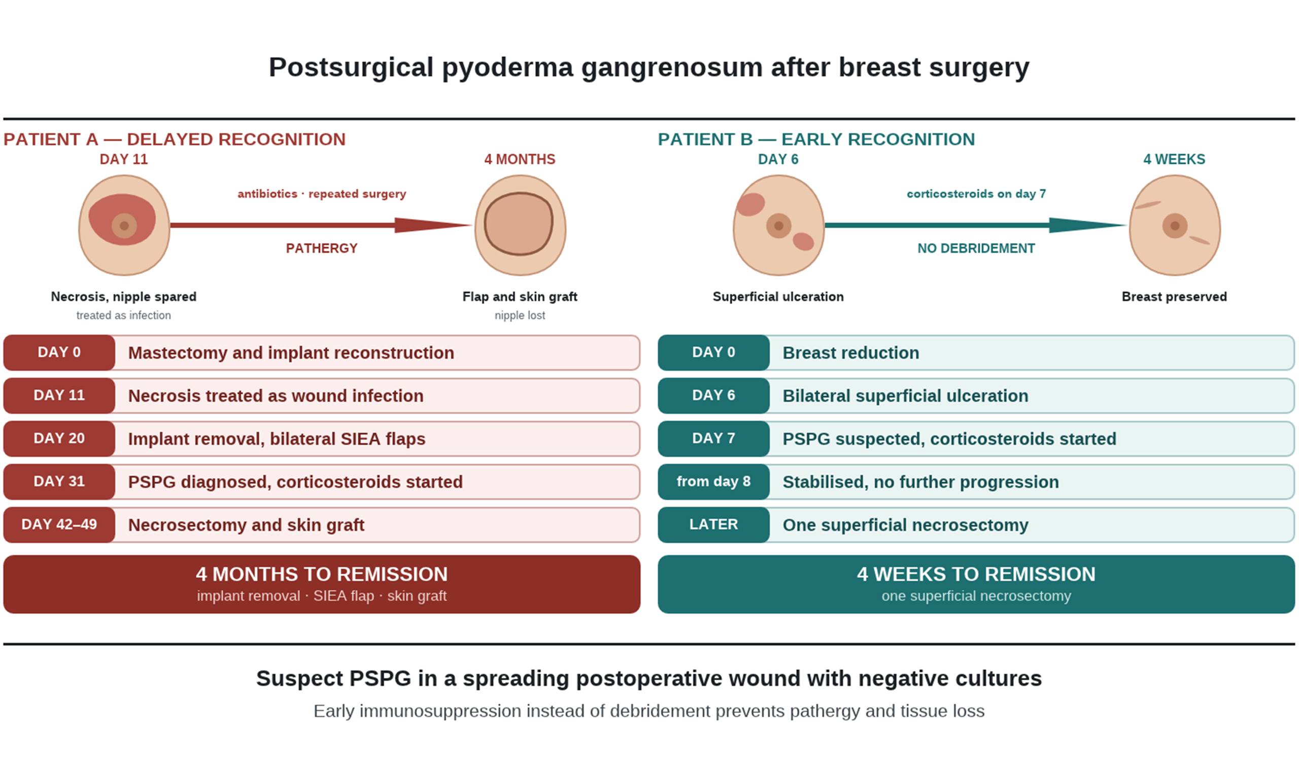
Graphical Abstract
Abstract
Postsurgical pyoderma gangrenosum (PSPG) is a rare, noninfectious neutrophilic dermatosis that can occur after breast surgery. Because it closely mimics a surgical site infection, the diagnosis is often delayed, and repeated surgical interventions may aggravate the disease through pathergy and result in extensive tissue loss and poor aesthetic outcomes. Therefore, early diagnosis is important. We report two cases of PSPG following breast surgery with contrasting clinical courses associated with delayed vs. early recognition. In Patient A, bilateral wound necrosis was misdiagnosed as an infection and treated accordingly, which led to multiple surgical interventions, including implant removal, flap reconstruction, and skin grafting. In Patient B, who presented with a similar picture, PSPG was suspected at presentation and systemic corticosteroid therapy was started one week after surgery, followed by rapid clinical improvement with the need for only one superficial necrosectomy after disease stabilization, which led to an acceptable aesthetic outcome. These cases show that atypical postoperative wound ulcerations or healing disturbances after breast surgery should raise suspicion of PSPG, particularly when cultures remain negative and wounds fail to respond to antibiotics. Increased awareness may allow earlier immunosuppressive treatment and help to avoid unnecessary surgical procedures.
Keywords


 Download PDF
INTRODUCTION
Pyoderma gangrenosum (PG) is a rare, noninfectious, neutrophilic dermatosis characterized by rapidly progressive, painful cutaneous ulcers with violaceous borders[1,2]. PG may occur idiopathically or in association with systemic diseases such as inflammatory bowel disease, rheumatologic disorders, or hematologic malignancies[1,3-5]. The incidence is estimated at approximately 3-10 cases per million people annually, primarily affecting adults between 40 and 60 years of age, with a slight female predominance[6-8]. Although PG most commonly affects the lower extremities, it may occur at any anatomical site[9].
The pathophysiology is not fully understood and is considered multifactorial, involving the dysregulation of the innate and adaptive immune system[8,10,11]. This leads to increased and abnormal neutrophil activation and migration, contributing to progressive tissue destruction and rapid peripheral extension of lesions. Minor trauma or a subsequent surgical procedure can exacerbate the exaggerated inflammatory response through the so-called pathergy phenomenon[10-12].
Postsurgical pyoderma gangrenosum has been reported to occur after breast surgery. It typically presents within the first postoperative week, sometimes with fever and leukocytosis. In a systematic review of 87 PSPG cases after aesthetic and reconstructive breast surgery, most cases occurred after breast reduction surgery (44%), and bilateral surgery was reported in 60% of cases[13].
Diagnosis is primarily clinical and based on exclusion of other causes. It remains challenging due to the absence of specific laboratory or pathognomonic findings and is frequently misdiagnosed as postoperative infection. Key features include disproportionate pain, sparing of the nipple-areola complex, negative cultures, and lack of response to antibiotic therapy[2,11,12,14-16]. Early recognition and prompt immunosuppressive treatment are essential to prevent disease progression, salvage tissue, and avoid devastating reconstructive outcomes[1,4,14].
In this case report, we present two cases of PSPG following breast surgery with different clinical outcomes, highlighting the importance of early diagnosis.
CASE PRESENTATION 1
Patient A is a 54-year-old woman with a medical history of hereditary breast and ovarian cancer syndrome, who presented at the plastic surgery department due to a significant breast asymmetry following breast conserving surgery and right-sided adjuvant radiotherapy in the late 1990s. Aside from the aforementioned comorbidities, Patient A had no other pre-existing conditions, no drug allergies and no regular medication. She is a nonsmoker.
Initially, she underwent a contralateral left-sided reduction mammoplasty, which healed without complications, followed six months later by a bilateral nipple-sparing mastectomy with immediate implant-based reconstruction using prepectoral anatomically shaped silicone implants (510 cc) [Figure 1A]. The latter represents the timepoint reference for the following case description.
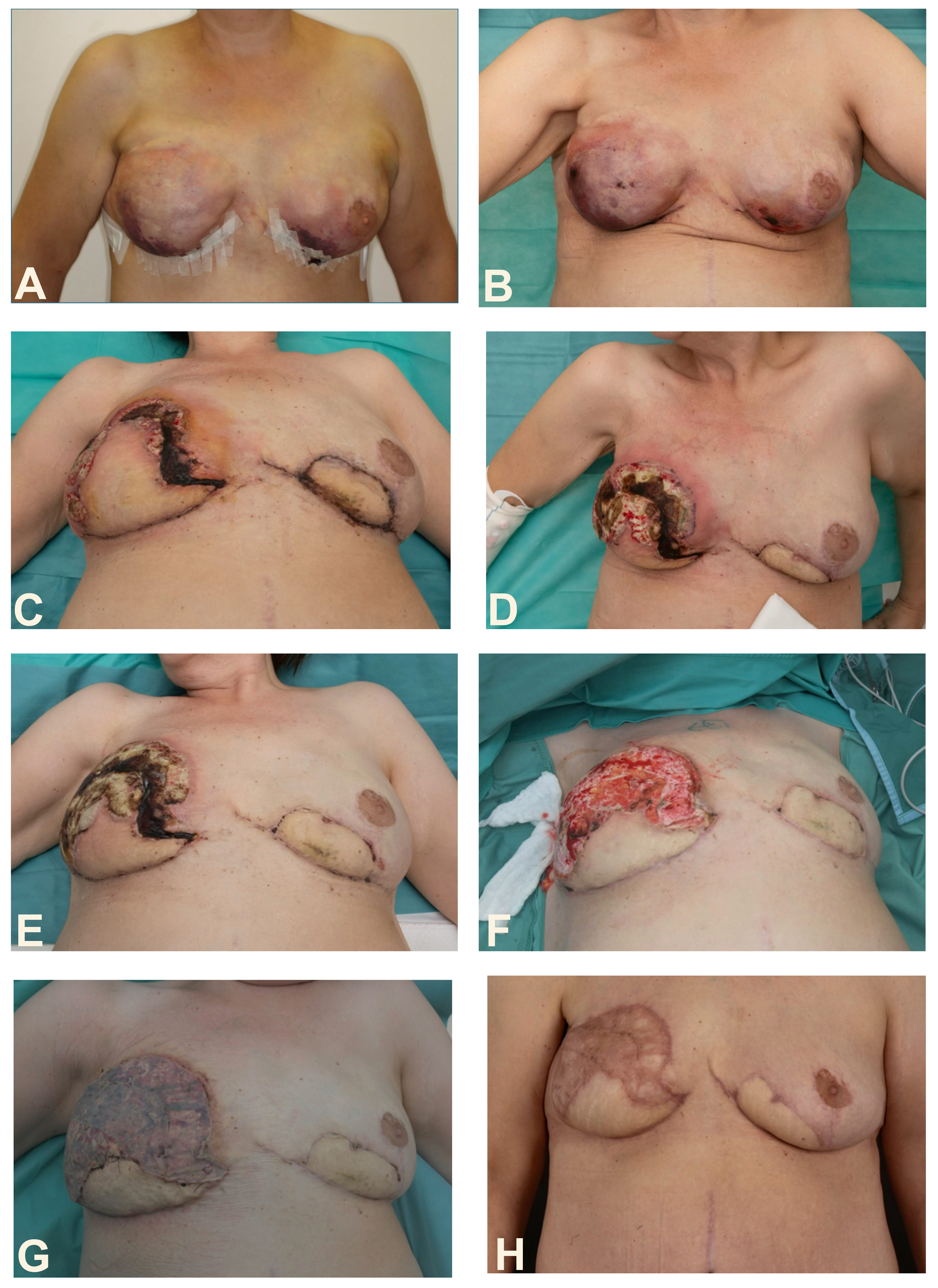
Figure 1. (A) Immediate postoperative appearance after implant-based breast reconstruction; (B) Postoperative wound necrosis at the inframammary fold on postoperative day 11 after implant-based breast reconstruction; (C) Clinical appearance 11 days after autologous breast reconstruction, showing postoperative necrosis, predominantly on the right side; (D) Clinical appearance on the day of diagnosis of PSPG and initiation of corticosteroid therapy; (E) Early clinical improvement two days after initiation of corticosteroid therapy; (F) Local wound appearance after necrosectomy of the right breast; (G) Final reconstructive procedure with split-thickness grafting; (H) Outcome after multiple surgical interventions following delayed diagnosis of PSPG. PSPG: Postsurgical pyoderma gangrenosum.
On postoperative day 11, Patient A presented with bilateral necrosis in the inframammary fold and along the vertical scar [Figure 1B]. A surgical site infection was suspected. Wounds were treated with povidone-iodine, and an empirical antibiotic therapy with amoxicillin/clavulanic acid (Augmentin® 1 g twice daily) was initiated.
Necrosis progressed bilaterally despite antibiotic treatment. By postoperative day 16, the right breast demonstrated a palm-sized necrosis reaching the nipple-areola complex, while the left showed a 5 cm × 3 cm lesion. There were no systemic signs of infection and wound swabs remained negative.
Due to the ongoing progression of bilateral skin necrosis, revision surgery with implant removal, a bilateral necrosectomy, and a reconstruction using bilateral superficial inferior epigastric artery (SIEA) flaps was performed on postoperative day 20. Inflammatory markers were only slightly elevated [C-reactive protein (CRP), 30.7 mg/L] at the time of surgery. Histopathological examination demonstrated extensive necrosis of the skin and subcutaneous tissue without evidence of malignancy.
Postoperatively, minor wound-healing disturbances with erythema at the flap margins occurred on the right breast. While the flaps remained clinically vital and perfused, a small wound-healing disturbance at the cranial flap margin, a small necrotic area medially, and a small blister at the upper outer pole with purulent discharge formed. Local wound care (antiseptic irrigation and iodine-based dressings), antibiotic therapy and close monitoring were continued. However, the wound margins continued to deteriorate despite treatment. Multiple wound swabs remained negative. Inflammatory markers stayed high, but the patient stayed afebrile without systemic signs of infection.
Eleven days after autologous breast reconstruction, given the atypical progression despite antibiotic therapy, absence of clinical signs of infection, and remaining negative cultures, PSPG was suspected [Figure 1C]. Dermatology was consulted, and punch biopsies were obtained. Four days later, histopathological examination showed a neutrophilic dermatosis. Therefore, systemic corticosteroid therapy with prednisolone 75 mg/day for six days was initiated, followed by 50 mg/day. Local wound care consisted of antiseptic compresses and topical corticosteroids. The ulcerations remained superficially extensive [Figure 1D]. Under systemic therapy, inflammatory markers gradually declined, and the violaceous borders diminished [Figure 1E].
An extensive laboratory workup was performed to exclude infectious, autoimmune, and hematological causes. They showed no significant results. Gastroenterological evaluation revealed no evidence of inflammatory bowel disease. Antibiotic treatment was switched to clindamycin 300 mg three times daily.
One week after initiation of systemic corticosteroid therapy, necrosectomy with wound edge revision, reduction of wound defect, excision of poorly perfused tissue, and open wound management were performed [Figure 1F]. The postoperative wound remained stable. Dermatological reassessment recommended a continuation of prednisolone 50 mg/day.
Seven days later, definitive wound management was performed, including debridement, unmeshed split-thickness skin grafting, and vacuum-assisted closure (VAC) therapy for three days [Figure 1G].
Postoperatively, the inflammatory activity had decreased, and the disease was considered clinically inactive under corticosteroid treatment. Given the patient’s oncologic history, tumor necrosis factor-alpha (TNF-α) was not considered, and instead, long-term dapsone was initiated. Prednisolone 50 mg/day was continued, followed by a tapering plan.
One week later, dermatology confirmed a clinically inactive PSPG under systemic corticosteroid therapy. The split-thickness skin graft was well integrated without tenderness, swelling, or any sign of infection. Prednisolone tapering was initiated, followed by stepwise weekly reductions. Dapsone 50 mg/day was continued as a steroid-sparing agent.
At the combined plastic surgical and dermatological follow-up four weeks after the final surgery, the graft remained viable with complete integration and no signs of recurrent inflammatory activity. Corticosteroids were further reduced.
At the following check-ups up to 20 weeks after diagnosis, PSPG remained inactive and showed no recurrence despite a slow tapering and subsequent discontinuation of corticosteroids and dapsone. The breast reconstruction remained stable, the skin graft was fully integrated, and both donor and recipient sites healed satisfactorily while inflammatory markers remained low [Figure 1H].
CASE PRESENTATION 2
Shortly after the management of the first case, a 48-year-old female patient (Patient B) presented to the department with bilateral wound healing disturbances one week after breast reduction surgery in a different clinic [Figure 2A]. At a postoperative follow-up visit with the treating surgeon on postoperative day six, bilateral superficial ulcerations with purple discoloration were noted, accompanied by chills, but without significant pain. Empirical antibiotic therapy with amoxicillin/clavulanic acid was initiated, and the chills resolved.
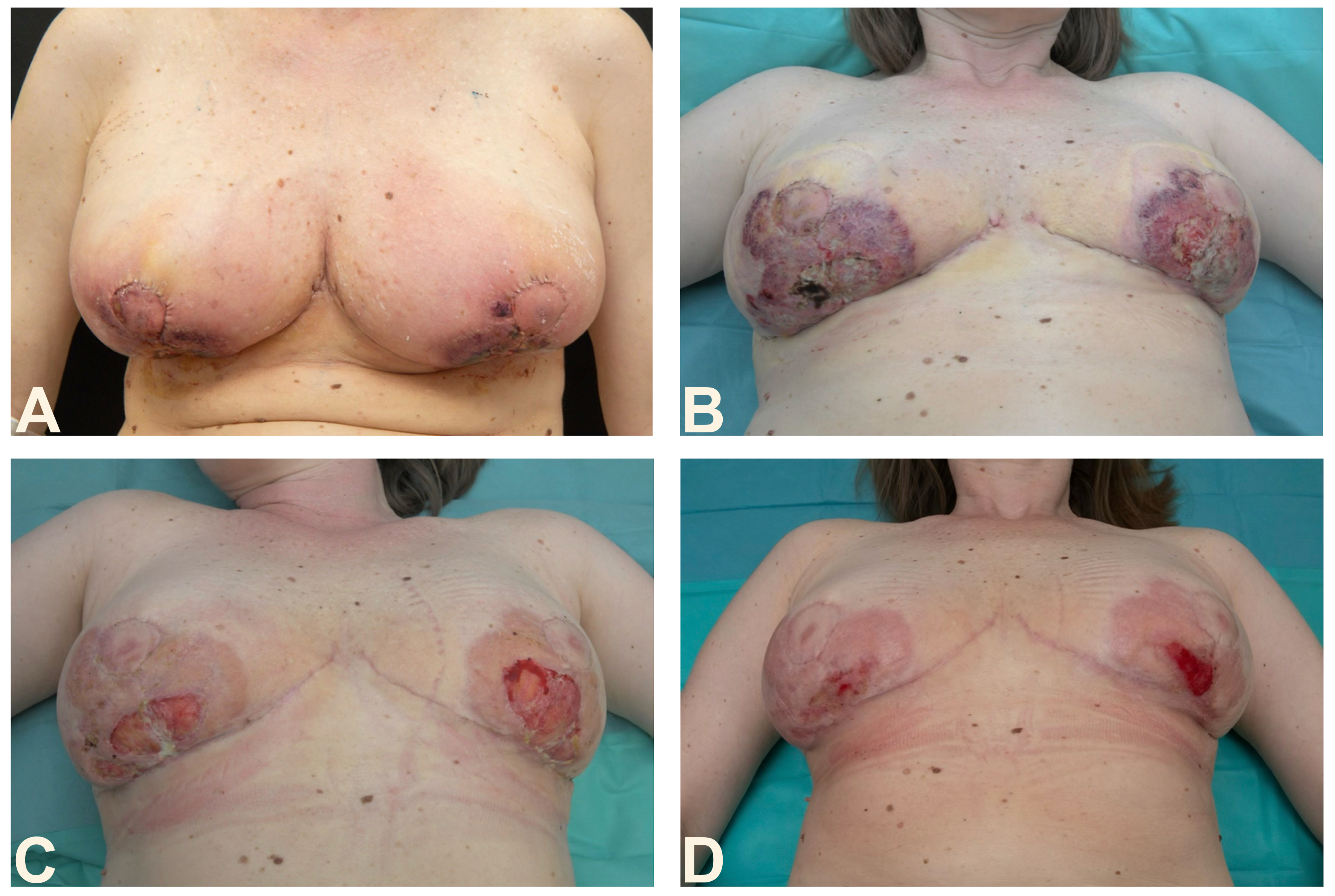
Figure 2. (A) One-week postoperative presentation showing bilateral superficial ulcerating lesions with livid discoloration; (B) Clinical presentation three days after initiation of corticosteroid therapy, demonstrating superficial fibrin-covered erosions without infiltration, undermined borders or further progression; (C) Clinical improvement after 17 days of corticosteroid therapy, combined with local wound care and regular HBO therapy; (D) Outcome after four weeks of treatment of PSPG. HBO: Hyperbaric oxygen; PSPG: postsurgical pyoderma gangrenosum.
The patient had a history of Hashimoto thyroiditis treated with levothyroxine. No other relevant comorbidities, prior surgeries, or medication allergies were reported. The patient was a nonsmoker.
On postoperative day seven, the patient presented at our department, where, given the previous experiences, an immediate dermatological consult was requested, and systemic therapy with corticosteroids (75 mg/day prednisolone) was initiated. Clinical examination showed bilateral superficial ulcerating lesions with livid discoloration and surrounding erythema in the inframammary region, accompanied by small pustules. The nipple-areola complex was spared bilaterally. Microbiological wound swabs were obtained and remained negative.
A lab work-up showed leukocyte counts within the normal range, with no elevation in procalcitonin and a slight elevation of CRP, which declined rapidly.
Within 24 h of treatment initiation, clinical improvement was observed, with a reduction in livid discoloration and ulcerative changes. The wounds appeared as superficial fibrin-covered erosions without infiltration or undermined borders, with no further progression on day 3 after corticosteroid initiation [Figure 2B]. Based on rapid response, the diagnosis of PSPG was supported.
Additionally, the patient received local wound care and systemic antibiotics for seven days. Furthermore, hyperbaric oxygen (HBO) therapy was initiated during the inpatient stay, after the commencement of systemic corticosteroid therapy, as part of the wound management strategy recommended by the consulting dermatologist and was subsequently continued on a daily outpatient basis.
Debridement was limited to a single superficial necrosectomy at outpatient follow-up after the PSPG had stabilized under systemic corticosteroid therapy. During treatment, the inflammatory markers declined rapidly. The wound remained stable without progression, so a tapering corticosteroid plan was established. Furthermore, local wound care and HBO therapy were continued throughout the healing period.
The patient was discharged with corticosteroid therapy, a tapering plan, regular follow-up appointments, antiseptic wound dressing changes every 2-3 days and with outpatient HBO daily [Figure 2C].
Over the following weeks, the wounds progressively healed, resulting in a pleasing aesthetic outcome [Figure 2D].
The second case of PSPG was diagnosed early, leading to timely initiation of systemic corticosteroid therapy. Consecutively, the outcome was favorable, emphasizing the importance of awareness of this condition despite its low incidence. Previous experience with PSPG may have contributed to the rapid initiation of treatment. The clinical courses of both patients are summarized and compared below [Table 1].
Comparison of the clinical course of PSPG in Patient A and Patient B
| Patient A | Patient B | |
| Surgery immediately preceding PSPG onset | Nipple-sparing mastectomy with implant-based reconstruction | Breast reduction surgery |
| Time from surgery to definitive diagnosis | Around 5 weeks | Around 1 week |
| Maximum leukocyte count (× 109/L) | 12.36 | Stayed within the normal range |
| Maximum CRP (mg/L) | 231.6 | Only slightly elevated and declined very quickly |
| Initial corticosteroid therapy regimen | Prednisolone - 75 mg/day for 6 days | Prednisolone - 75 mg/day for 4 days |
| Length of prednisolone intake | Around 17 weeks | Around 6 weeks |
| Additional treatments | Dapsone, topical corticosteroids, VAC therapy | HBO therapy, local wound care |
| Surgical interventions | 3 operations (implant removal with bilateral necrosectomy and flap reconstruction; necrosectomy; skin grafting) | 1 superficial necrosectomy during outpatient follow-up |
| Length of hospital stay | Three times for 5 days and one time for 8 days (total length of hospital stay was 23 days) | Three days |
| Hospital admissions | 4 | 1 |
| Time from triggering surgery to remission | Around 4 months | Around 4 weeks |
| Outcome | Stable reconstruction with integrated skin graft and no recurrence [Figure 1H] | Good aesthetic outcome without major tissue loss or revision surgery [Figure 2D] |
DISCUSSION
The two cases highlight the importance of early recognition and timely initiation of appropriate therapy in PSPG. In Patient A, the diagnosis was only made after repeated surgical interventions, possibly exacerbating disease progression through the pathergy phenomenon and contributing to substantial tissue loss requiring skin grafting. In contrast, early recognition in Patient B, due to experience gained from the first case, led to an early start of immunosuppressive therapy. Surgical treatment was limited to a single superficial necrosectomy after the disease had stabilized in this case, avoiding extensive debridement and reconstructive surgery.
PG is commonly associated with underlying conditions such as inflammatory bowel disease, rheumatologic disorders, and hematologic diseases in up to 50%-75% of cases[11]. In the present report, Patient A had no identifiable systemic disease, whereas Patient B had a history of Hashimoto thyroiditis. In Patient A, however, previous radiotherapy treatment to the right breast may have been a predisposing factor for the development of PSPG, as previous reports have shown involvement of previously irradiated breasts or chest wall tissue[17], which may also have contributed to the worse aesthetic outcome on the affected side.
Diagnosis remains challenging due to the absence of specific criteria and is primarily based on clinical presentation and exclusion of other causes. Histopathological findings are nonspecific but can be supportive: a dense neutrophilic inflammatory infiltrate beneath the ulcer border is characteristic of PG, as seen in the punch biopsies of Patient A, whereas an infectious cause must still be excluded clinically and microbiologically[5,11,12]. Though extremely rare, surgeons must be cognizant of it, as PSPG typically presents within the first postoperative week and can mimic surgical site infection. However, while surgical site infections rarely occur symmetrically, a hallmark of PSPG following breast surgery is the bilateral involvement with sparing of the nipple-areola complex[2,18]. The reason for sparing of the nipple-areola complex, also called the Bork-Baykal phenomenon, is not known, but a protective role of melanocytes and melanocyte-stimulating hormone has been proposed[16]. The sparing is not absolute: in Patient B the complex was spared, whereas in the previously irradiated breast of Patient A the necrosis reached it.
Notably, the two patients in this report presented with different clinical features, further demonstrating the variability of PSPG presentation. Patient A had previously undergone breast surgery within the same year without any complications. Six months later, following nipple-sparing mastectomy and implant reconstruction, lesions formed that even reached the right nipple-areola complex of the previously irradiated breast. Though it clinically resembled a wound infection, this might have been the beginning of PSPG. Only after autologous reconstruction, when bilateral lesions recurred, was PSPG suspected, and treatment initiated. It remains unclear why PSPG developed after the subsequent operation but not after the earlier surgery, underscoring that prior uneventful surgery does not preclude its later occurrence[16]. In Patient A, the procedure preceding PSPG involved previously irradiated tissue and implant placement, both of which may have altered the local inflammatory response and lowered the threshold for pathergy, although this remains speculative. Patient B, however, presented with more typical signs, including bilateral superficial ulcerations with livid discoloration and sparing of the nipple-areola complex, which, together with the increased awareness in our department, allowed early initiation of treatment. This variability can contribute to diagnostic uncertainty and delayed recognition.
The onset and presentation of PSPG can vary, ranging from slowly progressive lesions to rapidly developing ulcerations. Key features include disproportionate pain, negative microbiological cultures, violaceous wound borders, sparing of the nipple-areola complex, involvement of multiple sites, and rapid response to corticosteroid therapy. Deep tissue involvement is common, although deeper structures can remain unaffected. General symptoms such as fever, malaise, and myalgia can appear before the wound changes become visible. Owing to its clinical resemblance to wound infection, wound dehiscence, ischemia, and necrotizing fasciitis, it is frequently misdiagnosed[2,12,18,19]. Dermatology was consulted in both patients because the skin changes could not be definitively classified by the plastic surgical team alone. In Patient A, the diagnosis was established by punch biopsies showing a neutrophilic dermatosis, whereas in Patient B it was established clinically.
In contrast to surgical site infection, surgical intervention should be avoided where possible, due to the risk of pathergy[11,20,21]. First-line treatment consists of systemic corticosteroids, which typically result in rapid clinical improvement and stabilization of lesion progression[2,10,22]. In both patients, the corticosteroid dose and taper were guided by dermatology according to the clinical course rather than a fixed protocol. Additional immunosuppressive therapies may be considered in advanced cases, and reconstructive procedures may be required once disease activity is controlled. Implant removal is not recommended, as it may represent an additional traumatic trigger[13]. HBO therapy has been described as an adjunctive treatment in selected patients with PG, with proposed benefits related to improved tissue oxygenation and promotion of wound healing[23]. However, the available evidence is limited and HBO therapy is not considered an established standard treatment for PG[24]. In Patient B, HBO therapy was therefore used only as an adjunct to systemic corticosteroid therapy and local wound management. As these treatments were administered concurrently, the rapid clinical improvement cannot be attributed to corticosteroids, HBO therapy, or any other individual intervention alone.
Similar symptoms and clinical characteristics including rapid wound progression and frequent initial misdiagnosis have been described in numerous case reports and case series of PSPG after breast surgery highlighting the importance of early diagnosis[15,19,20,25-34]. In contrast, the present report demonstrates how increased clinical awareness may facilitate earlier diagnosis and targeted management, as demonstrated in Patient B, who achieved a favorable aesthetic outcome without flap reconstruction or skin grafting.
This case report should be interpreted within the limitations inherent to a retrospective case-based study, including a small sample size, its retrospective nature, and differences in the primary surgical procedures performed. Also, the treatment for PSPG differed between both patients, limiting direct comparability between these two cases and a causal relationship between diagnostic timing and outcome cannot be established. Nevertheless, the contrasting clinical courses provide a clinically relevant illustration of PSPG after breast surgery and support the importance of early recognition and timely interdisciplinary management.
CONCLUSION
PG remains a diagnostic challenge due to the absence of specific criteria and its variable clinical presentation. Postoperative infections are more common than PSPG. PSPG should be considered in atypical postoperative wounds with incongruent clinical findings. These two cases illustrate contrasting courses associated with delayed vs. early recognition of PSPG. Early recognition and initiation of immunosuppressive therapy may prevent pathergy-related damage and avoid extensive and repeated surgical procedures, thereby improving clinical and aesthetic outcomes.
DECLARATIONS
Acknowledgments
The authors thank the patients for consenting to the publication of their cases and the surgical and dermatological teams involved in their care. The Graphical Abstract was created by the authors in Canva using only basic drawing tools - plain geometric shapes, lines, and text boxes. No stock images, photographs, icons, illustrations, templates, or other library elements from Canva or any other source were used. All graphical elements, including the schematic breast illustrations, were drawn by the authors specifically for this manuscript.
Authors’ contributions
Conceptualised the study: Weixler J, Hutter MF, Nischwitz SP, Hubmer M
Contributed to the study design: Weixler J, Hutter MF, Nischwitz SP, Hubmer M
Performed data collection: Weixler J, Hutter MF
Performed data analysis and interpretation: Weixler J, Hutter MF
Performed the literature search: Weixler J
Led the drafting of the manuscript: Weixler J
Critically revised the manuscript for important intellectual content: Hutter MF, Smolle C, Hubmer M, Nischwitz SP, Horn M, Kamolz LP
Supervised the study: Kamolz LP
Availability of data and materials
Not applicable.
AI and AI-assisted tools statement
Not applicable.
Financial support and sponsorship
None.
Conflicts of interest
All authors declared that there are no conflicts of interest.
Ethical approval and consent to participate
Ethical approval was obtained from the Ethics Committee of the Medical University of Graz (approval number: 1272/2026). Written informed consent to participate was obtained from both patients.
Consent for publication
Written informed consent for the publication of their clinical details and clinical photographs was obtained from both patients.
Copyright
© The Author(s) 2026.
REFERENCES
1. Maronese CA, Pimentel MA, Li MM, Genovese G, Ortega-Loayza AG, Marzano AV. Pyoderma gangrenosum: an updated literature review on established and emerging pharmacological treatments. Am J Clin Dermatol. 2022;23:615-34.
2. Guliyeva G, Janis JE. Postsurgical pyoderma gangrenosum requiring plastic surgical intervention: a practical review. Plast Reconstr Surg Glob Open. 2024;12:e5505.
3. Marzano AV, Borghi A, Meroni PL, Cugno M. Pyoderma gangrenosum and its syndromic forms: evidence for a link with autoinflammation. Br J Dermatol. 2016;175:882-91.
4. Miller J, Yentzer BA, Clark A, Jorizzo JL, Feldman SR. Pyoderma gangrenosum: a review and update on new therapies. J Am Acad Dermatol. 2010;62:646-54.
5. Zaino ML, Schadt CR, Callen JP, Owen LG. Pyoderma gangrenosum: diagnostic criteria, subtypes, systemic associations, and workup. Dermatol Clin. 2024;42:157-70.
6. Langan SM, Groves RW, Card TR, Gulliford MC. Incidence, mortality, and disease associations of pyoderma gangrenosum in the United Kingdom: a retrospective cohort study. J Invest Dermatol. 2012;132:2166-70.
7. Ruocco E, Sangiuliano S, Gravina AG, Miranda A, Nicoletti G. Pyoderma gangrenosum: an updated review. J Eur Acad Dermatol Venereol. 2009;23:1008-17.
8. Park AN, Raj A, Bajda J, Gorantla VR. Narrative review: pyoderma gangrenosum. Cureus. 2024;16:e51805.
9. Ahronowitz I, Harp J, Shinkai K. Etiology and management of pyoderma gangrenosum: a comprehensive review. Am J Clin Dermatol. 2012;13:191-211.
10. Ahn C, Negus D, Huang W. Pyoderma gangrenosum: a review of pathogenesis and treatment. Expert Rev Clin Immunol. 2018;14:225-33.
11. Moltrasio C, Romagnuolo M, Tavoletti G, Maronese CA, Marzano AV. Pyoderma gangrenosum: pathogenetic mechanisms and their implications for treatment. Semin Immunopathol. 2025;47:38.
12. Zuo KJ, Fung E, Tredget EE, Lin AN. A systematic review of post-surgical pyoderma gangrenosum: identification of risk factors and proposed management strategy. J Plast Reconstr Aesthet Surg. 2015;68:295-303.
13. Ehrl DC, Heidekrueger PI, Broer PN. Pyoderma gangrenosum after breast surgery: a systematic review. J Plast Reconstr Aesthet Surg. 2018;71:1023-32.
14. Tuffaha SH, Sarhane KA, Mundinger GS, et al. Pyoderma gangrenosum after breast surgery: diagnostic pearls and treatment recommendations based on a systematic literature review. Ann Plast Surg. 2016;77:e39-44.
15. Mella JR, Maselli AM, Guo L. A deceptive diagnosis: pyoderma gangrenosum after breast surgery-a case series and literature review. Ann Plast Surg. 2019;83:S21-30.
16. Caddia G, Voulliaume D, Dettori L, Rubino C, Delay E. Postsurgical pyoderma gangrenosum in breast surgery: an updated systematic review, takeaways, and the 6 commandments. Aesthet Surg J. 2025;45:NP142-53.
17. Hammond JB, Pflibsen LR, Kruger EA, et al. Pyoderma gangrenosum confined to the irradiated chest wall of the reconstructed breast. Clin Case Rep. 2021;9:445-9.
18. Dahy AA, Hassan RM, Altramsy A, et al. Outcomes and risk factors of pyoderma gangrenosum after reduction mammoplasty: a systematic review of the literature and case report. Aesthetic Plast Surg. 2025;49:5480-97.
19. Li WY, Andersen JC, Jung J, Andersen JS. Pyoderma gangrenosum after abdominal free tissue transfer for breast reconstruction: case series and management guidelines. Ann Plast Surg. 2019;83:63-8.
20. Edinger KM, Rao VK. The management of postsurgical pyoderma gangrenosum following breast surgery. Plast Reconstr Surg Glob Open. 2022;10:e4282.
21. Tolkachjov SN, Fahy AS, Wetter DA, et al. Postoperative pyoderma gangrenosum (PG): the Mayo Clinic experience of 20 years from 1994 through 2014. J Am Acad Dermatol. 2015;73:615-22.
22. Patel F, Fitzmaurice S, Duong C, et al. Effective strategies for the management of pyoderma gangrenosum: a comprehensive review. Acta Derm Venereol. 2015;95:525-31.
23. Vieira WA, Barbosa LR, Martin LM. Hyperbaric oxygen therapy as an adjuvant treatment for pyoderma gangrenosum. An Bras Dermatol. 2011;86:1193-6.
24. Kaur M, Diaz MJ, Anthony M, et al. Treatments for pyoderma gangrenosum: a systematic review and single-arm meta-analysis of systemic therapies. Int Wound J. 2025;22:e70733.
25. Grillo MA, Cavalheiro TT, da Silva Mulazani M, Rocha JL, Semchechen D, da Cunha CA. Postsurgical pyoderma gangrenosum complicating reduction mammaplasty. Aesthetic Plast Surg. 2012;36:1347-52.
26. Pop IC, Ilies RA, Baican C, Strilciuc S, Muntean V, Muntean M. Pyoderma gangrenosum post-breast surgery: a case report and comprehensive review of management strategies. J Clin Med. 2024;13:3800.
27. Ouazzani A, Berthe JV, de Fontaine S. Post-surgical pyoderma gangrenosum: a clinical entity. Acta Chir Belg. 2007;107:424-8.
28. Hammond D, Chaudhry A, Anderson D, Alameddine KO, Tajran J. Postsurgical pyoderma gangrenosum after breast surgery: a plea for early suspicion, diagnosis, and treatment. Aesthetic Plast Surg. 2020;44:2032-40.
29. Duval A, Boissel N, Servant JM, Santini C, Petit A, Vignon-Pennamen MD. Pyoderma gangrenosum of the breast: a diagnosis not to be missed. J Plast Reconstr Aesthet Surg. 2011;64:e17-20.
30. Costa J, Monteiro D, Valença-Filipe R, Reis J, Silva Á. Pyoderma gangrenosum after breast reduction: a rare complication. J Plast Reconstr Aesthet Surg. 2013;66:e336-7.
31. Rajapakse Y, Bunker CB, Ghattaura A, et al. Case report: pyoderma gangrenosum following Deep Inferior Epigastric Perforator (Diep) free flap breast reconstruction. J Plast Reconstr Aesthet Surg. 2010;63:e395-6.
32. Diaz R, Allievi R, Cuniolo L, et al. Post-surgical pyoderma gangrenosum after breast cancer surgery: a multidisciplinary case report. Curr Oncol. 2025;32:701.
33. Rogers CC, Nepper J, Holzem KE, Cortina CS. Postsurgical pyoderma gangrenosum after mastectomy with a familial component. J Surg Case Rep. 2024;2024:rjae667.
Cite This Article
 Open Access
How to Cite
Weixler J, Hutter MF, Hubmer M, Nischwitz SP, Smolle C, Horn M, Kamolz LP. Postsurgical pyoderma gangrenosum following breast surgery: a case report of two patients with delayed versus early diagnosis. Plast Aesthet Res. 2026;13:30. https://dx.doi.org/10.20517/2347-9264.2026.51
Download Citation
If you have the appropriate software installed, you can download article citation data to the citation manager of your choice. Simply select your manager software from the list below and click on download.
Export Citation File
Type of Import
Tips on Downloading Citation
Citation Manager File Format
Type of Import
Direct Import: When the Direct Import option is selected (the default state), a dialogue box will give you the option to Save or Open the downloaded citation data. Choosing Open will either launch your citation manager or give you a choice of applications with which to use the metadata. The Save option saves the file locally for later use.
Indirect Import: When the Indirect Import option is selected, the metadata is displayed and may be copied and pasted as needed.
About This Article
Special Topic
Copyright

Data & Comments
Data
0










Comments
Comments must be written in English. Spam, offensive content, impersonation, and private information will not be permitted. If any comment is reported and identified as inappropriate content by OAE staff, the comment will be removed without notice. If you have any queries or need any help, please contact us at [email protected].